Radiology Spotters Collection

Radiology Signs

The Radiology signs collection on RadioGyan is a high‑yield, searchable library of classic and contemporary imaging signs designed for rapid pattern recognition, streamlined reporting, and exam‑ready learning across modalities and subspecialties. Built for residents, fellows, and attendings, it links hallmark appearances to practical differentials, pitfalls, and clinical relevance to accelerate diagnosis in daily practice. Some of the cases have images while some don’t. I am updating these. Feel free to contribute images. The are arranged in the order they are posted. You can also check out the alphabetical list of radiological signs below.
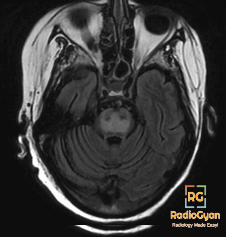
Axial FLAIR MRI showing hyperintensity in the central pontine region, indicative of osmotic demyelination syndrome.

Trident Sign (Pons) | Radiology Signs

Hurricane sign | Radiology Signs

Stocking-glove sign | Radiology Signs

Six-point star sign | Radiology Signs

Page kidney | Radiology Signs

Soap-bubble nephrogram | Radiology Signs

Flower vase ureter | Radiology Signs

Spinning top urethra | Radiology Signs

Christmas tree bladder | Radiology Signs

Northern exposure sign | Radiology Signs

Ram’s horn sign | Radiology Signs

Fat Halo Sign (Bowel) | Radiology Signs

Coiled spring sign | Radiology Signs

Chain of lakes sign | Radiology Signs

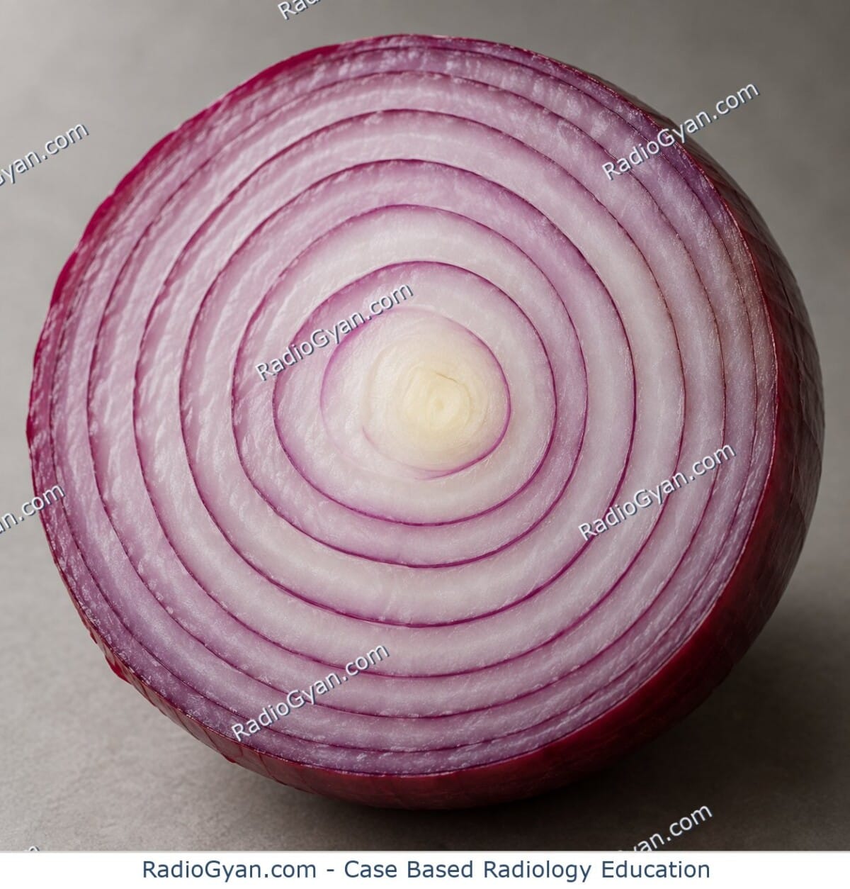
Onion Skin Sign (Testicular Epidermoid Cyst) | Radiology Signs

Wheel within a wheel | Radiology Signs

Bat sign | Radiology Signs

Quad sign | Radiology Signs

Curtain Sign (Lung Ultrasound) | Radiology Signs

Cogwheel sign | Radiology Signs

Intradecidual sign | Radiology Signs

Cortical Rim Sign (Kidney) | Radiology Signs

Sonographic Murphy sign | Radiology Signs

Seashore sign | Radiology Signs

Lung point sign | Radiology Signs

Double decidual sac sign | Radiology Signs

Twin-peak sign | Radiology Signs

Ring of fire sign | Radiology Signs

Starry sky liver | Radiology Signs

WES sign | Radiology Signs

Secondary cleft sign | Radiology Signs

Serpiginous tract sign | Radiology Signs

Asterisk sign | Radiology Signs

Double spinous process sign | Radiology Signs

Fat C2 sign | Radiology Signs

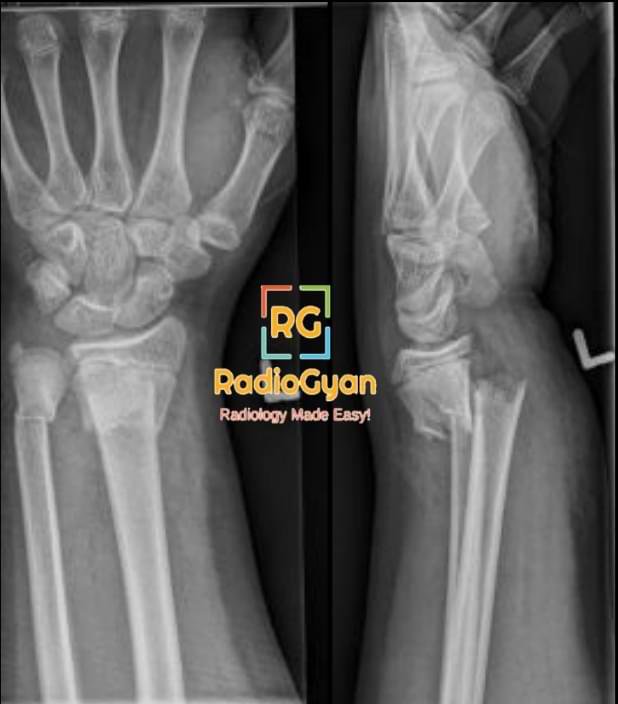
Anteroposterior and lateral radiographs of the distal left radius demonstrate a transverse fracture through the distal metaphysis with dorsal angulation and impaction in keeping with a Colle fracture.

Dinner Fork (Silver Fork) Deformity | Radiology Signs

Kissing contusion | Radiology Signs

Split fat sign | Radiology Signs

Rim Sign (Hip and Shoulder) | Radiology Signs

Crossover sign | Radiology Signs

Empty notch sign | Radiology Signs

Flipped meniscus sign | Radiology Signs

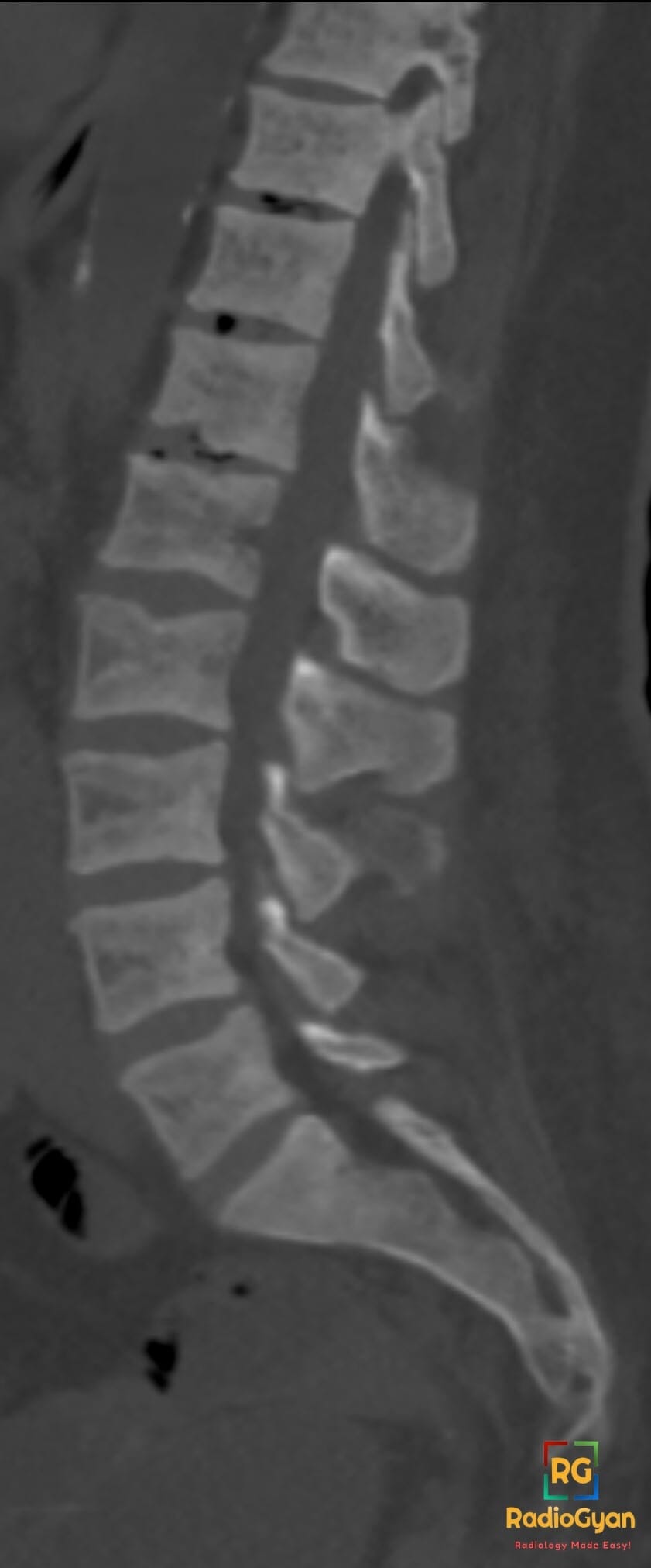
Intravertebral vacuum cleft | Radiology Signs

Penumbra sign | Radiology Signs

Sagging rope sign | Radiology Signs

Arcuate sign | Radiology Signs

Andersson lesion | Radiology Signs

Shiny corner sign | Radiology Signs

Cyclops lesion | Radiology Signs

Segond fracture | Radiology Signs

Melting ice cube sign | Radiology Signs

Interface sign | Radiology Signs

Ginkgo leaf sign | Radiology Signs

Dark bronchus sign | Radiology Signs

Positive bronchus sign | Radiology Signs

Oreo cookie sign | Radiology Signs

Monod sign | Radiology Signs

Polo mint sign | Radiology Signs

Fleischner sign | Radiology Signs

Feeding vessel sign | Radiology Signs

Cheerio sign | Radiology Signs

Halo Sign (Lung) | Radiology Signs

Harlequin appearance | Radiology Signs

Tau sign | Radiology Signs

Tigroid pattern | Radiology Signs

Trident Sign (Spinal Cord) | Radiology Signs

Hockey stick sign | Radiology Signs

Pulvinar sign | Radiology Signs

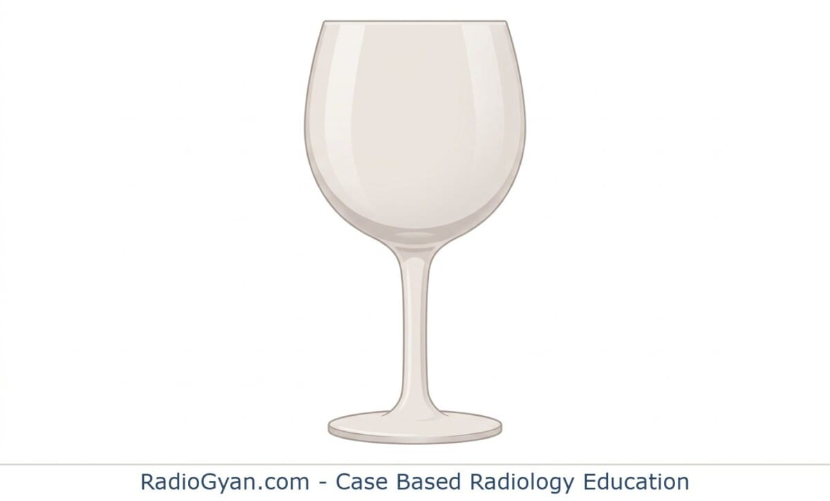
Wine glass sign | Radiology Signs

Pancake brain | Radiology Signs

Reversal sign | Radiology Signs

White cerebellum sign | Radiology Signs

Ivy sign | Radiology Signs

Ice-cream cone sign | Radiology Signs

Medusa head sign | Radiology Signs

Owl eye sign | Radiology Signs

Face of the giant panda | Radiology Signs

Hummingbird sign | Radiology Signs

Spot sign | Radiology Signs

Swirl Sign (Brain) | Radiology Signs

Dural tail sign | Radiology Signs

Cord sign | Radiology Signs

Empty delta sign | Radiology Signs

Flame-shaped breast | Radiology Signs

Rim Sign (Hepatobiliary Scintigraphy) | Radiology Signs

Pedestal sign | Radiology Signs

Stipple sign | Radiology Signs

Pseudo-Rigler’s sign | Radiology Signs

Tooth Sign (Patella) | Radiology Signs

Inside-out liver sign | Radiology Signs

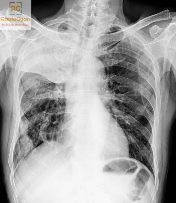
Pie-in-the-sky sign | Radiology Signs

Rolled edge sign | Radiology Signs

Pencil pointing | Radiology Signs

Fishmouthing | Radiology Signs

Champagne glass pelvis | Radiology Signs

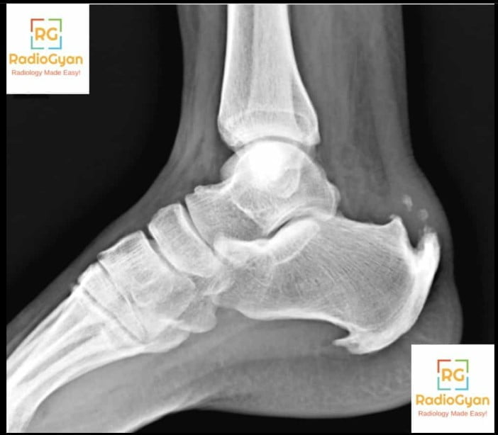
Heel Pad Sign (Calcaneus) | Radiology Signs

Kissing ulcers | Radiology Signs

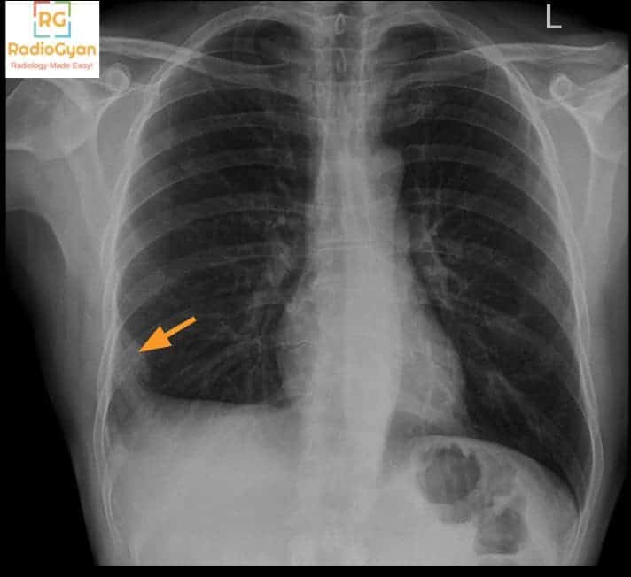
Fallen lung sign | Radiology Signs

Bite sign | Radiology Signs

Crescent Sign (Femoral Head) | Radiology Signs

Lambda sign | Radiology Signs

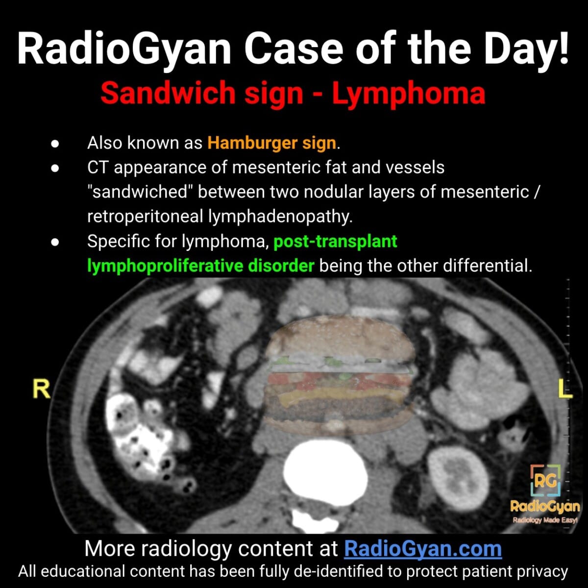
Sandwich Sign (Mesentery) | Radiology Signs

Soap-bubbly appearance | Radiology Signs

Swiss cheese appearance | Radiology Signs

Puff of smoke sign | Radiology Signs

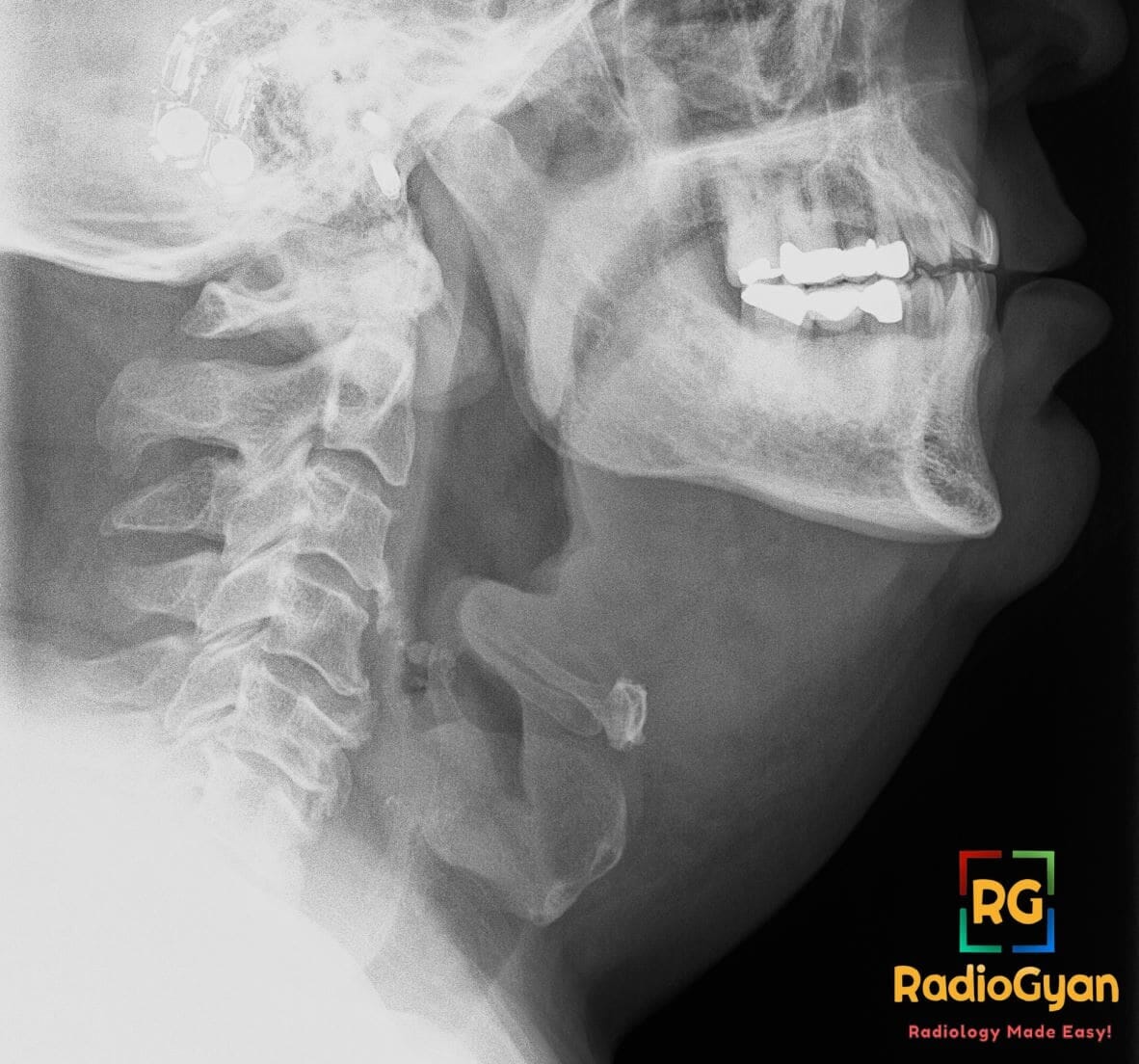
Lateral neck radiograph showing a swollen epiglottis with the classic thumb sign of epiglottitis

Thumb sign | Radiology Signs

Teacup Sign (Breast) | Radiology Signs

Sack of marbles sign | Radiology Signs

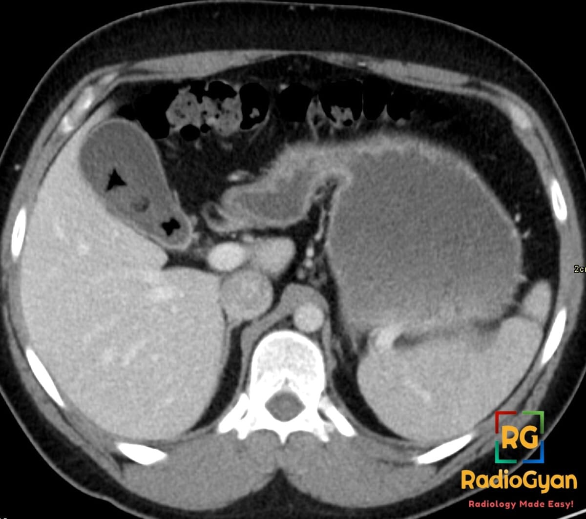
Axial CT showing a gallstone within the gallbladder with a triradiate Mercedes-Benz sign from gas-filled fissures

Mercedes-Benz sign | Radiology Signs

Y Sign (Spine) | Radiology Signs

Winking owl sign | Radiology Signs

Molar tooth sign | Radiology Signs

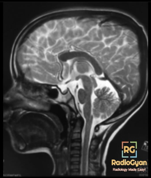
J – Shaped Sella Radiology Case.

J-shaped sella | Radiology Signs

Eye-of-the-tiger sign | Radiology Signs

Blend sign | Radiology Signs

Banana sign | Radiology Signs

Wimberger Sign (Congenital Syphilis) | Radiology Signs

Wimberger Ring Sign (Scurvy) | Radiology Signs

Trolley track sign | Radiology Signs

Shepherd’s crook deformity | Radiology Signs

Scottie dog | Radiology Signs

Sausage digit (dactylitis) | Radiology Signs

Rugger jersey spine | Radiology Signs

Ribbon ribs | Radiology Signs

Rachitic rosary | Radiology Signs

Nutcracker fracture | Radiology Signs

Light bulb sign | Radiology Signs

Ivory phalanx | Radiology Signs

Hill-Sachs deformity | Radiology Signs

Hair-on-end | Radiology Signs

H-shaped vertebrae | Radiology Signs

Ghost Sign (Foot) | Radiology Signs

Fragment-in-notch sign | Radiology Signs

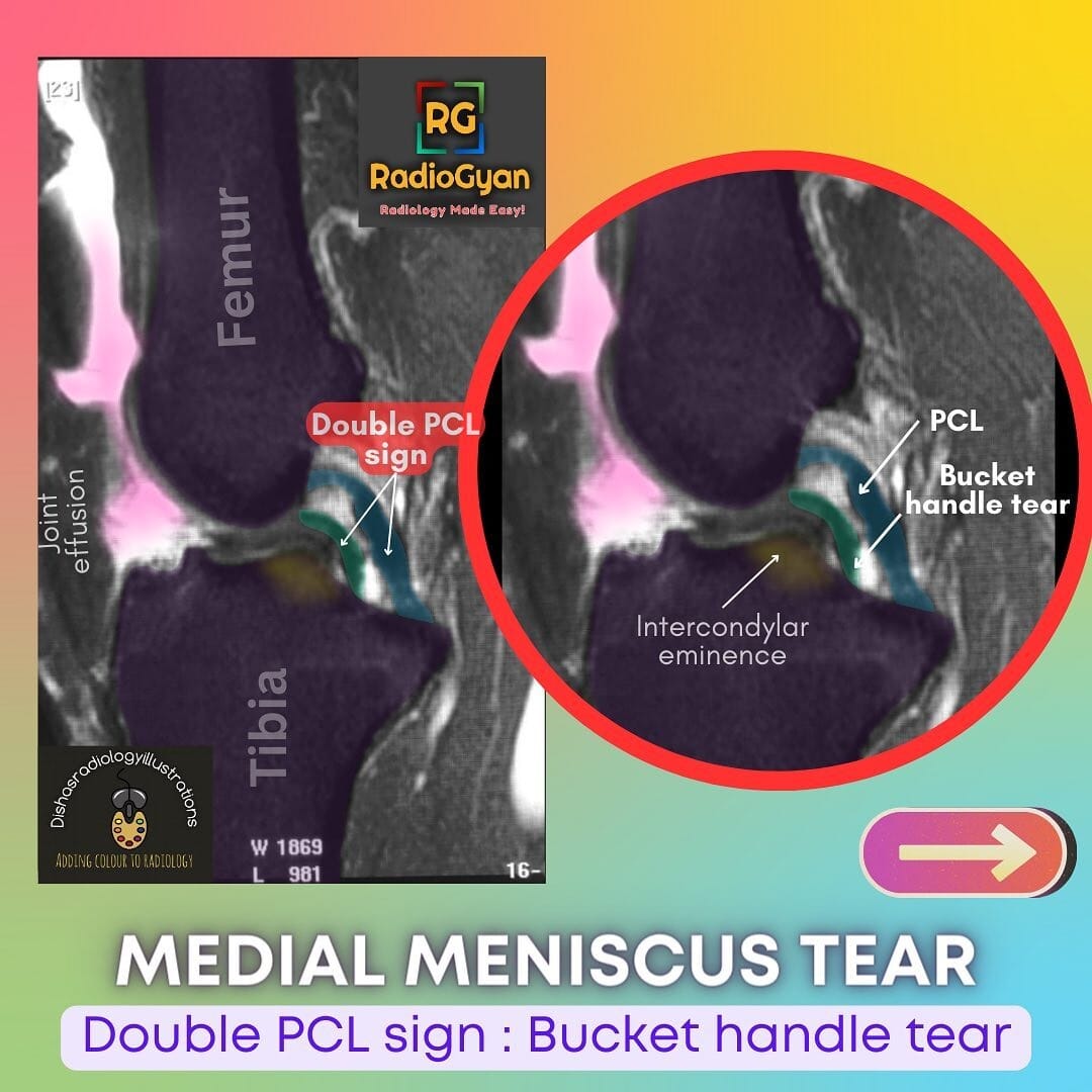
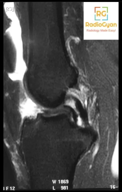
Sagittal MRI image of the knee demonstrates a bucket handle tear of the medial meniscus with the “double PCL sign”. The image is annotated to show the femur, tibia, joint effusion, intercondylar eminence, PCL, and bucket handle tear. A red circle surrounds the bucket handle tear.

Double PCL Sign (Medial Meniscus) | Radiology Signs

Dagger sign | Radiology Signs

Crowded carpal sign | Radiology Signs

Coned epiphyses | Radiology Signs

Boutonnière deformity | Radiology Signs

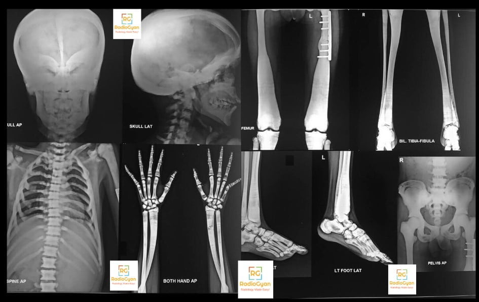
Osteopetrosis Radiology Case

Bone within a bone | Radiology Signs

Blade of grass sign | Radiology Signs

Beveled edge sign | Radiology Signs

Banana fracture | Radiology Signs

Vanishing tumour | Radiology Signs

Spinnaker sail sign | Radiology Signs

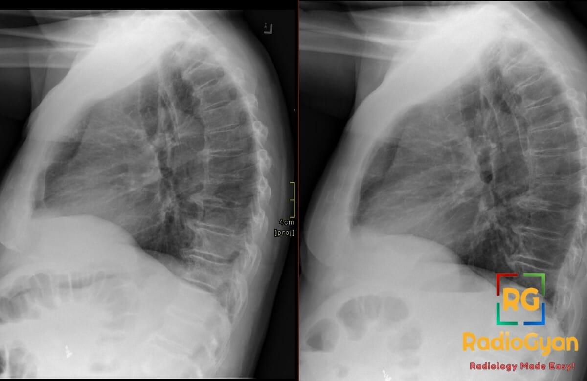
Paired lateral chest X-rays showing increased lower lobe density, lost spine lucency, consistent with pneumonia

Spine sign | Radiology Signs

Signet Ring Sign (Lung) | Radiology Signs

Knuckle sign | Radiology Signs

Spoke wheel appearance | Radiology Signs

Spaghetti sign | Radiology Signs

Pancake kidney | Radiology Signs

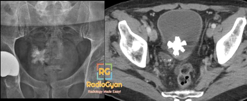
Jackstone Urinary bladder calculus on CT

Jackstone calculus | Radiology Signs

Fishhook ureter | Radiology Signs

Elephant on a flagpole | Radiology Signs

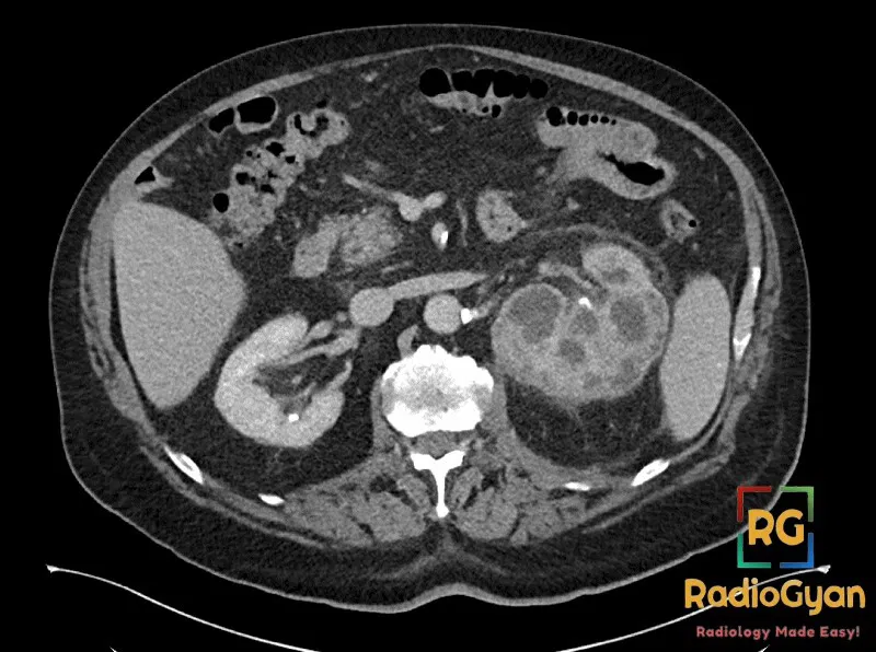
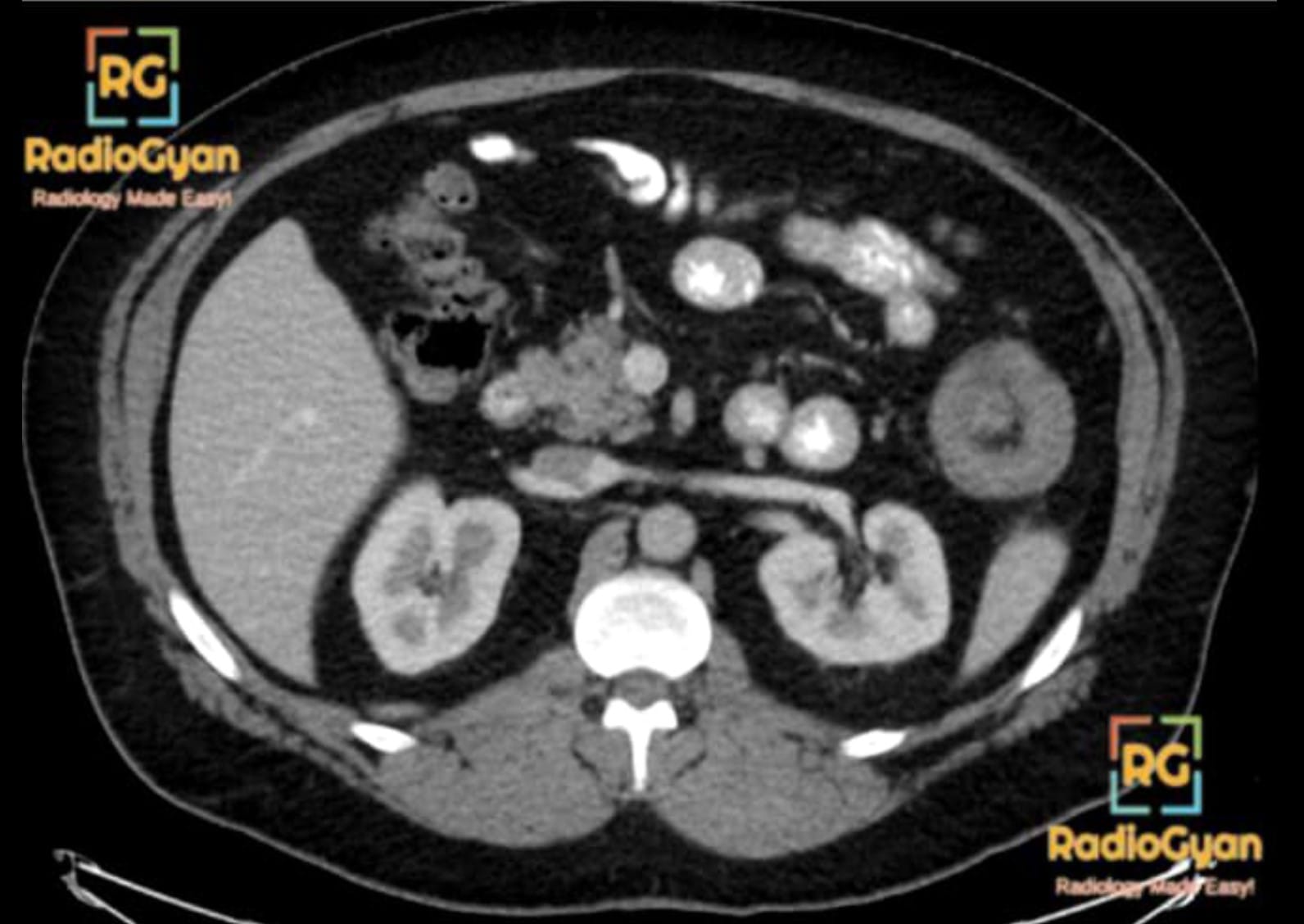
Axial CT scan of the abdomen showing a left kidney with xanthogranulomatous pyelonephritis, characteristic 'bear paw' sign.

Bear Paw Sign (Xanthogranulomatous Pyelonephritis) | Radiology Signs

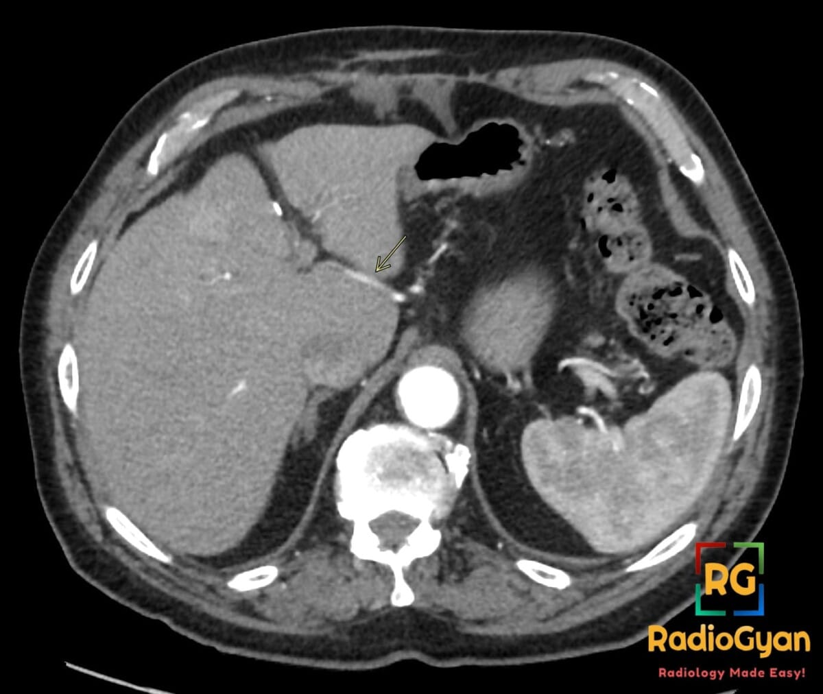
Axial contrast-enhanced CT showing a replaced left hepatic artery coursing through the fissure for ligamentum venosum

Vessel through Strait sign | Radiology Signs

Tumbling bullet | Radiology Signs

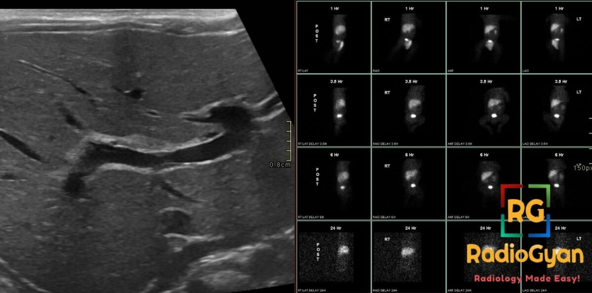
Ultrasound showing triangular cord sign and hepatobiliary scan indicating lack of hepatic excretion, diagnostic for biliary atresia.

Triangular cord sign | Radiology Signs

Sentinel loop sign | Radiology Signs

Sawtooth sign | Radiology Signs

Rat tail sign | Radiology Signs

Gut signature sign | Radiology Signs

Double track sign | Radiology Signs

Double bubble sign | Radiology Signs

Cupola sign | Radiology Signs

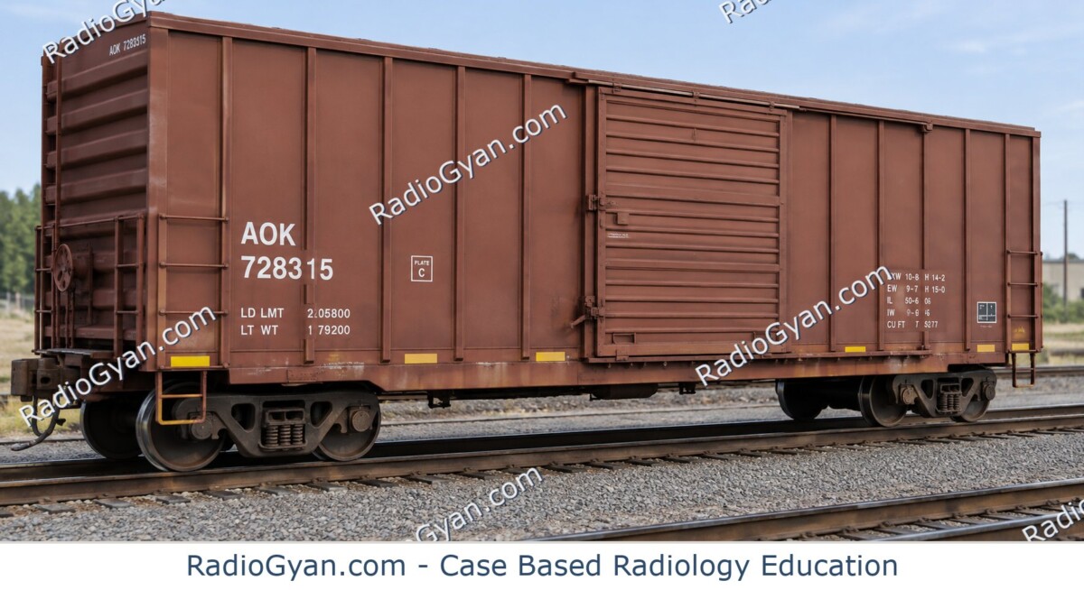
Cottage loaf sign | Radiology Signs

Corkscrew oesophagus | Radiology Signs

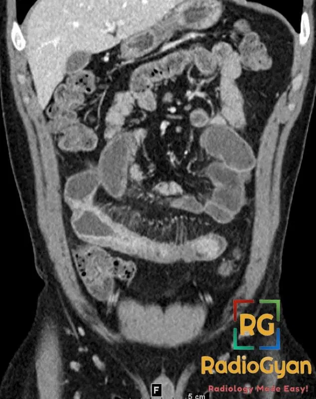
Coronal CT image of the abdomen showing the 'comb sign' indicative of Crohn's disease in the small bowel.

Comb sign | Radiology Signs

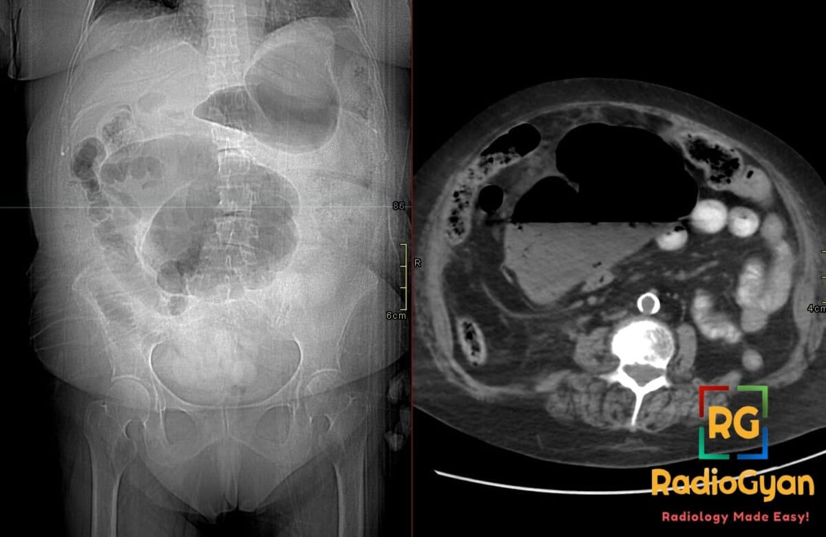
Abdominal radiograph and axial CT showing cecal volvulus with dilated displaced cecum in left upper quadrant and whirl sign.

Coffee-bean sign | Radiology Signs

Cobblestone appearance | Radiology Signs

Central dot sign | Radiology Signs

Antral nipple sign | Radiology Signs

Accordion sign | Radiology Signs

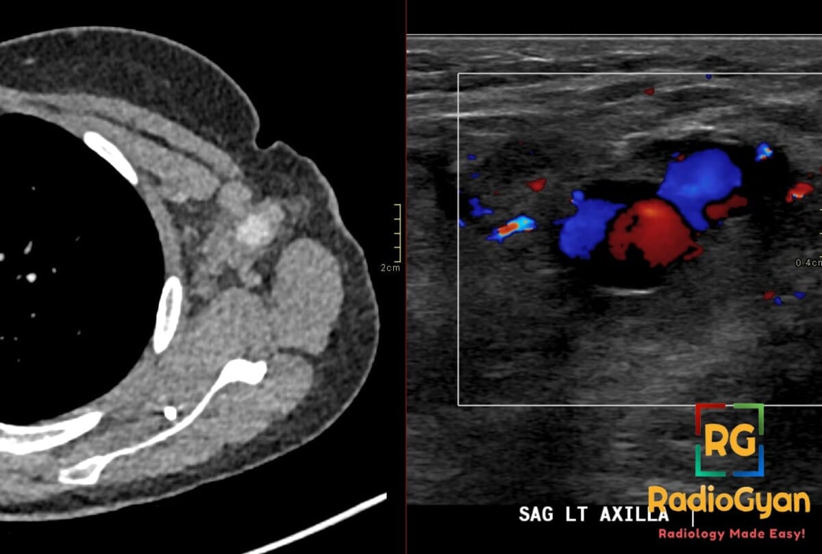
Axial CT and sagittal Doppler ultrasound show a left axillary nodal pseudoaneurysm with central enhancement, yin-yang flow

Yin-yang sign | Radiology Signs

Tulip bulb sign | Radiology Signs

Hairpin turn sign | Radiology Signs

Floating viscera sign | Radiology Signs

Figure 3 sign | Radiology Signs

Egg-on-a-string sign | Radiology Signs

Draped aorta sign | Radiology Signs

Cobweb sign | Radiology Signs

Aortic nipple | Radiology Signs

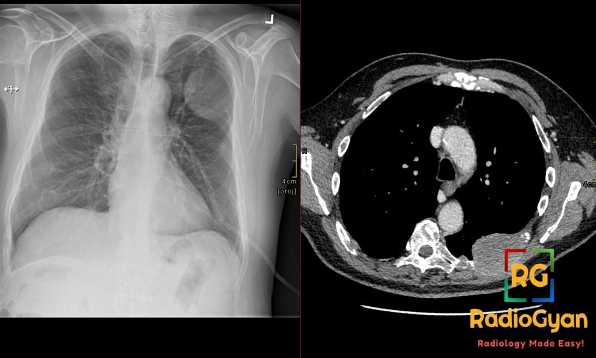
Frontal chest radiograph and axial CT show an expansile left upper rib lesion with the incomplete-border sign.

Incomplete border sign | Radiology Signs

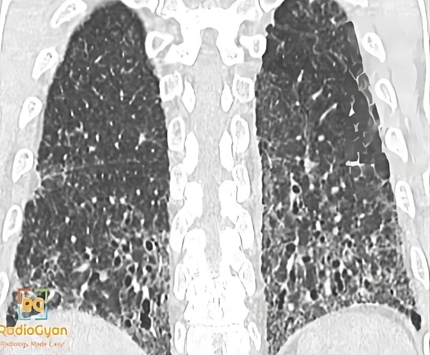
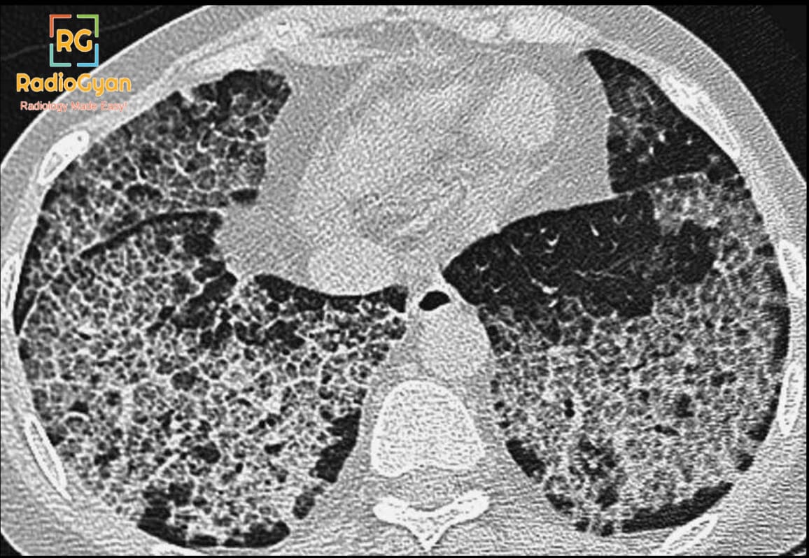
CT scan of lungs showing Usual Interstitial Pneumonia (UIP) pattern with honeycombing and traction bronchiectasis.

Honeycombing | Radiology Signs

Galaxy sign | Radiology Signs

Eggshell calcification | Radiology Signs

Double bronchial wall sign | Radiology Signs

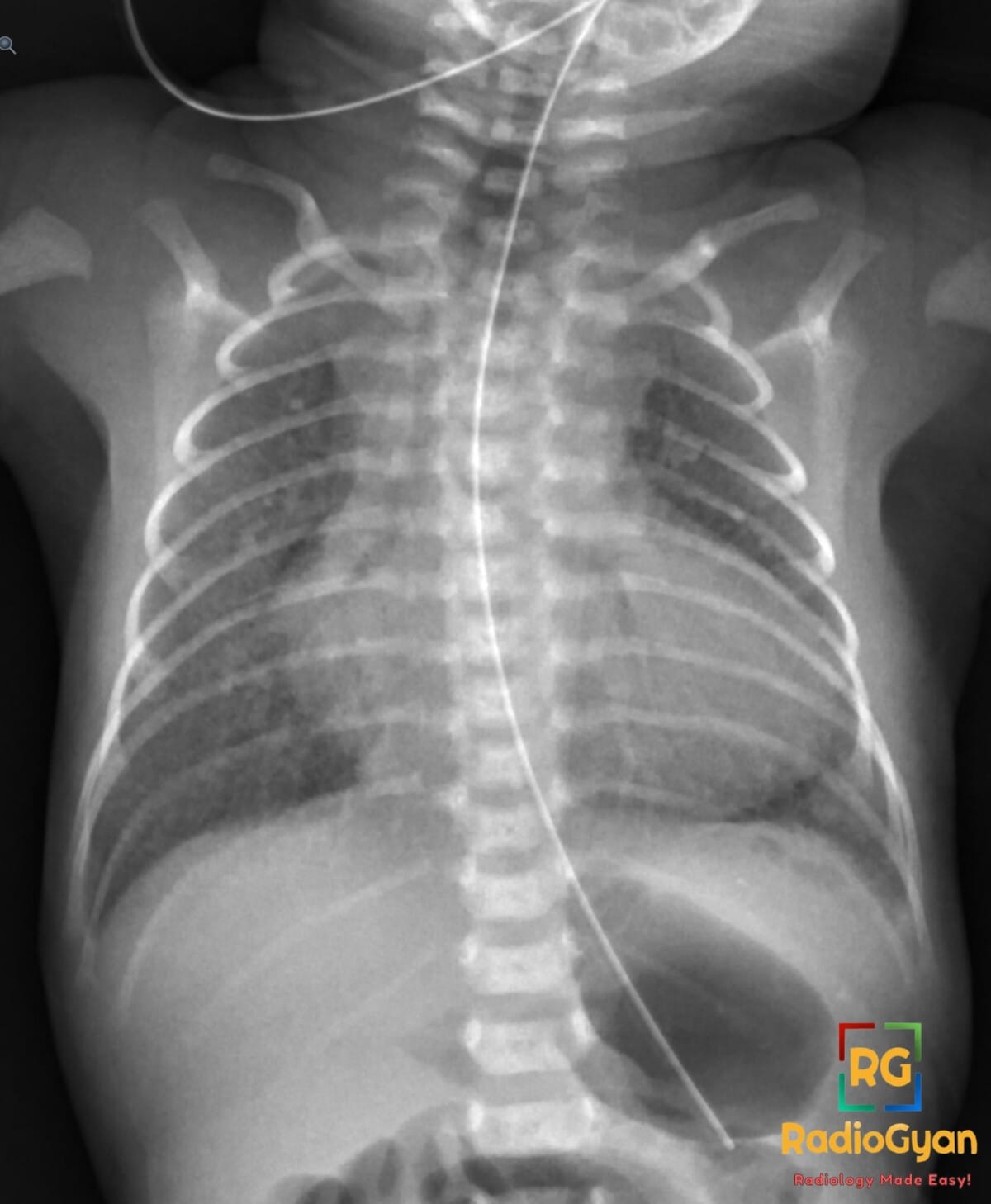
Chest X-ray showing left deep sulcus sign from moderate left pneumothorax with midline shift in neonate

Deep sulcus sign | Radiology Signs

Coin lesion | Radiology Signs

Cannonball metastases | Radiology Signs

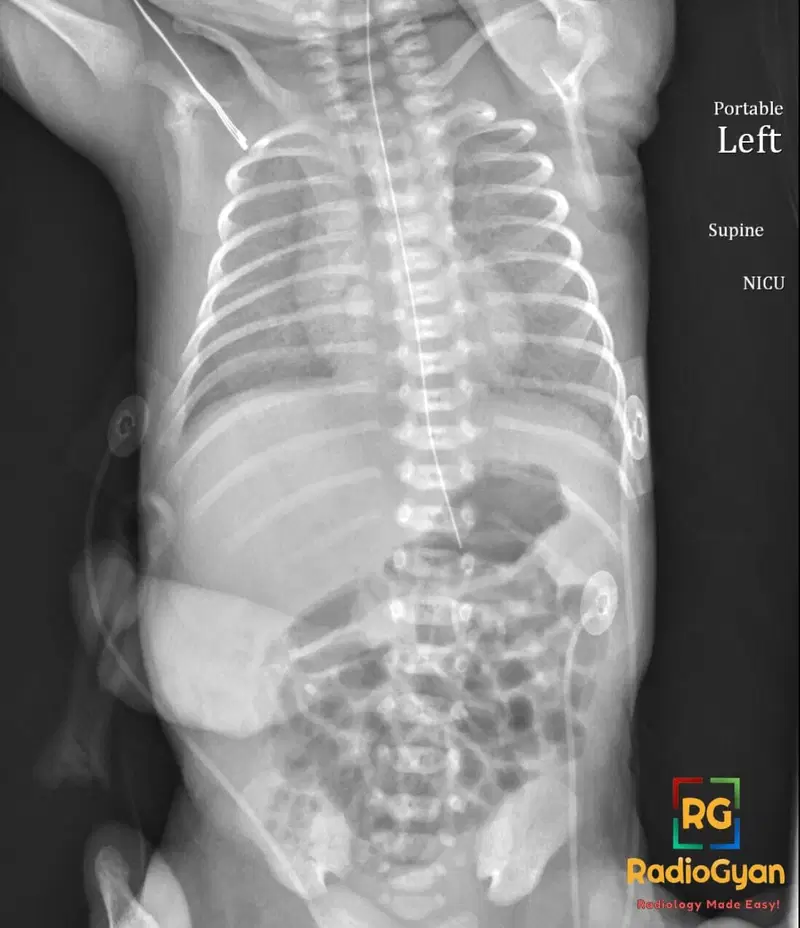
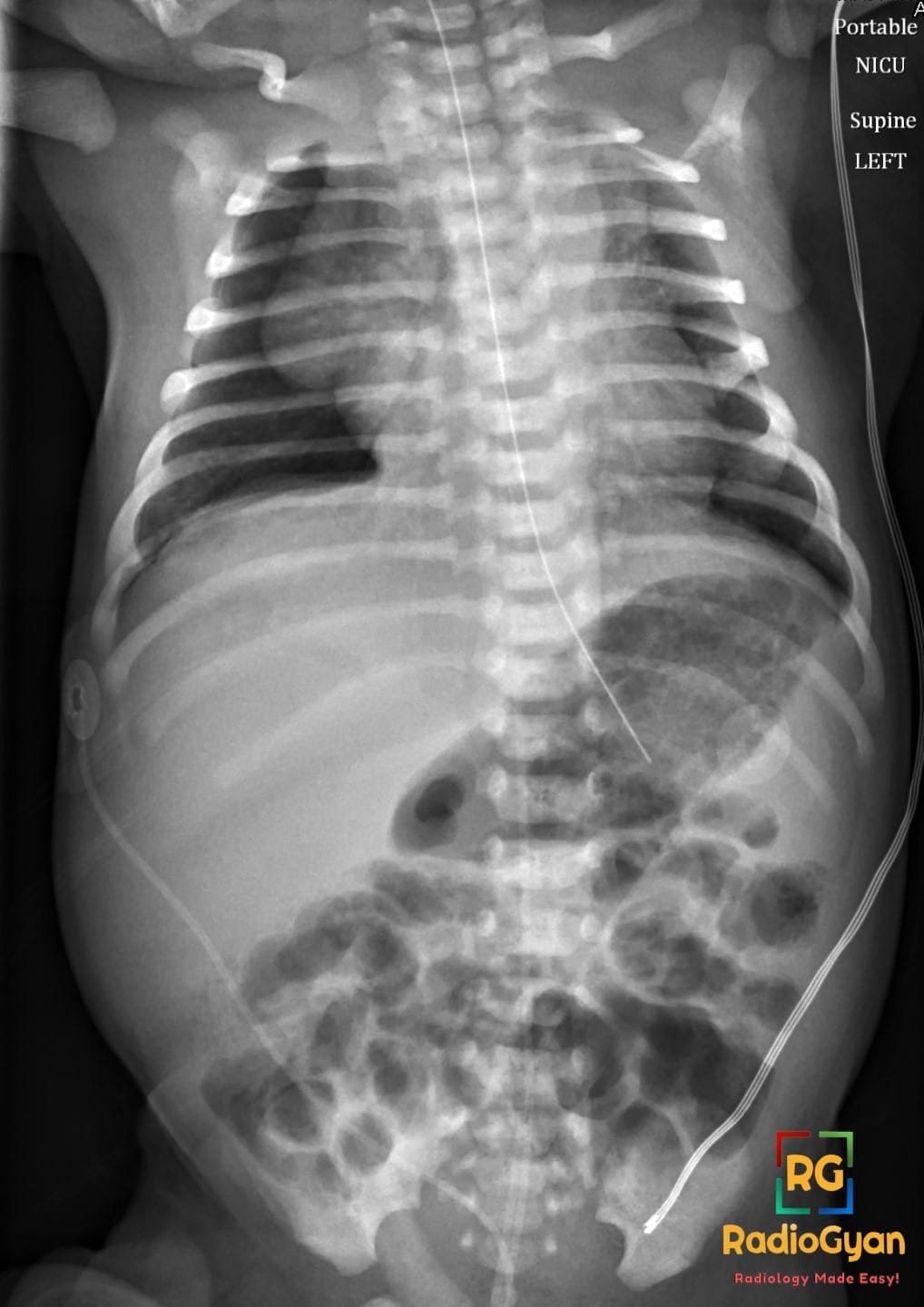
Chest X-ray of a premature neonate in supine position with respiratory distress syndrome, showing granular opacities and air bronchograms.

Air bronchogram | Radiology Signs

Rice grain calcification | Radiology Signs

Ulcerative Colitis- Lead Pipe Colon Radiology Case

Lead-pipe colon | Radiology Signs

Ghost Meniscus Sign (Knee) | Radiology Signs

Whirlpool Sign (Abdomen) | Radiology Signs

Double Delta Sign (Lateral Meniscus) | Radiology Signs

Sarcoidosis Chest Radiograph showing garland sign pawnbroker sign and 1-2-3 sign

1-2-3 Sign | Radiology Signs

Half Moon Sign (Subphrenic Free Gas) | Radiology Signs

Juxtaphrenic peak sign | Radiology Signs

Spilled Teacup Sign (Wrist) | Radiology Signs

Acorn cyst | Radiology Signs

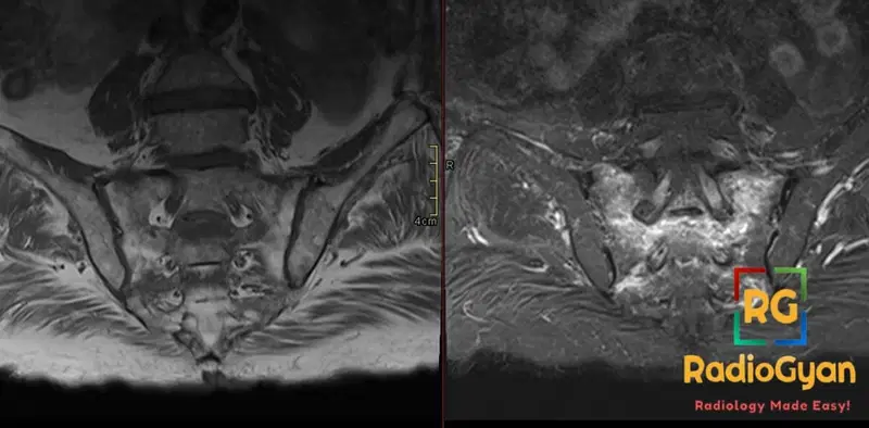
MRI pelvic bone shows sacral insufficiency fracture with Honda sign (H-sign) indicated by distinct bone marrow edema.

Honda sign | Radiology Signs

Snowman sign | Radiology Signs

Incomplete fissure sign | Radiology Signs

Moulage sign | Radiology Signs

Falciform ligament sign | Radiology Signs

Piece-of-pie sign | Radiology Signs

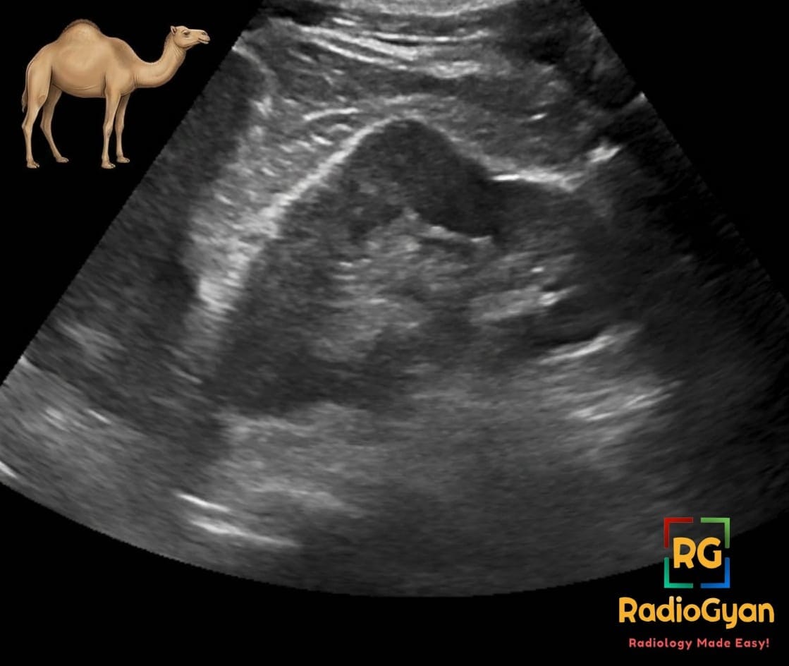
Ultrasound image of a left kidney showing a dromedary hump, a normal kidney variant, with a camel icon for illustration.

Dromedary hump | Radiology Signs

Mini Brain Appearance | Radiology Signs

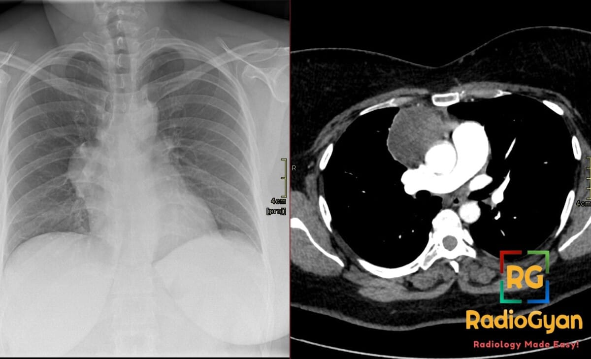
Chest X-ray and CT scan showing a large anterior mediastinal mass with hilum overlay sign, consistent with mature cystic teratoma.

Hilum overlay sign | Radiology Signs

CT showing an intussusception in the left upper quadrant of the abdomen

Target Sign (Bowel) | Radiology Signs

Ring-around-artery sign | Radiology Signs

Snow-capping | Radiology Signs

Cockade Sign | Radiology Signs

Head cheese sign | Radiology Signs

Whiskering | Radiology Signs

Marching Cleft Sign | Radiology Signs

Cotton wool appearance | Radiology Signs

Teardrop sign | Radiology Signs

Scalloped vertebra | Radiology Signs

Bowler hat sign | Radiology Signs

Bird of prey sign | Radiology Signs

Dense hilum sign | Radiology Signs

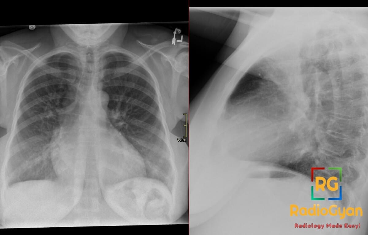
PA and lateral chest radiograph showing severe left atrial enlargement with a prominent double density sign.

Double density sign | Radiology Signs

Floating teeth | Radiology Signs

Reverse pulmonary edema | Radiology Signs

Naked facet sign | Radiology Signs

Football sign | Radiology Signs

Hilum convergence sign | Radiology Signs

Stepladder appearance | Radiology Signs

Shmoo sign | Radiology Signs

Cervicothoracic sign | Radiology Signs

Tree-in-bud sign | Radiology Signs

Salt and pepper skull Radiology Signs

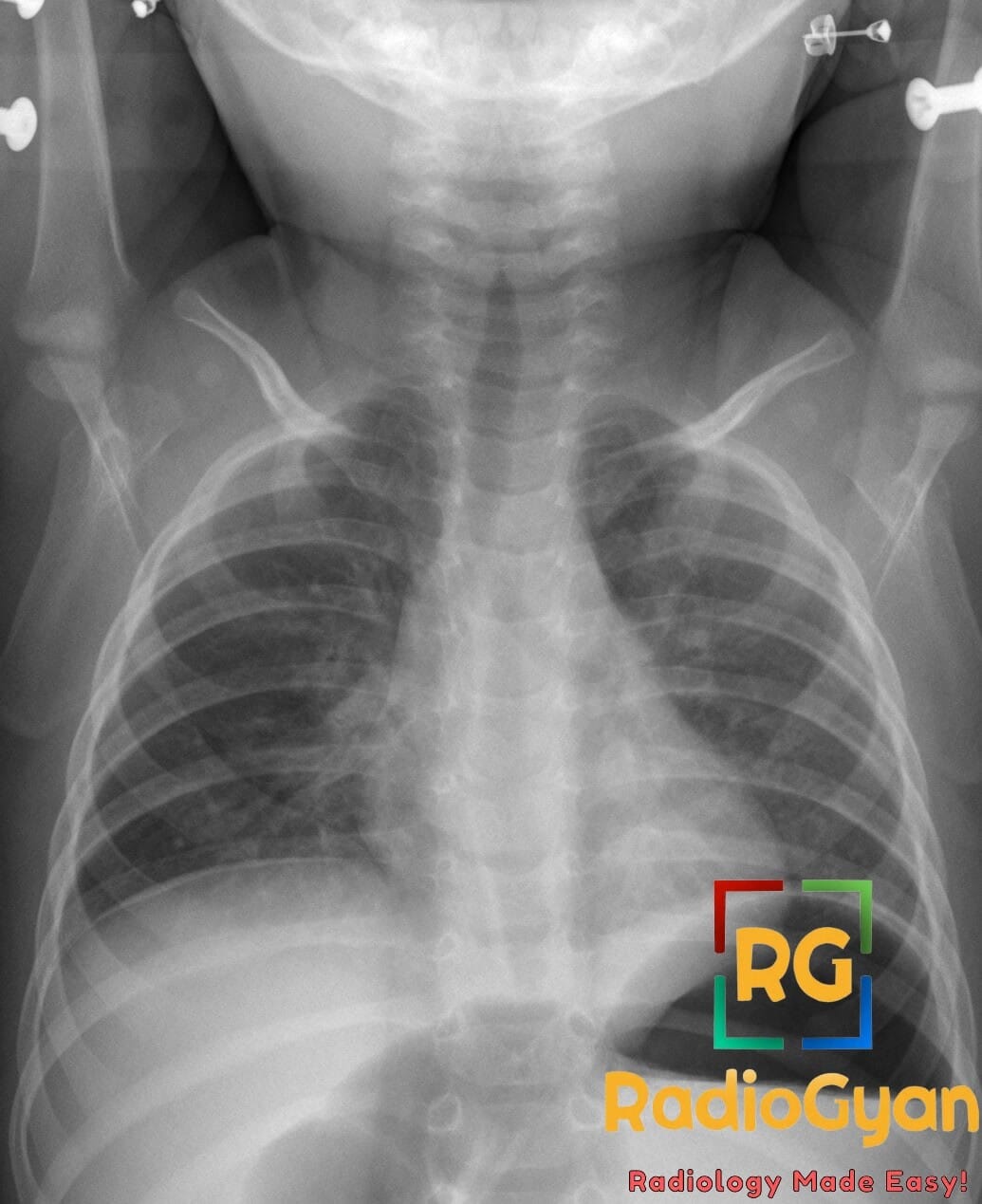
Chest X-ray showing steeple sign, indicative of croup in a pediatric patient.

Steeple Sign in Croup: X-ray Findings and Differentials

Flat waist sign | Radiology Signs

Water bottle heart Radiology Signs

Berry aneurysm sign | Radiology Signs

CT Leontiasis Ossea: Radiological Signs and Features

Oil Droplet Sign CT: Radiological Appearance and Significance

Satellite sign | Radiology Signs

Westermark’s sign | Radiology Signs

Mickey Mouse Ears (Liver) | Radiology Signs

Anteater nose sign Radiology Signs

Island sign Radiology Signs

Omental caking | Radiology Signs

Saber shin deformity Radiology Signs

C Sign (Foot) | Radiology Signs

Empty sella | Radiology Signs

Bowtie appearance Radiology Signs

Thymic sail sign | Radiology Signs

Overview

Curated, image‑first explainers standardize how signs are recognized and reported across X‑ray, CT, MRI, ultrasound, and nuclear medicine. Each entry is concise yet comprehensive, balancing quick reference utility with the depth expected by medical professionals.

Clinical relevance

Radiologic signs translate visual patterns into shared clinical language, improving communication, triage, and decision‑making across multidisciplinary teams. Consistent use enhances board preparation, viva performance, and structured reporting by anchoring observations to well‑defined entities.

What’s inside

Entries follow a consistent blueprint: definition, imaging appearance, key differentials, common pitfalls, and diagnostic pearls. Coverage spans thoracic, abdominal, MSK, neuroradiology, and GU/GYN systems, emphasizing entities frequently encountered on call and in assessments.

How to use

Integrate sign pages into rotation‑specific study plans and point‑of‑care lookups during case review to narrow differentials and refine report language. Pair with mnemonics and case discussions on RadioGyan to build durable pattern recognition and reinforce clinical application.

Alphabetical List of Radiology Signs
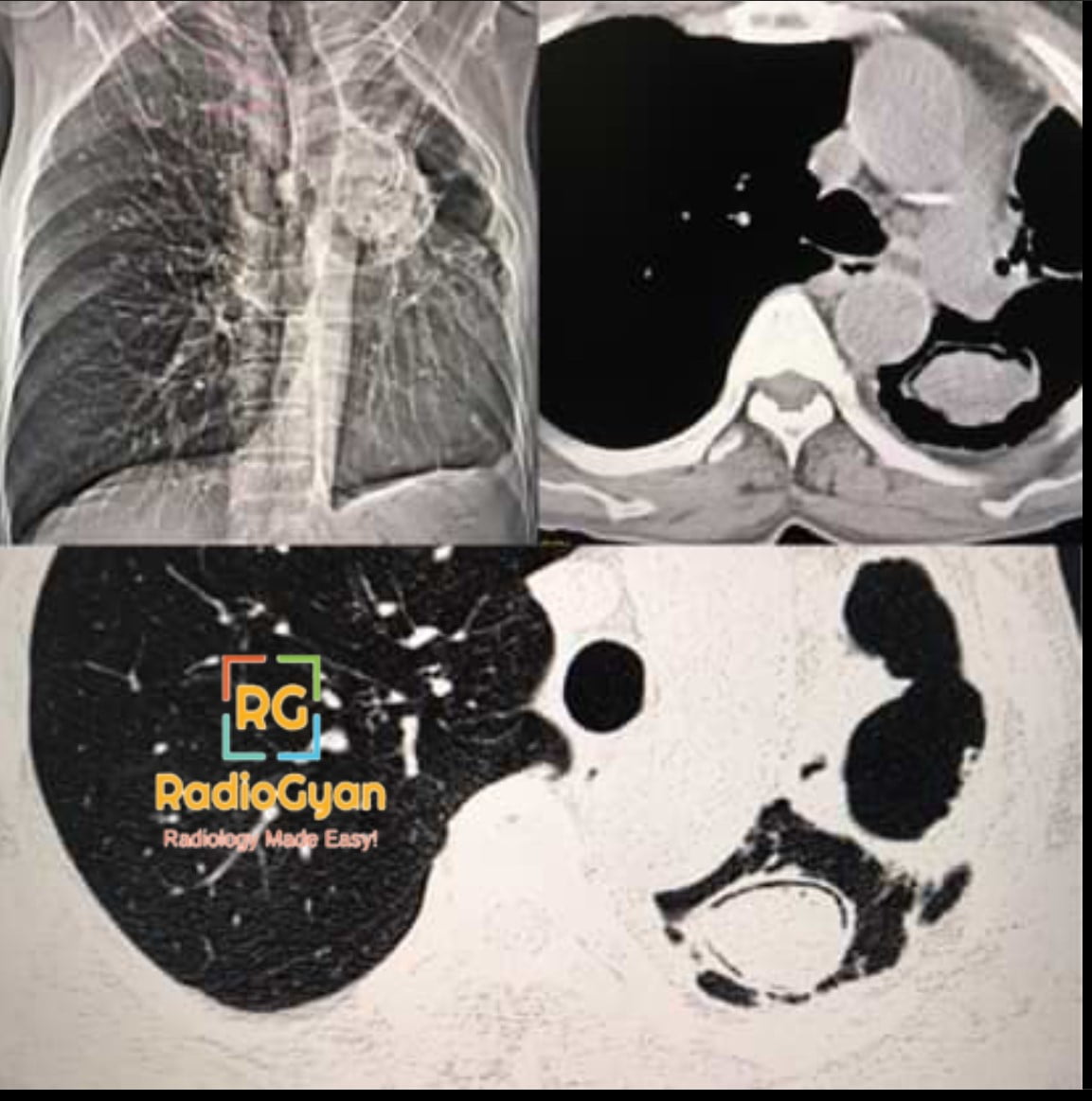
Sarcoidosis Chest Radiograph showing garland sign pawnbroker sign and 1-2-3 sign

1-2-3 Sign | Radiology Signs
Abdominal Cocoon Sign | Radiology Signs
Absent bow-tie sign | Radiology Signs
Accordion sign | Radiology Signs
Acorn cyst | Radiology Signs

Chest X-ray of a premature neonate in supine position with respiratory distress syndrome, showing granular opacities and air bronchograms.

Air bronchogram | Radiology Signs
Aspergilloma With Fibro-Cavitary Changes In The Left Upper Lobe showing Air Crescent sign

Air Crescent Sign | Radiology Signs
Andersson lesion | Radiology Signs
Anteater nose sign Radiology Signs

Antral nipple sign | Radiology Signs
Antral Pad Sign (Stomach) | Radiology Signs
Aortic nipple | Radiology Signs
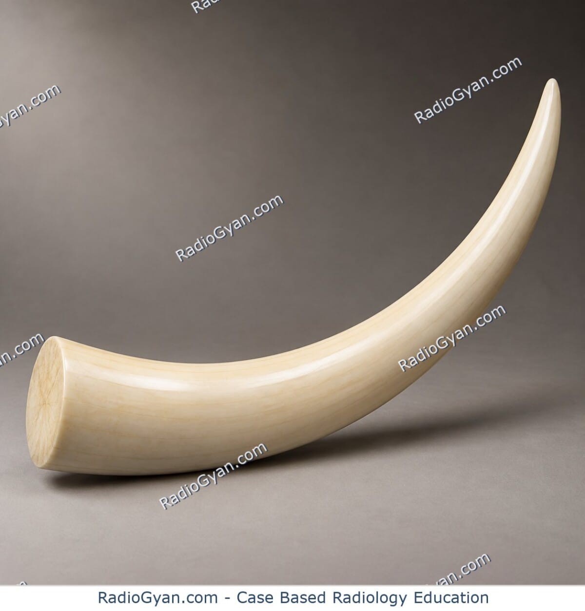
Apple Core Sign: Causes, Imaging and Differentials

Arcuate sign | Radiology Signs
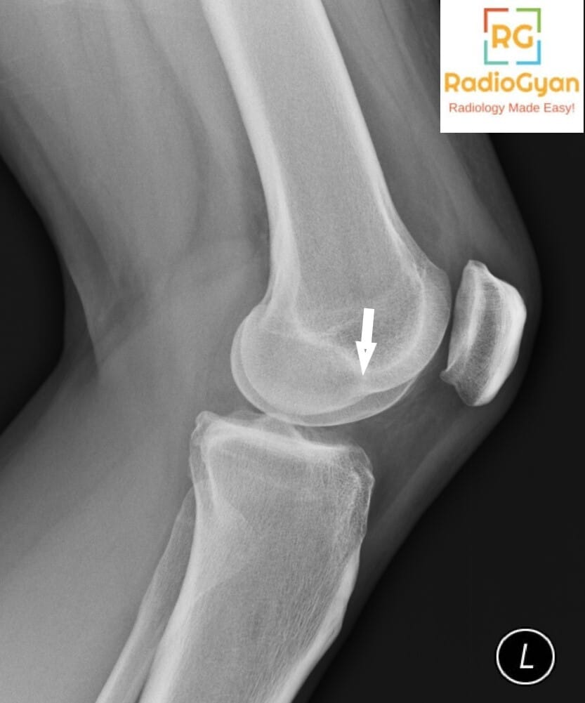
Asterisk sign | Radiology Signs
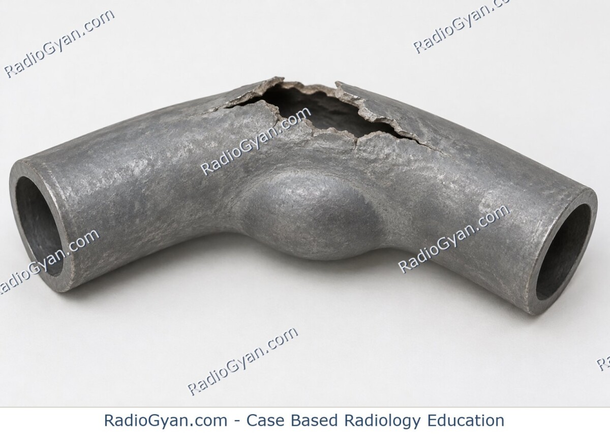
Bamboo spine | Radiology Signs
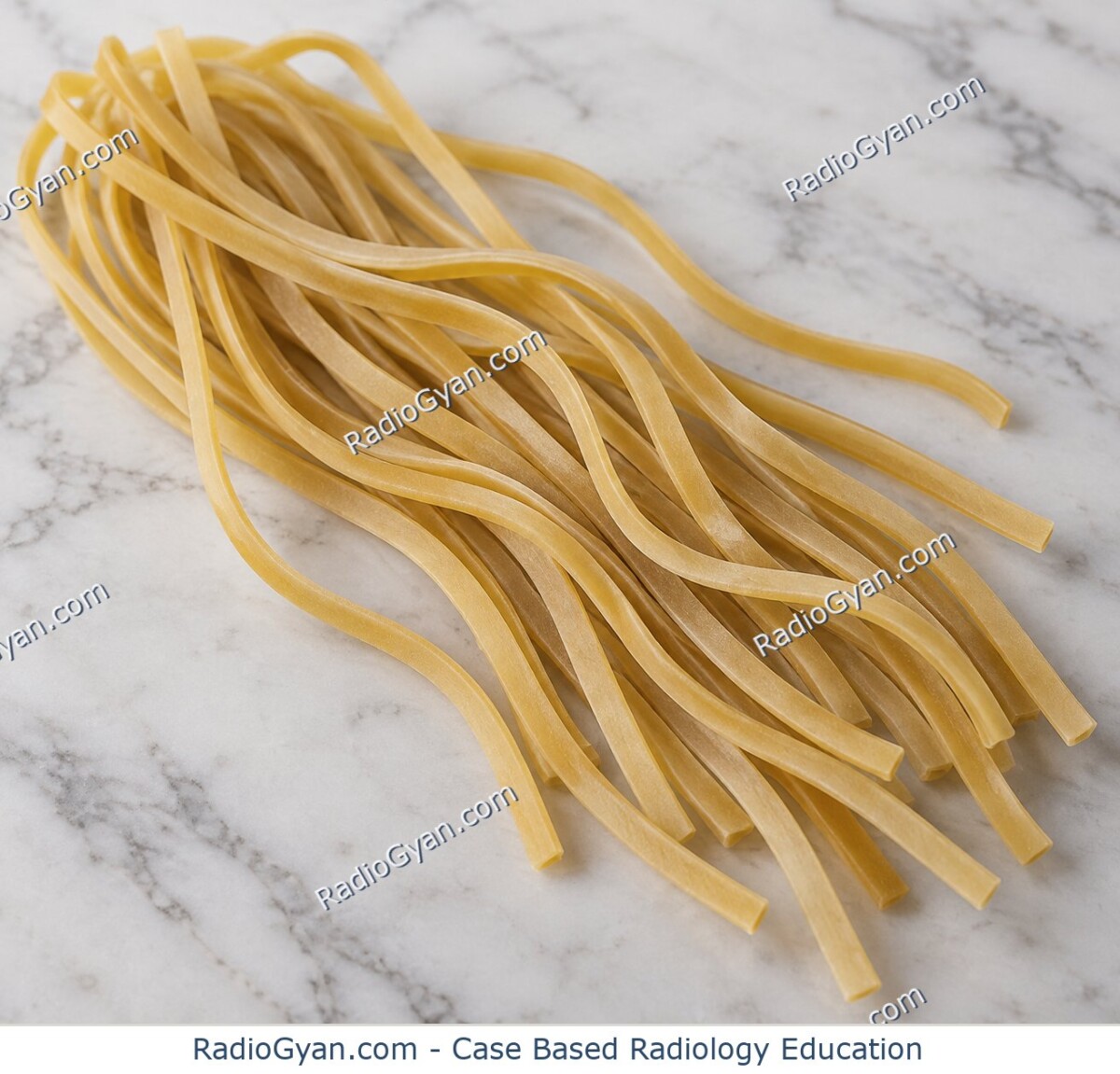
Banana fracture | Radiology Signs

Banana sign | Radiology Signs

Bare Orbit Sign | Radiology Signs

Bat sign | Radiology Signs

Bat-Wing Appearance (Butterfly Sign): Causes and Imaging

Axial contrast-enhanced CT image of the chest demonstrating Type A aortic dissection with an intimal flap separating the true and false lumens in the descending thoracic aorta.

Beak Sign (Aorta) | Radiology Signs

Axial CT scan of the abdomen showing a left kidney with xanthogranulomatous pyelonephritis, characteristic 'bear paw' sign.

Bear Paw Sign (Xanthogranulomatous Pyelonephritis) | Radiology Signs

Berry aneurysm sign | Radiology Signs

Beveled edge sign | Radiology Signs

Bird of prey sign | Radiology Signs

Bite sign | Radiology Signs

Black hole sign | Radiology Signs

Black pleural line | Radiology Signs

Blade of grass sign | Radiology Signs

Blend sign | Radiology Signs

Osteopetrosis Radiology Case

Bone within a bone | Radiology Signs

Boomerang sign | Radiology Signs

Pediatric chest X-ray showing the classic boot-shaped heart (coeur en sabot) of Tetralogy of Fallot with upturned apex

Boot-Shaped Heart on X-ray: Tetralogy of Fallot and Mimics

Boutonnière deformity | Radiology Signs

Bowl of Grapes Sign | Radiology Signs

Bowler hat sign | Radiology Signs

Bowtie appearance Radiology Signs

Box-shaped heart | Radiology Signs

Boxcar ventricles | Radiology Signs

Breast in a breast | Radiology Signs

Brim sign | Radiology Signs

Double PCL Sign–Bucket Handle Tear MRI

Bucket handle tear | Radiology Signs

Bulging fissure sign | Radiology Signs

Butterfly vertebra | Radiology Signs

C Sign (Foot) | Radiology Signs

Cannonball metastases | Radiology Signs

Carman Meniscus Sign | Radiology Signs

Celery stalking | Radiology Signs

Central dot sign | Radiology Signs

Cervicothoracic sign | Radiology Signs

Chain of lakes sign | Radiology Signs

Champagne glass pelvis | Radiology Signs

Cheerio sign | Radiology Signs

Christmas tree bladder | Radiology Signs

Cluster of grapes sign | Radiology Signs

Cobblestone appearance | Radiology Signs

Cobra head sign | Radiology Signs

Cobweb sign | Radiology Signs

Cockade Sign | Radiology Signs

Codfish vertebrae | Radiology Signs

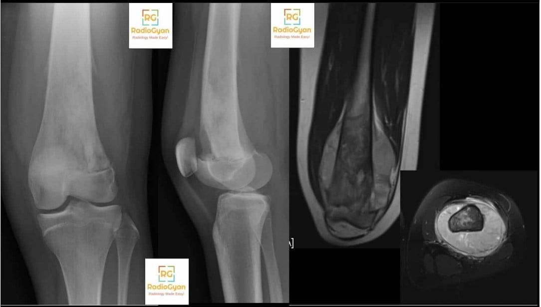
Osteogenic Sarcoma Distal Femur With Codman’s Triangle Radiology Case

Codman Triangle on X-ray: Causes, Mimics and Next Steps

Abdominal radiograph and axial CT showing cecal volvulus with dilated displaced cecum in left upper quadrant and whirl sign.

Coffee-bean sign | Radiology Signs

Cogwheel sign | Radiology Signs

Coiled spring sign | Radiology Signs

Coin lesion | Radiology Signs

Collar button ulcer | Radiology Signs

Colon cut-off sign | Radiology Signs

Coronal CT image of the abdomen showing the 'comb sign' indicative of Crohn's disease in the small bowel.

Comb sign | Radiology Signs

Comet Sign (Ureter) | Radiology Signs

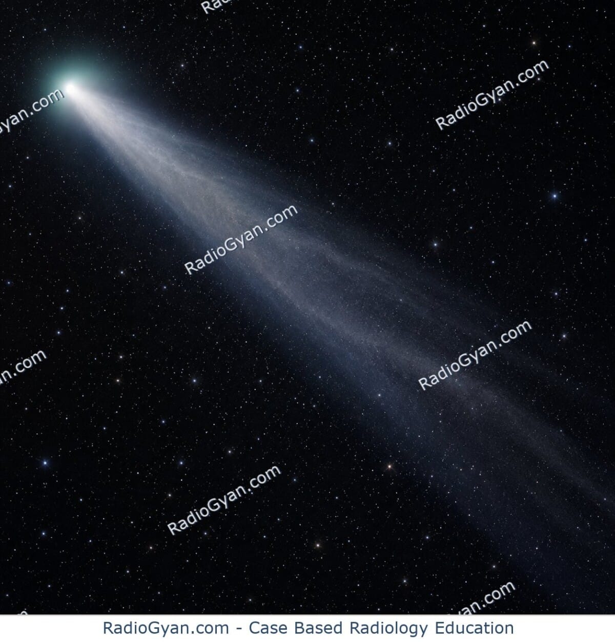
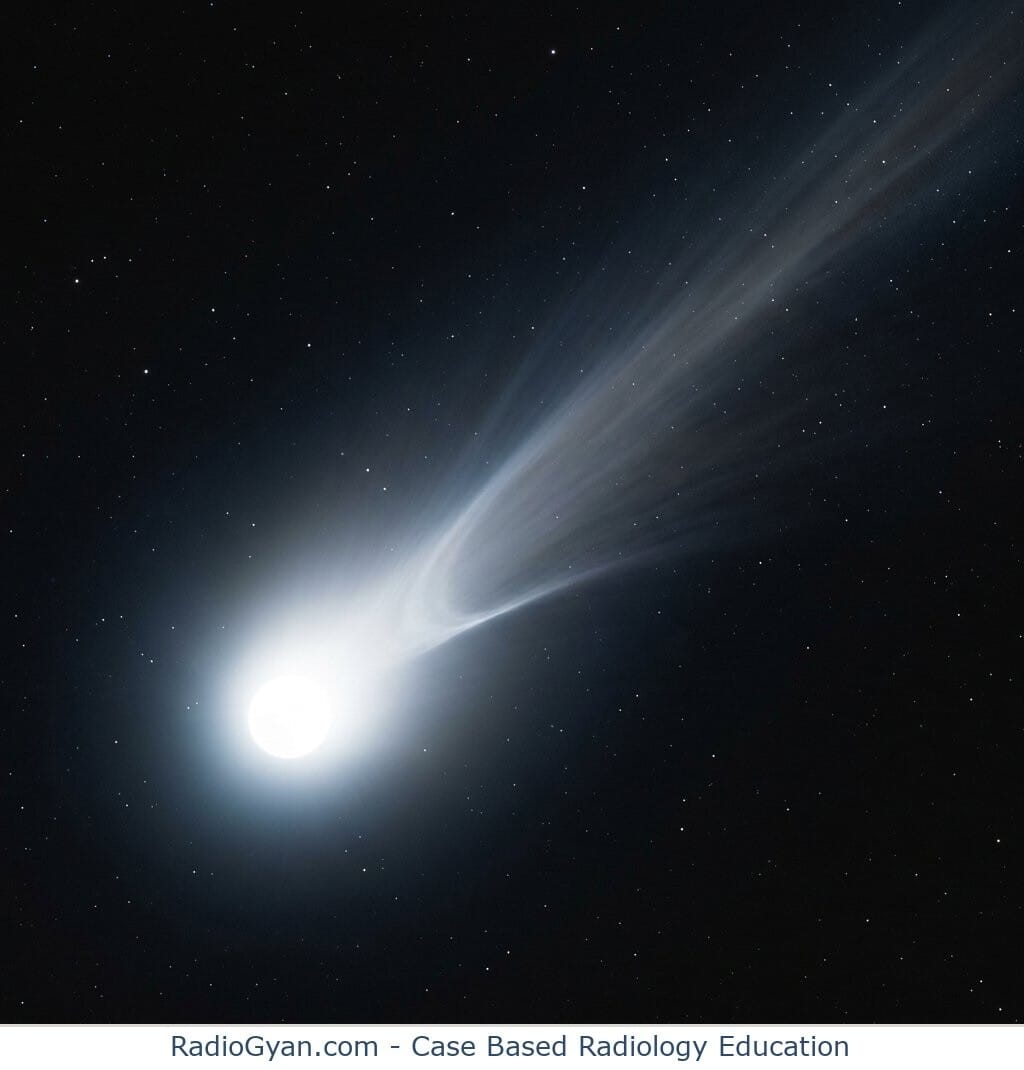
A comet with a bright nucleus in the lower left and a wide faint tail streaming toward the upper right, illustrating the comet tail sign.

Comet Tail Sign (Lung) | Radiology Signs

Coned epiphyses | Radiology Signs

Continuous diaphragm sign | Radiology Signs

Cord sign | Radiology Signs

Corduroy sign | Radiology Signs

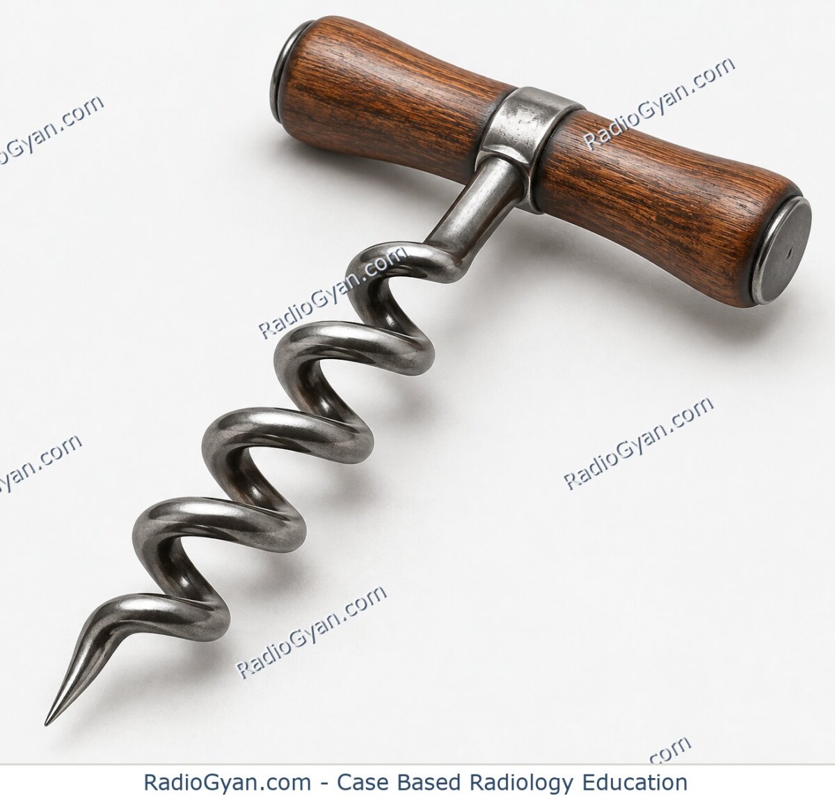
Corkscrew oesophagus | Radiology Signs

Corkscrew Sign (Small Bowel) | Radiology Signs

Cortical Rim Sign (Kidney) | Radiology Signs

Cortical Ring Sign (Wrist) | Radiology Signs

Cottage loaf sign | Radiology Signs

Cotton wool appearance | Radiology Signs

CT scan of lungs showing crazy paving pattern indicative of pulmonary alveolar proteinosis.

Crazy paving | Radiology Signs

Crescent Sign (Femoral Head) | Radiology Signs

Crossover sign | Radiology Signs

Crowded carpal sign | Radiology Signs

CT angiogram sign | Radiology Signs

CT Leontiasis Ossea: Radiological Signs and Features

Cupola sign | Radiology Signs

Curtain Sign (Lung Ultrasound) | Radiology Signs

Cyclops lesion | Radiology Signs

Dagger sign | Radiology Signs

Dark bronchus sign | Radiology Signs

Chest X-ray showing left deep sulcus sign from moderate left pneumothorax with midline shift in neonate

Deep sulcus sign | Radiology Signs

Dense hilum sign | Radiology Signs

Axial non-contrast CT of the brain demonstrates hyperdense left MCA.

Dense MCA sign | Radiology Signs

Anteroposterior and lateral radiographs of the distal left radius demonstrate a transverse fracture through the distal metaphysis with dorsal angulation and impaction in keeping with a Colle fracture.

Dinner Fork (Silver Fork) Deformity | Radiology Signs

Double bronchial wall sign | Radiology Signs

Double bubble sign | Radiology Signs

Double decidual sac sign | Radiology Signs

Double Delta Sign (Lateral Meniscus) | Radiology Signs

PA and lateral chest radiograph showing severe left atrial enlargement with a prominent double density sign.

Double density sign | Radiology Signs

Coronal CT scan showing dilated common bile duct and pancreatic duct, indicating a double duct sign.

Double Duct sign | Radiology Signs

Double Line Sign | Radiology Signs

Sagittal MRI image of the knee demonstrates a bucket handle tear of the medial meniscus with the “double PCL sign”. The image is annotated to show the femur, tibia, joint effusion, intercondylar eminence, PCL, and bucket handle tear. A red circle surrounds the bucket handle tear.

Double PCL Sign (Medial Meniscus) | Radiology Signs

Double spinous process sign | Radiology Signs

Double track sign | Radiology Signs

Draped aorta sign | Radiology Signs

Dripping Candle Wax Sign | Radiology Signs

Ultrasound image of a left kidney showing a dromedary hump, a normal kidney variant, with a camel icon for illustration.

Dromedary hump | Radiology Signs

Drooping lily sign | Radiology Signs

Drooping shoulder sign | Radiology Signs

Dural tail sign | Radiology Signs

Egg-on-a-string sign | Radiology Signs

Eggshell calcification | Radiology Signs

Elephant on a flagpole | Radiology Signs

Empty delta sign | Radiology Signs

Empty notch sign | Radiology Signs

Empty sella | Radiology Signs

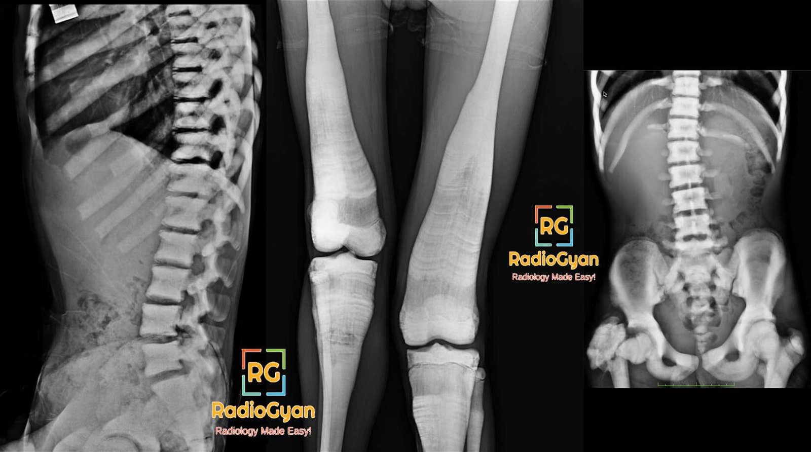
Radiograph showing osteopetrosis marble bone disease Albers-Schönberg disease interesting radiology case

Erlenmeyer flask deformity | Radiology Signs

Eye-of-the-tiger sign | Radiology Signs

Face of the giant panda | Radiology Signs

Faceless kidney | Radiology Signs

Falciform ligament sign | Radiology Signs

Fallen fragment sign | Radiology Signs

Fallen lung sign | Radiology Signs

Fat C2 sign | Radiology Signs

Fat Halo Sign (Bowel) | Radiology Signs

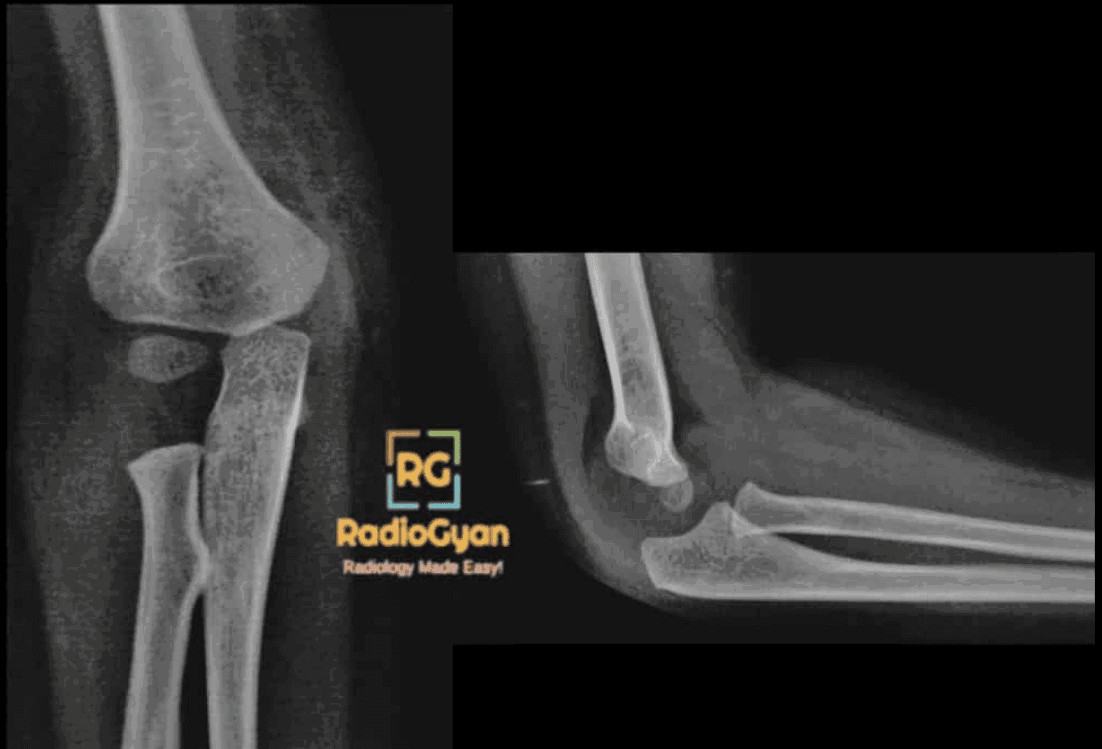
Supracondylar Fracture Humerus – Positive Fat Pad Sign And Displaced Anterior Humeral Line Radiology Case

Fat Pad Sign (Elbow) | Radiology Signs

Feeding vessel sign | Radiology Signs

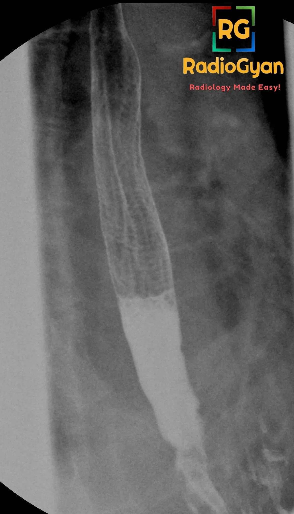
Feline Esophagus Barium esophagogram in patient with gastroesophageal reflux

Feline esophagus | Radiology Signs

Figure 3 sign | Radiology Signs

Fishhook ureter | Radiology Signs

Fishmouthing | Radiology Signs

Flame-shaped breast | Radiology Signs

Flat waist sign | Radiology Signs

Fleischner sign | Radiology Signs

Flipped meniscus sign | Radiology Signs

Floating teeth | Radiology Signs

Floating viscera sign | Radiology Signs

Flower vase ureter | Radiology Signs

Football sign | Radiology Signs

Fragment-in-notch sign | Radiology Signs

Galaxy sign | Radiology Signs

Ghost Meniscus Sign (Knee) | Radiology Signs

Ghost Sign (Foot) | Radiology Signs

Ginkgo leaf sign | Radiology Signs

Chest X-ray and CT scans showing finger-in-glove appearance in allergic bronchopulmonary aspergillosis (ABPA)

Gloved finger sign | Radiology Signs

Clean medical-style vector illustration of a classic stemmed drinking goblet, the namesake of the goblet sign in uroradiology.

Goblet sign | Radiology Signs

AP chest radiograph demonstrates right upper lobe collapse with an associated hilar mass forming the "Golden S" sign.

Golden’s S sign | Radiology Signs

Golf-ball-on-a-tee sign | Radiology Signs

Gooseneck sign | Radiology Signs

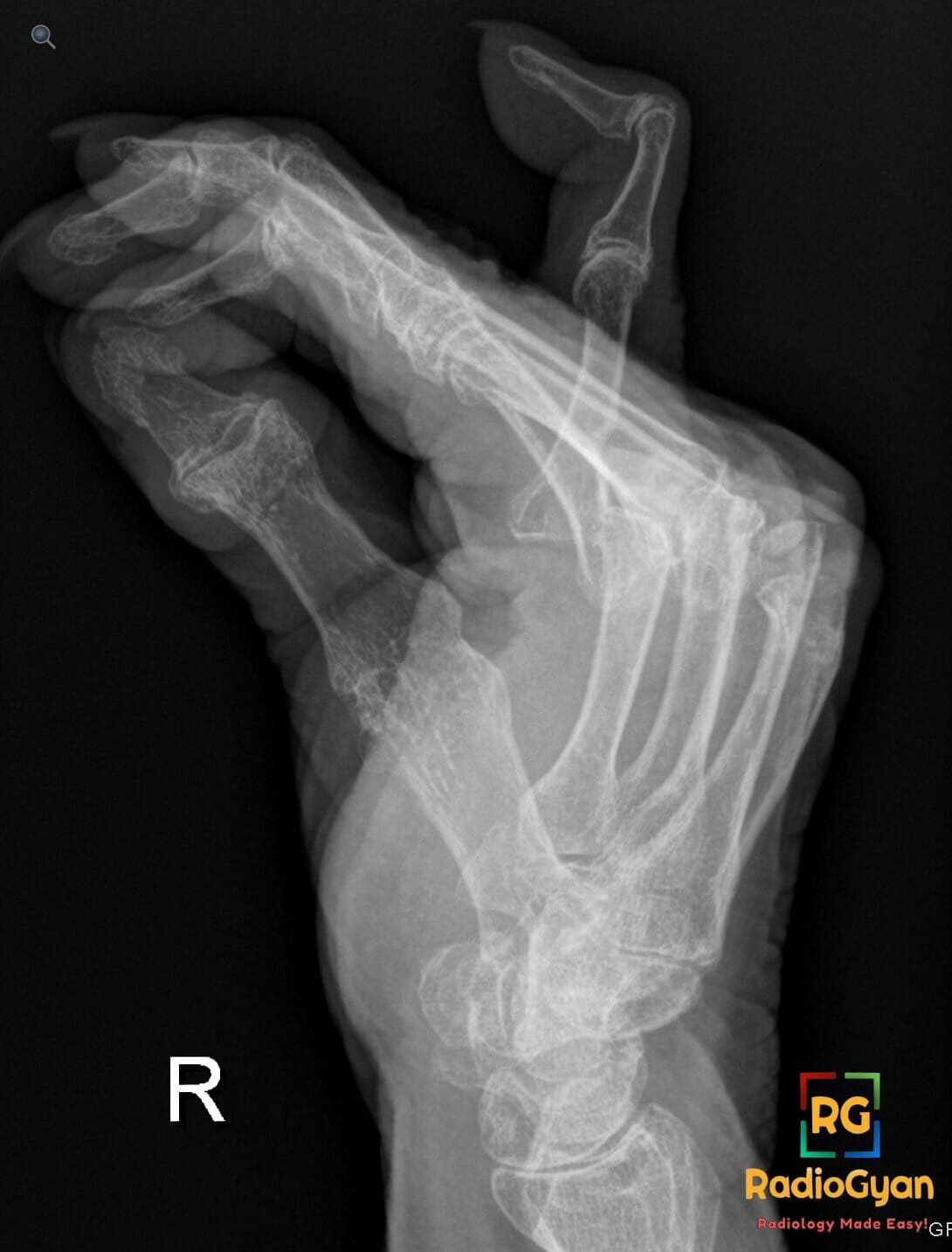
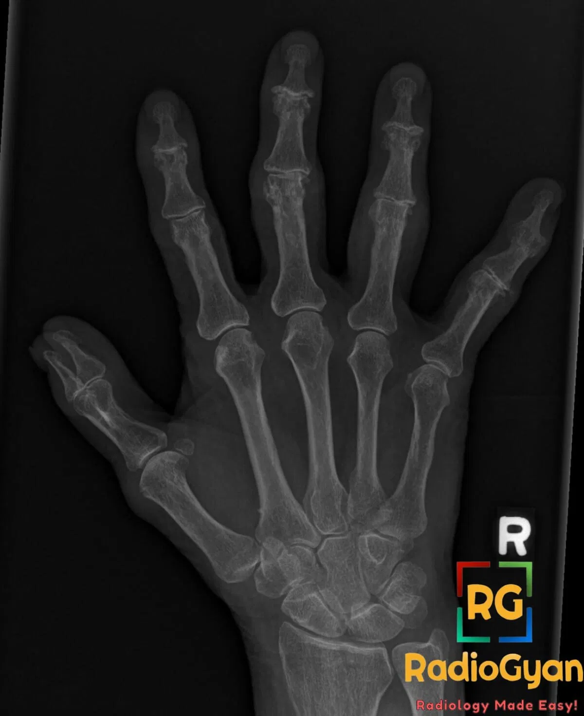
X-ray of the right hand showing erosive changes in the interphalangeal joints and polydactyly of the thumb.

Gull Wing (Seagull) Appearance | Radiology Signs

Gut signature sign | Radiology Signs

H-shaped vertebrae | Radiology Signs

Insertional Calcific Tendinitis Involving Achilles Tendon and Haglund deformity.

Haglund’s deformity | Radiology Signs

Hair-on-end | Radiology Signs

Hairpin turn sign | Radiology Signs

Half Moon Sign (Subphrenic Free Gas) | Radiology Signs

Halo Sign (Lung) | Radiology Signs

Axial abdominal CT showing the hamburger sandwich sign with mesenteric lymphadenopathy in lymphoma

Hamburger bun sign | Radiology Signs

Pulmonary embolism diagnosis : Hampton Hump

Hampton’s hump | Radiology Signs

Hampton’s Line | Radiology Signs

Harlequin appearance | Radiology Signs

Hatchet head | Radiology Signs

Hawkins Sign | Radiology Signs

Head cheese sign | Radiology Signs

Heel Pad Sign (Calcaneus) | Radiology Signs

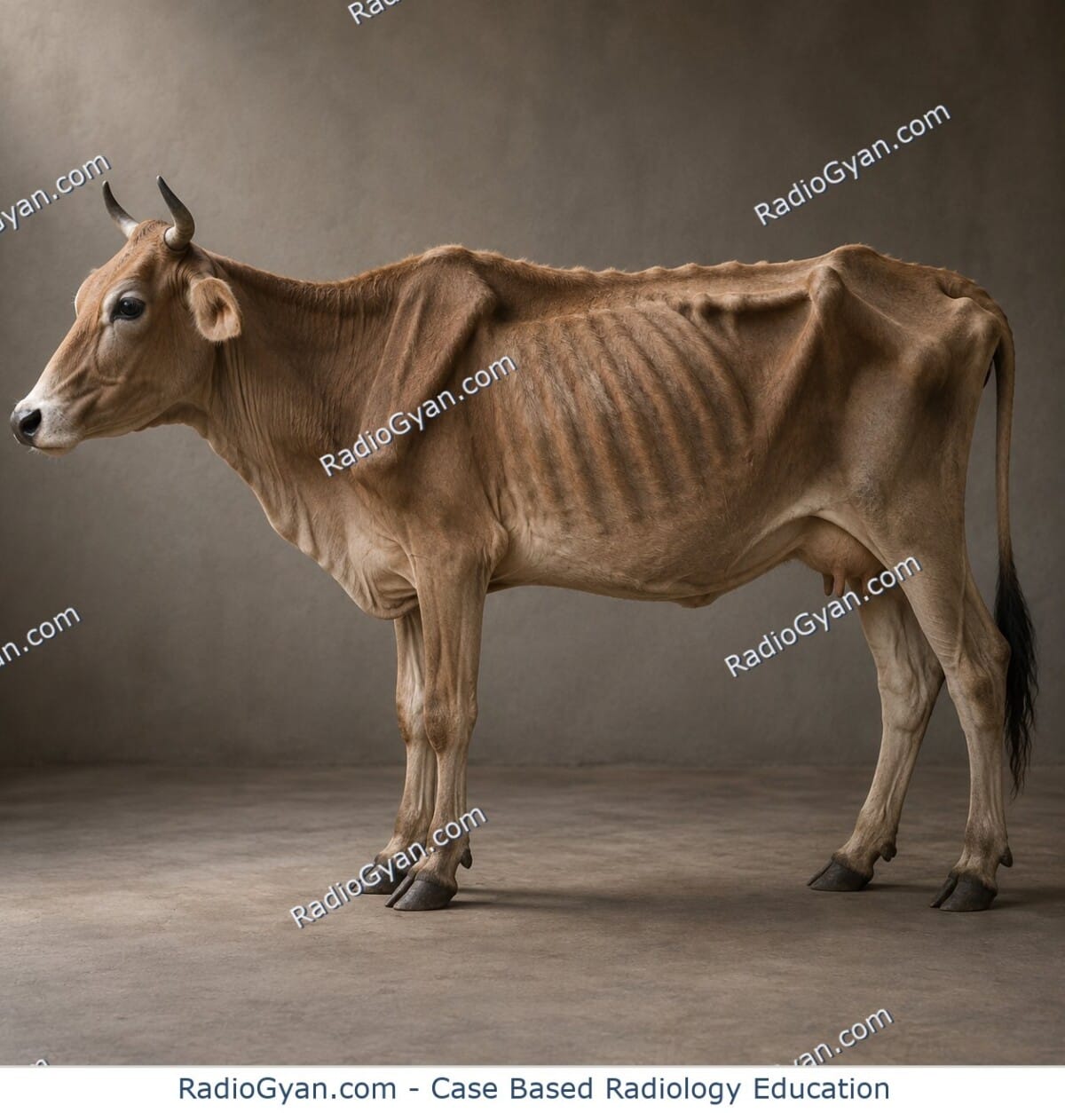
Hide-bound appearance | Radiology Signs

Hill-Sachs deformity | Radiology Signs

Hilum convergence sign | Radiology Signs

Chest X-ray and CT scan showing a large anterior mediastinal mass with hilum overlay sign, consistent with mature cystic teratoma.

Hilum overlay sign | Radiology Signs

Hockey stick sign | Radiology Signs

Holly leaf sign | Radiology Signs

MRI pelvic bone shows sacral insufficiency fracture with Honda sign (H-sign) indicated by distinct bone marrow edema.

Honda sign | Radiology Signs

CT scan of lungs showing Usual Interstitial Pneumonia (UIP) pattern with honeycombing and traction bronchiectasis.

Honeycombing | Radiology Signs

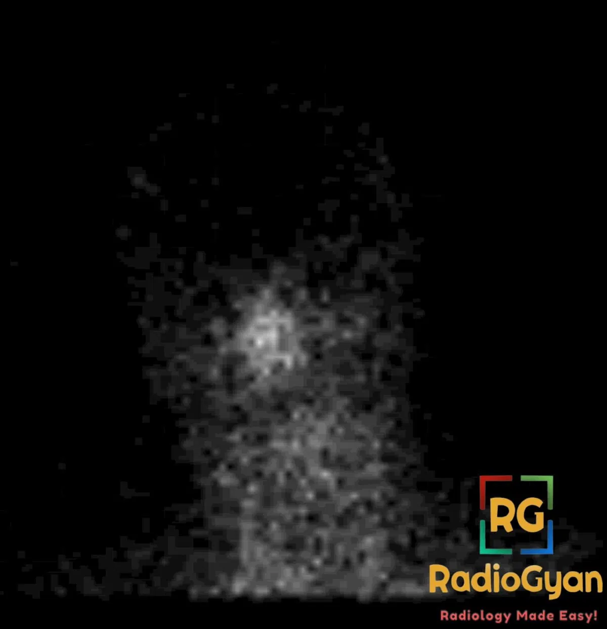
Nuclear medicine scan showing increased radiotracer uptake in the nasopharynx, consistent with the hot nose sign.

Hot Nose sign | Radiology Signs

Hot quadrate lobe sign focal hepatic hot spot in SVC obstruction on CT

Hot Quadrate Lobe Sign

Hot-cross bun sign | Radiology Signs

Hummingbird sign | Radiology Signs

Hurricane sign | Radiology Signs

Ice-cream cone sign | Radiology Signs

Frontal chest radiograph and axial CT show an expansile left upper rib lesion with the incomplete-border sign.

Incomplete border sign | Radiology Signs

Incomplete fissure sign | Radiology Signs

Inside-out liver sign | Radiology Signs

CT of insular ribbon sign

Insular Ribbon Sign: Early CT Sign of MCA Infarction

Interface sign | Radiology Signs

Intradecidual sign | Radiology Signs

Intravertebral vacuum cleft | Radiology Signs

Island sign Radiology Signs

Ivory phalanx | Radiology Signs

Ivory vertebra | Radiology Signs

Ivy sign | Radiology Signs

J – Shaped Sella Radiology Case.

J-shaped sella | Radiology Signs

Jackstone Urinary bladder calculus on CT

Jackstone calculus | Radiology Signs

Right John Thomas Sign/ Thockmorton sign in a patient with Right Osteomyelitis

John Thomas Sign / Throckmorton Sign | Radiology Signs

Juxtaphrenic peak sign | Radiology Signs

Keyhole sign | Radiology Signs

Abdominal radiograph and axial CT showing cecal volvulus with dilated displaced cecum in left upper quadrant and whirl sign.

Kidney bean sign | Radiology Signs

Kissing contusion | Radiology Signs

Kissing ulcers | Radiology Signs

Knuckle sign | Radiology Signs

Lambda sign | Radiology Signs

Lateral femoral notch sign lateral radiograph

Lateral Femoral Notch Sign | Radiology Signs

Lead Pipe Fracture | Radiology Signs

Ulcerative Colitis- Lead Pipe Colon Radiology Case

Lead-pipe colon | Radiology Signs

Lemon Sign | Radiology Signs

Licked candy stick appearance | Radiology Signs

Light bulb sign | Radiology Signs

Linguine sign | Radiology Signs

PA chest radiograph demonstrates a large opacity in the left upper lobe with associated superior mediastinal shift to the right and a crescent of lucency between the opacity and the mediastinum, representing the Luftsichel sign.

Luftsichel sign | Radiology Signs

Lung point sign | Radiology Signs

Maiden waist deformity | Radiology Signs

Marching Cleft Sign | Radiology Signs

Medusa head sign | Radiology Signs

Melting ice cube sign | Radiology Signs

Axial CT showing a gallstone within the gallbladder with a triradiate Mercedes-Benz sign from gas-filled fissures

Mercedes-Benz sign | Radiology Signs

Mickey Mouse Ears (Liver) | Radiology Signs

Mickey Mouse pelvis | Radiology Signs

Mini Brain Appearance | Radiology Signs

Misty Mesentery ( Sclerosing Mesenteritis )

Misty mesentery sign | Radiology Signs

Molar tooth sign | Radiology Signs

Monod sign | Radiology Signs

Moulage sign | Radiology Signs

Naked facet sign | Radiology Signs

AP X-ray of lumbar spine demonstrating Inverted Napoleon Hat Sign in L5-S1 spondylolisthesis.

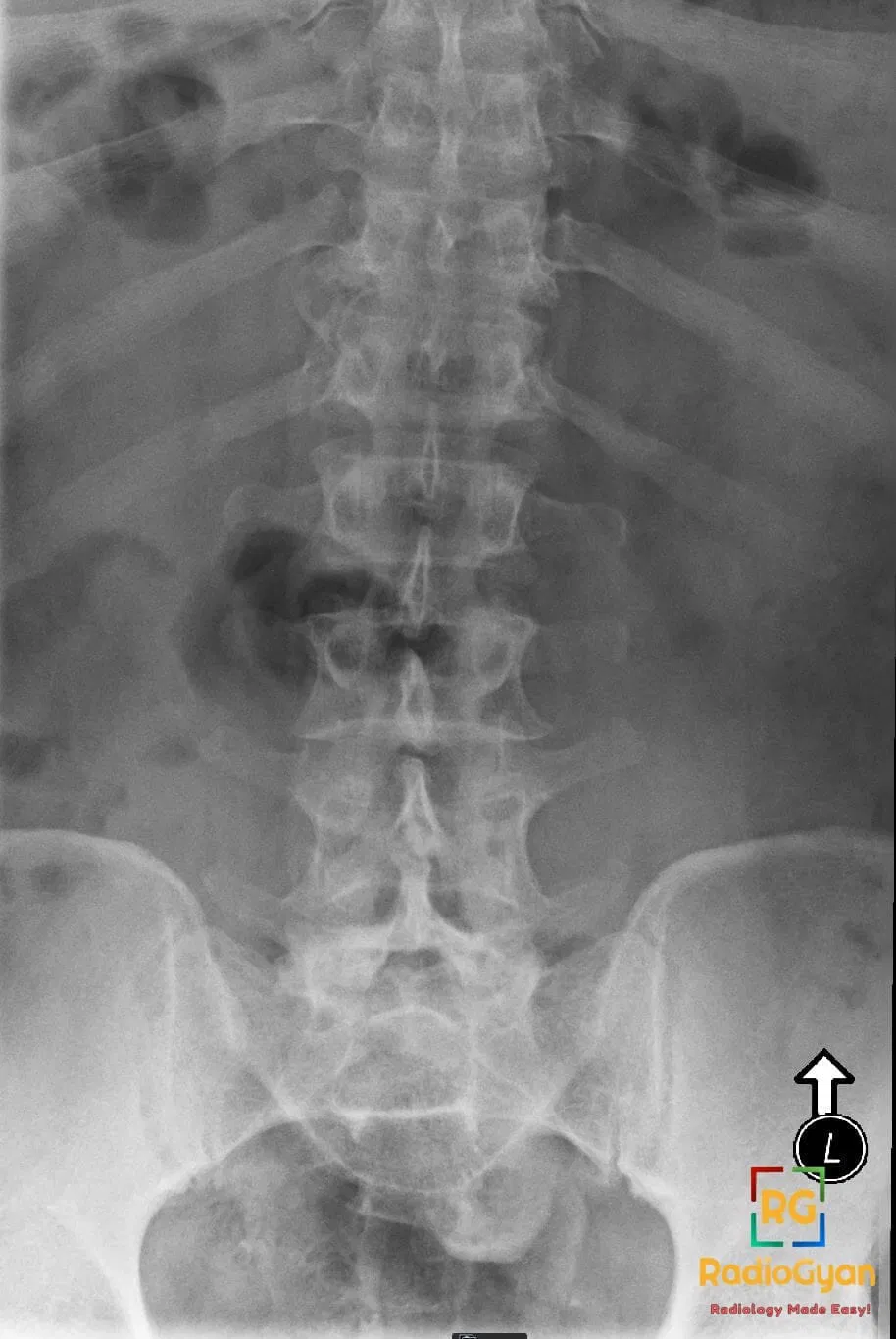
Napoleon hat sign | Radiology Signs

Northern exposure sign | Radiology Signs

Nubbin Sign (Hepatobiliary Scintigraphy) | Radiology Signs

Nutcracker fracture | Radiology Signs

Oil Droplet Sign CT: Radiological Appearance and Significance

Omental caking | Radiology Signs

Onion Skin Periosteal Reaction (Bone) | Radiology Signs

Onion Skin Sign (Testicular Epidermoid Cyst) | Radiology Signs

Oreo cookie sign | Radiology Signs

Owl eye sign | Radiology Signs

Page kidney | Radiology Signs

Pancake brain | Radiology Signs

Pancake kidney | Radiology Signs

Pancake vertebra | Radiology Signs

Sarcoidosis Chest Radiograph showing garland sign pawnbroker sign and 1-2-3 sign

Pawnbroker’s sign | Radiology Signs

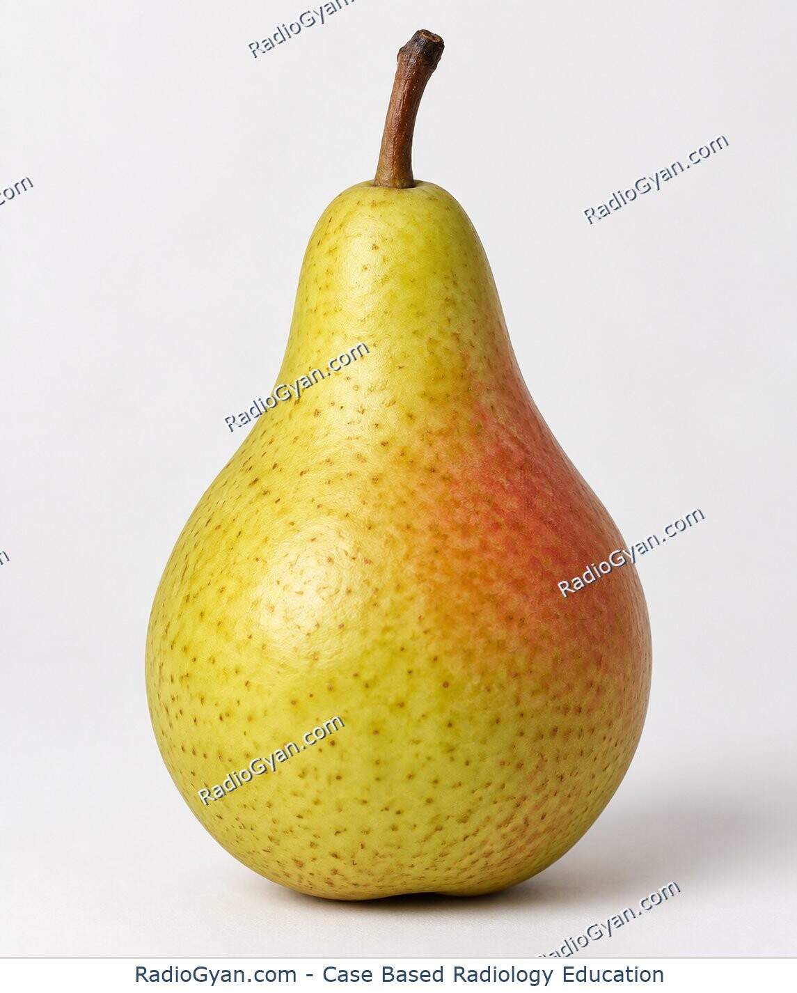
Pear-shaped Bladder | Radiology Signs

Pedestal sign | Radiology Signs

Pencil in Cup Deformity | Radiology Signs

Pencil pointing | Radiology Signs

Penumbra sign | Radiology Signs

Phantom Calyx Sign | Radiology Signs

Picture Frame Vertebral Body | Radiology Signs

Pie-in-the-sky sign | Radiology Signs

Piece-of-pie sign | Radiology Signs

Polka Dot sign | Radiology Signs

Polo mint sign | Radiology Signs

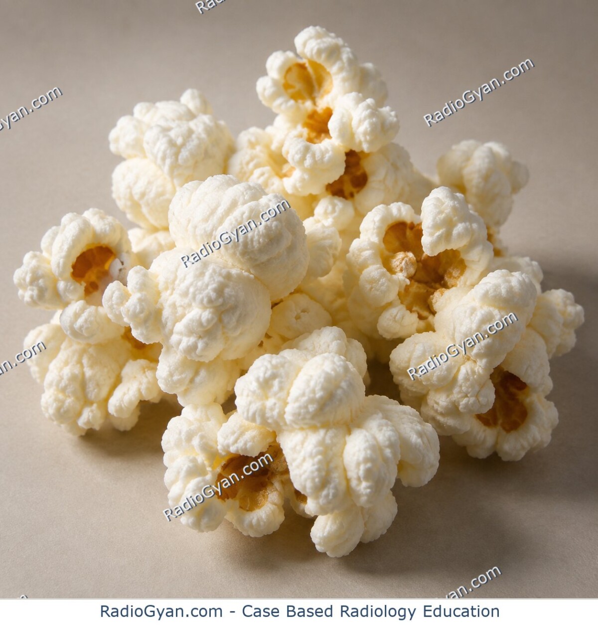
Popcorn Calcification | Radiology Signs

Positive bronchus sign | Radiology Signs

Pseudo-kidney Sign | Radiology Signs

Pseudo-Rigler’s sign | Radiology Signs

Puckered Panniculus Sign | Radiology Signs

Puff of smoke sign | Radiology Signs

Pulvinar sign | Radiology Signs

Putty kidney | Radiology Signs

Pyloric Tit sign | Radiology Signs

Quad sign | Radiology Signs

Rachitic rosary | Radiology Signs

Ram’s horn sign | Radiology Signs

Rat bite sign | Radiology Signs

Rat tail sign | Radiology Signs

Reversal sign | Radiology Signs

Reverse 3 sign | Radiology Signs

Reverse pulmonary edema | Radiology Signs

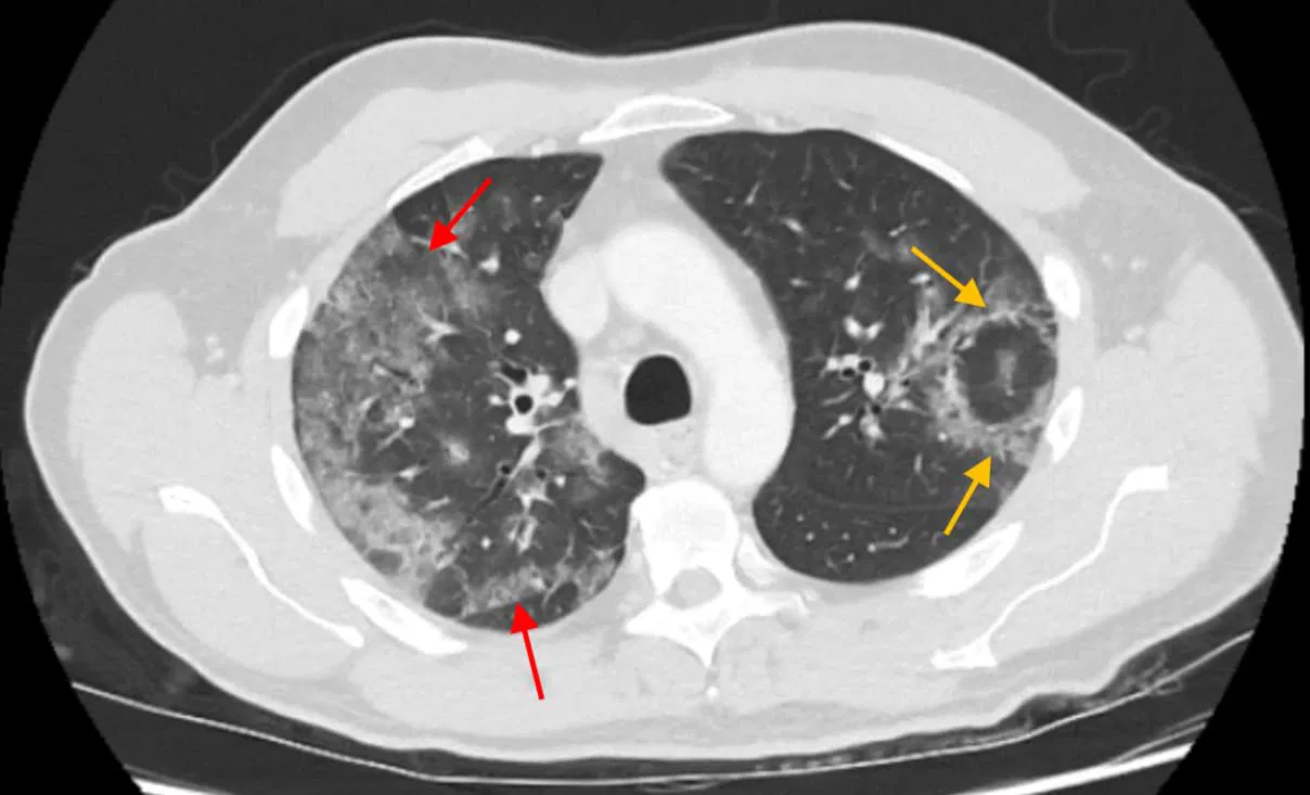
Coronavirus radiology CT imaging features atoll sign reverse halo sign

Reversed Halo Sign | Radiology Signs

Ribbon ribs | Radiology Signs

Rice grain calcification | Radiology Signs

Abdominal X-rays showing Rigler's sign, indicating pneumoperitoneum with air on both sides of bowel walls.

Rigler’s sign | Radiology Signs

Rim Sign (Hepatobiliary Scintigraphy) | Radiology Signs

Rim Sign (Hip and Shoulder) | Radiology Signs

Ring of fire sign | Radiology Signs

Ring Sign (Bone Tumours) | Radiology Signs

Ring-around-artery sign | Radiology Signs

Rolled edge sign | Radiology Signs

Rugger jersey spine | Radiology Signs

Saber sheath trachea | Radiology Signs

Saber shin deformity Radiology Signs

Sack of marbles sign | Radiology Signs

Sagging rope sign | Radiology Signs

Salt and pepper skull Radiology Signs

Sandwich Sign (Mesentery) | Radiology Signs

Sandwich Vertebra (Spine) | Radiology Signs

Satellite sign | Radiology Signs

Sausage digit (dactylitis) | Radiology Signs

Sawtooth sign | Radiology Signs

Scalloped vertebra | Radiology Signs

Scimitar sign | Radiology Signs

Scottie dog | Radiology Signs

Seashore sign | Radiology Signs

Secondary cleft sign | Radiology Signs

Segond fracture | Radiology Signs

Sentinel loop sign | Radiology Signs

Serpiginous tract sign | Radiology Signs

Shaggy esophagus | Radiology Signs

Shepherd’s crook deformity | Radiology Signs

Shiny corner sign | Radiology Signs

Shmoo sign | Radiology Signs

Signet Ring Sign (Lung) | Radiology Signs

Silhouette Sign | Radiology Signs

Axial and sagittal CT images showing an umbilical nodule and widespread abdominal and pelvic carcinomatosis.

Sister Mary Joseph sign | Radiology Signs

Six-point star sign | Radiology Signs

CT scan showing small bowel feces sign and an adhesion in a patient with bowel obstruction

Small bowel feces sign | Radiology Signs

Snow-capping | Radiology Signs

Snowman sign | Radiology Signs

Sagittal ultrasound image of the uterus showing a complete molar pregnancy with hydropic villi.

Snowstorm appearance | Radiology Signs

Soap-bubble nephrogram | Radiology Signs

Soap-bubbly appearance | Radiology Signs

Soft Tissue Rim Sign (Ureter) | Radiology Signs

Sonographic Murphy sign | Radiology Signs

Spaghetti sign | Radiology Signs

Spilled Teacup Sign (Wrist) | Radiology Signs

Paired lateral chest X-rays showing increased lower lobe density, lost spine lucency, consistent with pneumonia

Spine sign | Radiology Signs

Spinnaker sail sign | Radiology Signs

Spinning top urethra | Radiology Signs

Split fat sign | Radiology Signs

Split Pleura Sign – Empyema Radiology Case.

Split pleura sign | Radiology Signs

Spoke wheel appearance | Radiology Signs

Spot sign | Radiology Signs

Spotted nephrogram | Radiology Signs

Stack-of-coins appearance | Radiology Signs

Starry sky liver | Radiology Signs

Chest X-ray showing steeple sign, indicative of croup in a pediatric patient.

Steeple Sign in Croup: X-ray Findings and Differentials

Stepladder appearance | Radiology Signs

Stipple sign | Radiology Signs

Stocking-glove sign | Radiology Signs

String of Pearls Sign (Small Bowel) | Radiology Signs

String sign | Radiology Signs

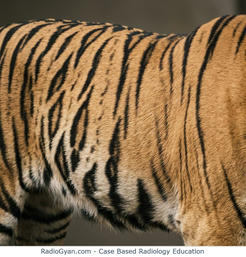
Close-up of a tiger's flank with bold black stripes running across orange-gold fur, illustrating the tigroid stripe sign.

Stripe sign | Radiology Signs

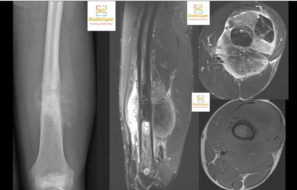
AP radiograph of the distal femur and MRI images show a geographic lesion with cortical destruction and soft tissue component in keeping with an osteogenic sarcoma

Sunburst periosteal reaction | Radiology Signs

X-ray of the right hand showing swan-neck deformity of the fifth digit and palmar subluxation at the MCP joint.

Swan neck deformity | Radiology Signs

Swirl Sign (Brain) | Radiology Signs

Swiss cheese appearance | Radiology Signs

Talar beak | Radiology Signs

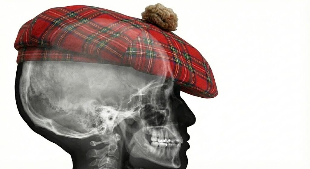
Lateral skull X-ray displaying the Tam O'Shanter Hat sign, characteristic of Paget's disease of the bone.

Tam O’ Shanter Sign | Radiology Signs

Target Calcification (Breast) | Radiology Signs

CT showing an intussusception in the left upper quadrant of the abdomen

Target Sign (Bowel) | Radiology Signs

Tau sign | Radiology Signs

Teacup Sign (Breast) | Radiology Signs

Teardrop sign | Radiology Signs

Terry Thomas Sign Scapholunate dislocation

Terry Thomas Sign | Radiology Signs

Threads and streaks sign | Radiology Signs

Three Territory sign | Radiology Signs

Lateral neck radiograph showing a swollen epiglottis with the classic thumb sign of epiglottitis

Thumb sign | Radiology Signs

Thumbprinting of colon on abdominal radiograph due to colitis

Thumbprinting Sign on Abdominal X-ray: Causes and Differentials

Thymic sail sign | Radiology Signs

Tigroid pattern | Radiology Signs

Tooth Sign (Patella) | Radiology Signs

Tram-tracking | Radiology Signs

Tree-in-bud sign | Radiology Signs

Ultrasound showing triangular cord sign and hepatobiliary scan indicating lack of hepatic excretion, diagnostic for biliary atresia.

Triangular cord sign | Radiology Signs

Axial FLAIR MRI showing hyperintensity in the central pontine region, indicative of osmotic demyelination syndrome.

Trident Sign (Pons) | Radiology Signs

Trident Sign (Spinal Cord) | Radiology Signs

Triple Sign | Radiology Signs

Trolley track sign | Radiology Signs

X-ray of a right shoulder showing posterior dislocation, reverse Bankart, and reverse Hill-Sachs lesions with a trough sign.

Trough sign | Radiology Signs

Tubular artery sign | Radiology Signs

Tulip bulb sign | Radiology Signs

Tumbling bullet | Radiology Signs

Twin-peak sign | Radiology Signs

Upside down stomach sign | Radiology Signs

Vacuum disk sign | Radiology Signs

Vacuum Joint | Radiology Signs

Vanishing tumour | Radiology Signs

Axial contrast-enhanced CT showing a replaced left hepatic artery coursing through the fissure for ligamentum venosum

Vessel through Strait sign | Radiology Signs

Virchow's nodes and Troisier sign

Virchow’s Nodes and Troisier Sign | Radiology Capsule

Water bottle heart Radiology Signs

Water lily sign | Radiology Signs

WES sign | Radiology Signs

Westermark’s sign | Radiology Signs

Wheel within a wheel | Radiology Signs

Whirlpool Sign (Abdomen) | Radiology Signs

Whiskering | Radiology Signs

White cerebellum sign | Radiology Signs

Wimberger Ring Sign (Scurvy) | Radiology Signs

Wimberger Sign (Congenital Syphilis) | Radiology Signs

Wine glass sign | Radiology Signs

Winking owl sign | Radiology Signs

Y Sign (Spine) | Radiology Signs

Axial CT and sagittal Doppler ultrasound show a left axillary nodal pseudoaneurysm with central enhancement, yin-yang flow

Yin-yang sign | Radiology Signs

This page was last updated on Sep 4, 2026 @ 1:28 pm

About the Author


Dr. Amar Udare, MD, DNB

Dr Amar Udare Dr. Amar Udare is a board-certified radiologist (MD,DNB) with over 10 years of experience and a special interest in body imaging. He currently serves as a Clinical Associate Professor in Diagnostic Imaging (Radiology) at the University of Calgary . With a passion for teaching, he has been a semi-finalist for the Aunt-Minnie Most effective Radiology Educator Awards in 2018 and 2020.

Dr. Udare holds an MBBS and MD degree, and his expertise lies in the field of radiology. He has authored multiple peer-reviewed publications, contributing significantly to the medical field. His works can be accessed on PubMed and Google Scholar.

In addition to his academic and professional achievements, Dr. Udare is an avid reader and enjoys exploring the latest advancements in medical technology. His commitment to making complex medical knowledge accessible to patients and the general public aligns with our mission at RadioGyan.com.

For any further questions or clarifications, feel free to reach out to Dr. Udare via the contact form.


Wish to be a BETTER Radiologist? Join 15000 Radiology Colleagues !

Enter your email address below to access HIGH YIELD radiology content, updates, and resources.

Email Newsletter Subscription Pop Up

No spam, only VALUE! Unsubscribe anytime with a single click.

Scroll to Top