


What causes the insular ribbon sign in the brain on computed tomography?
Let me know in the comments.
Click to reveal the answer
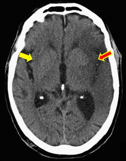
Answer: The insular ribbon sign (loss of the insular ribbon) is caused by acute middle cerebral artery (MCA) infarction. It represents early ischemic edema affecting the insular cortex, a region supplied by the insular segment of the MCA. This ischemic insult leads to loss of the normal gray-white matter differentiation at the lateral margin of the insula, reflecting cytotoxic edema in the brain tissue. Clinically, this sign is associated with acute ischemic stroke in the MCA territory, seen on non-contrast CT.
Why is it called so?
It is named the “insular ribbon” because the insular cortex normally appears as a thin ribbon of gray matter along the lateral margin visible on CT scans. The sign refers specifically to the loss of this distinct gray-white matter interface, hence the term “loss of the insular ribbon sign.”


Pathophysiology
Following occlusion of the middle cerebral artery, blood flow cessation leads to ischemia and subsequent cytotoxic edema in the insular cortex. The insula lies in a vascular “watershed” zone most distal from collateral flow from the anterior and posterior cerebral arteries, making it highly vulnerable to ischemia. Edema causes the gray and white matter to lose their normal density contrast on CT, resulting in loss of the insular ribbon sign visibility. This early edema is one of the first radiologic signs of acute MCA territory stroke.
Alternative names: None
Other associated named signs: Obscuration of the lentiform nucleus, hyperdense MCA sign, hemispheric sulcal effacement (other early CT signs of MCA infarction)
The insular ribbon sign (more precisely, loss of the insular ribbon) is loss of the normal grey-white matter definition along the lateral margin of the insular cortex on non-contrast CT. It is an early, hyperacute-to-acute sign of middle cerebral artery (MCA) territory infarction, first described by Truwit and colleagues in 1990. The insula is the MCA territory most distant from anterior and posterior cerebral collaterals, so it is the first cortex to show cytotoxic oedema when MCA flow stops.
What is the insular ribbon sign?
On a normal non-contrast CT, the insular cortex is seen as a thin band (“ribbon”) of grey matter along the lateral wall of the Sylvian fissure, sharply separated from the subjacent white matter of the extreme capsule and claustrum. In acute infarction, cytotoxic oedema increases tissue water and lowers attenuation, blurring this grey-white boundary so the ribbon becomes indistinct or isodense with adjacent white matter. That blurring is the loss of the insular ribbon.
- Modality: non-contrast CT (the sign is defined on NCCT; it is not needed once DWI shows the infarct).
- Location: lateral insular cortex, deep to the Sylvian fissure.
- Finding: loss of the normal grey-white differentiation, best judged by side-to-side comparison with the normal hemisphere.
Why the insula is vulnerable: pathophysiology
The insular cortex is supplied by the insular segment (M2) of the MCA and its claustral branches. Unlike the convexity cortex, the insula receives essentially no leptomeningeal collateral flow from the anterior or posterior cerebral arteries, so it sits at the far end of the collateral network, in effect an internal watershed. When MCA flow ceases, the insula is the region most quickly deprived of perfusion and the first to develop cytotoxic oedema, which is why loss of the insular ribbon is one of the earliest parenchymal signs of MCA stroke.
How to look for it on CT
- Compare sides. Scroll to the ganglionic level and directly compare the two insular cortices; the sign is a relative loss of contrast on one side.
- Use narrow (stroke) windows. A narrow window width (around 30-40 HU) exaggerates the small attenuation difference between oedematous and normal grey matter and makes subtle early ischaemia visible.
- Confirm on consecutive slices. A true change should be visible on at least two consecutive cuts, which helps separate real ischaemia from partial-volume averaging over the Sylvian fissure.
- Correlate with the vessels. Look for a companion hyperdense MCA sign and always proceed to CT angiography to localise the occlusion.
Insular ribbon loss among the early CT signs of MCA infarction
Loss of the insular ribbon is one of a family of early ischaemic changes. Recognising the group, and what each implies about the occlusion site, sharpens the read.
| Early CT sign | What you see | Basis | Occlusion clue |
|---|---|---|---|
| Loss of the insular ribbon | Blurred grey-white interface at the lateral insula | Cytotoxic oedema in a collateral-poor watershed cortex | Proximal MCA or ICA occlusion |
| Obscuration of the lentiform nucleus | Faint, ill-defined basal ganglia | Early oedema of lentiform nucleus | M1 (sphenoidal) MCA occlusion |
| Hyperdense MCA sign | Dense M1 segment on NCCT | Intraluminal thrombus | Confirms MCA clot; does not localise cortex |
| MCA “dot” sign | Dense dot in the Sylvian fissure | Thrombus in an M2/M3 branch | Distal branch occlusion |
| Loss of grey-white differentiation | Cortical ribbon blurring over the convexity | Cytotoxic oedema | Territorial MCA ischaemia |
| Cortical sulcal effacement | Loss of sulci from swelling | Vasogenic and cytotoxic oedema, mass effect | Branch or territorial occlusion |
Relationship to ASPECTS
The insula (“I”) is one of the ten regions of the Alberta Stroke Program Early CT Score (ASPECTS). Early ischaemic change in any part of the insular ribbon subtracts one point from the score of 10, so loss of the insular ribbon directly lowers ASPECTS. Because ASPECTS is used to select patients for reperfusion, reliably recognising insular change matters for triage, not just for the spot diagnosis. Estimate ASPECTS quickly with our ASPECTS score calculator, and revise the relevant vascular territories with our case on brain arterial vascular territories.
Timing, sensitivity and reliability
- Timing: loss of the insular ribbon can appear within the first few hours of onset and, in the original series, was seen in MCA strokes less than 6 hours old.
- Sensitivity is limited: across a systematic review of early CT infarction signs, mean sensitivity was about 66% with specificity about 87%, meaning the signs are often missed but, when confidently seen, are usually genuine.
- Reader dependent: interobserver agreement for parenchymal early ischaemic changes is only fair to moderate and improves with experience and with narrow review windows.
- CT is a first pass: diffusion-weighted MRI is far more sensitive and specific (both above 90%) for hyperacute infarction; a normal insula on CT never excludes stroke.
Differential diagnosis and mimics
Insular cortical hypoattenuation or ribbon loss is most often ischaemic, but it is not specific. Consider the clinical context and the pattern of involvement.
| Cause | Clue that favours it |
|---|---|
| Acute MCA infarction | Sudden focal deficit; unilateral, wedge-shaped territorial pattern; companion hyperdense MCA sign; matching CTA occlusion |
| Herpes simplex encephalitis | Fever, confusion, seizures; insular and medial temporal or limbic involvement, often bilateral and asymmetric; not confined to one vascular territory |
| Seizure or status epilepticus | Cortical swelling and blurring that crosses territories, may be transient and reversible; clinical seizure history |
| Hypoglycaemia | Symmetric cortical and basal ganglia changes; documented low glucose; deficit reverses with correction |
| MELAS | Younger patient; stroke-like lesions crossing vascular territories, often parieto-occipital and insular; lactate elevation |
Clinical significance and pitfalls
- Trigger, not endpoint: the sign should prompt urgent CT angiography, perfusion imaging where available, and a time-critical decision on thrombolysis and thrombectomy, not a wait-and-watch approach.
- Do not overcall. Beam-hardening artefact at the skull base, an old infarct, or partial-volume averaging over the Sylvian fissure can mimic the sign; require the change on consecutive slices and correlate with symptoms.
- Absence proves nothing. A normal insular ribbon does not exclude hyperacute stroke; if suspicion is high, proceed to CTA, CT perfusion or MRI.
- Extent matters. Widespread early ischaemic change (low ASPECTS) flags a large established core and a higher haemorrhagic risk with reperfusion.
Frequently asked questions
What is the insular ribbon sign?
The insular ribbon sign, or loss of the insular ribbon, is loss of the normal grey-white matter definition along the lateral margin of the insular cortex on non-contrast CT. It is an early sign of acute middle cerebral artery territory infarction caused by cytotoxic oedema of the insular cortex.
What causes loss of the insular ribbon?
It is caused by acute middle cerebral artery infarction. The insula is the MCA territory most distant from anterior and posterior cerebral collaterals, so it is the first cortex to develop ischaemic cytotoxic oedema when MCA flow stops, blurring the grey-white interface.
How early can the insular ribbon sign be seen after stroke onset?
It is a hyperacute-to-acute sign that can appear within the first few hours and, in the original description, was seen in MCA strokes less than 6 hours old. However, sensitivity is limited and a normal insula does not exclude stroke.
Is the insular ribbon part of the ASPECTS score?
Yes. The insula is one of the ten ASPECTS regions. Early ischaemic change in the insular ribbon subtracts one point from the score of 10, so recognising it directly affects ASPECTS and reperfusion triage.
What can mimic loss of the insular ribbon?
Mimics include herpes simplex encephalitis, seizure or status epilepticus, hypoglycaemia and MELAS, as well as technical pitfalls such as beam-hardening artefact, old infarct and partial-volume averaging. Clinical context and whether the change respects a vascular territory help distinguish them.
References
- Truwit CL, Barkovich AJ, Gean-Marton A, Hibri N, Norman D. Loss of the insular ribbon: another early CT sign of acute middle cerebral artery infarction. Radiology. 1990;176(3):801-806. PMID: 2389039.
- Nakano S, Iseda T, Kawano H, Yoneyama T, Ikeda T, Wakisaka S. Reappraisal of early CT signs to predict the arterial occlusion site in acute embolic stroke. J Neurol Neurosurg Psychiatry. 2003;74(5):649-653. PMID: 12700311.
- Wardlaw JM, Mielke O. Early signs of brain infarction at CT: observer reliability and outcome after thrombolytic treatment – systematic review. Radiology. 2005;235(2):444-453. PMID: 15858087.
- Barber PA, Demchuk AM, Zhang J, Buchan AM. Validity and reliability of a quantitative computed tomography score in predicting outcome of hyperacute stroke before thrombolytic therapy. ASPECTS Study Group. Lancet. 2000;355(9216):1670-1674. PMID: 10905241.
Access all radiology signs posted so far: https://radiogyan.com/radiology-signs/

Improve Content And Help Your Colleagues!
Found an error in the post? Please let us know using our contact page and we will update it with due credits!
If you wish to contribute radiological images for this case (or any other article on the website), you can submit them here and you will be duly credited: Submit radiology case