


What Causes Boot-Shaped Heart in the Cardiovascular System on Chest X-Ray?
Let me know in the comments.
Click to reveal the answer
The boot-shaped heart appearance on chest X-ray is caused by Tetralogy of Fallot, a cyanotic congenital heart disease characterized by four anomalies: pulmonary stenosis (right ventricular outflow tract obstruction), ventricular septal defect, overriding aorta, and right ventricular hypertrophy. This condition leads to reduced pulmonary blood flow and right ventricular enlargement.
Why is it called so?
It is named “boot-shaped heart” or “cœur en sabot” (French for “clog-shaped heart” or “boot-shaped heart”) because the cardiac silhouette on frontal chest radiograph resembles the shape of a boot or shoe, with an elevated apex and a concave main pulmonary artery segment.

Pathophysiology
The sign develops due to right ventricular hypertrophy causing the cardiac apex to be uplifted and pointing upwards, forming the “toe” of the boot. Simultaneously, infundibular pulmonary stenosis or pulmonary trunk hypoplasia leads to a diminished, concave main pulmonary artery segment, creating the “instep” of the boot. This characteristic appearance reflects the altered cardiac anatomy and hemodynamics from the obstructed right ventricular outflow and compensatory ventricular growth.
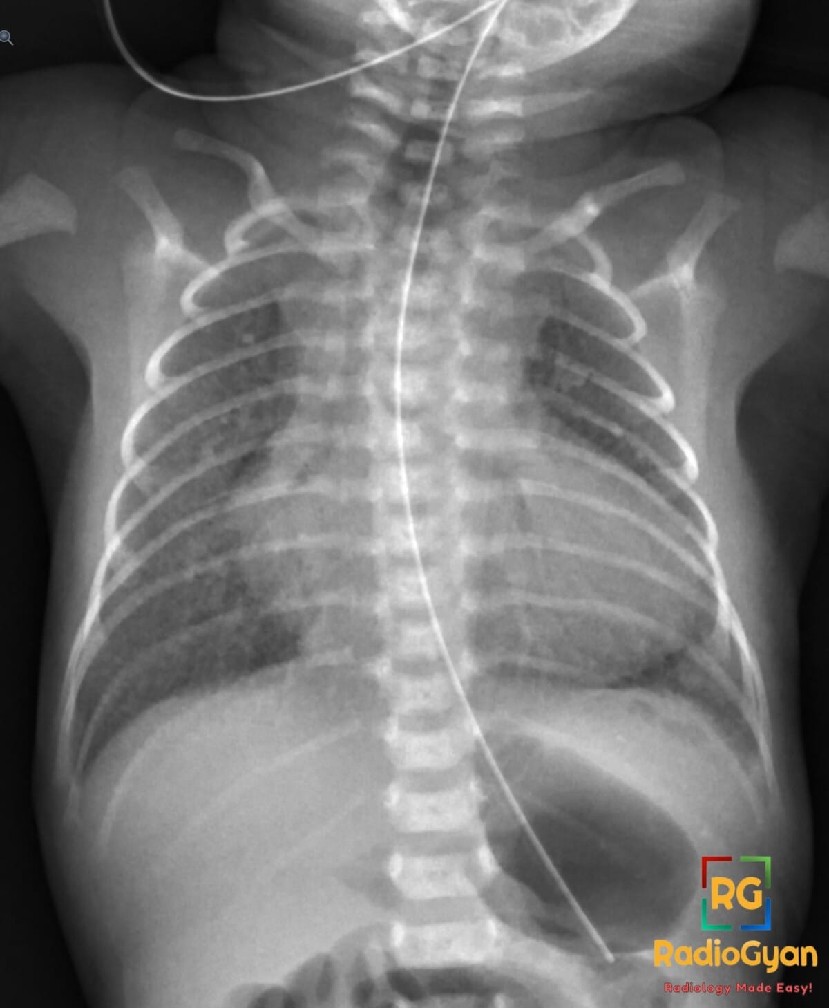
The boot-shaped heart – coeur en sabot – is a cardiac silhouette on the frontal chest radiograph in which the apex is lifted off the diaphragm and the left heart border is concave where the main pulmonary artery segment should bulge. The classic cause is tetralogy of Fallot. Two things matter more than the pattern itself. First, it is a late sign: it reflects right ventricular hypertrophy that develops over months, so the film of a newborn with tetralogy of Fallot is usually unremarkable. Second, the chest radiograph is a weak test for congenital heart disease in general – sensitivity of 26 to 59 percent in neonates, and about 71 percent accuracy for naming a specific lesion. A boot shape is a prompt to get an echocardiogram. Its absence is not reassurance.
What makes the heart look like a boot?
The silhouette has two components, and both are needed before the word boot is justified.
- The toe – the cardiac apex is uplifted and rounded, sitting above the left hemidiaphragm rather than resting on it. In tetralogy of Fallot the apex is formed by a hypertrophied right ventricle, which rotates the apex upwards and laterally.
- The instep – the left heart border immediately below the aortic knuckle is concave instead of convex, because the pulmonary trunk and right ventricular outflow tract are small. This is the segment that normally produces the pulmonary bay.
Two negatives are just as important. The heart is not enlarged in uncomplicated tetralogy of Fallot: the right ventricle hypertrophies against a fixed obstruction rather than dilating, and there is no left-to-right shunt to volume-load it. A genuinely big heart should push you towards a different lesion. And the lungs are oligaemic, because pulmonary blood flow is reduced – although, as the evidence below shows, this is the part of the gestalt readers are least reliable at seeing.
The four lesions and what each contributes to the film
| Component of the tetralogy | What it is | What it does on the chest X-ray |
|---|---|---|
| Right ventricular outflow tract obstruction | Infundibular (subvalvar) narrowing, often with valvar pulmonary stenosis and a hypoplastic pulmonary trunk | Concave main pulmonary artery segment – the instep – plus reduced pulmonary blood flow and oligaemic lungs |
| Ventricular septal defect | Large, non-restrictive, malalignment-type defect that equalises ventricular pressures | No chamber-specific radiographic sign of its own; it is why the right ventricle hypertrophies rather than fails |
| Overriding aorta | Aortic root straddles the septal defect and receives blood from both ventricles | Aortic arch and ascending aorta may look prominent; a right-sided arch is present in about a quarter of patients |
| Right ventricular hypertrophy | Concentric hypertrophy in response to the fixed outflow obstruction | Uplifted, rounded apex – the toe – with the overall cardiac size remaining normal |
Why is it called coeur en sabot?
Coeur en sabot is French for heart in a clog. A sabot is the traditional wooden shoe with a blunt, upturned toe – which is exactly what the uplifted apex and scooped-out pulmonary bay reproduce in outline. The malformation itself is named for Etienne-Louis Arthur Fallot, the Marseille physician who in 1888 set out the four components as a single entity and linked them to the clinical picture then called la maladie bleue. The clog descriptor belongs to the later radiographic era, once the cardiac silhouette could be seen in life.
One point of housekeeping, because it is a common search: there is no separate boat-shaped heart sign. Boat-shaped is a mishearing of boot-shaped, and the two refer to the same silhouette. If you meet the phrase in a report or a question bank, read it as coeur en sabot.
How good a test is the boot-shaped heart, really?
This is the part that classical teaching leaves out. The boot is taught as though it settles the diagnosis; the published performance of the chest radiograph in congenital heart disease says otherwise.
| Study | Design | Result |
|---|---|---|
| Fonseca et al., 2005 | 128 consecutive neonates with suspected heart disease; three blinded readers, echocardiography as reference | Sensitivity for structural heart disease only 26 to 59 percent, negative predictive value 46 to 52 percent. Agreement between the film and the echocardiogram was poor (kappa 0.15 to 0.34). Sensitivity was lower still below 2 kg or 35 weeks |
| Laya et al., 2006 | 281 children under 12 years; five paediatric radiologists, 12 categories of congenital heart disease plus controls | Accuracy for distinguishing normal from congenital heart disease 78 percent; accuracy for naming the specific lesion 71 percent. The authors concluded that the radiograph alone is not diagnostic of specific cardiac lesions |
| Tumkosit et al., 2012 | 120 children, pulmonary-to-systemic flow ratio at catheterisation as reference | Sensitivity for decreased pulmonary vascularity – the pattern of tetralogy of Fallot – was only 24 to 68 percent, against 84 to 94 percent for normal or increased vascularity |
Read together, these results explain why the sign behaves the way it does in practice. The half of the boot gestalt that depends on lung vascularity – oligaemia – is the half readers are worst at seeing. The silhouette itself takes months of hypertrophy to develop, so a newborn with a duct still open and a well-formed chest film can have a severe tetralogy. And even the coarse question is there heart disease at all is answered correctly by an experienced reader only about three-quarters of the time.
The sign is also becoming rarer in the settings where imaging is most available, simply because the diagnosis is now made earlier. In a US series, 63 percent of infants with tetralogy of Fallot were diagnosed prenatally, and universal newborn pulse oximetry screening catches a further share before any silhouette has had time to change. A florid boot in a school-age child today usually means the diagnosis was missed, or that access to care was limited.
What else produces a boot-shaped heart?
The pattern is a description of shape, not a diagnosis. Anything that combines a small pulmonary outflow segment with a right-ventricular apex will reproduce it, and several things imitate it for reasons that have nothing to do with the heart.
| Entity | How the film looks | What separates it |
|---|---|---|
| Tetralogy of Fallot | Normal heart size, uplifted apex, concave pulmonary bay, oligaemic lungs | Right aortic arch in about a quarter of cases; cyanosis with hypercyanotic spells; the reference diagnosis for this sign |
| Pulmonary atresia with ventricular septal defect | The same silhouette, usually with more marked oligaemia or a coarse reticular pattern from aortopulmonary collaterals | Radiographically indistinguishable from severe tetralogy; separated by echocardiography and CT angiography of the collateral supply |
| Tricuspid atresia | Can look boot-shaped with reduced pulmonary flow | Left-axis deviation with left ventricular dominance on the ECG – the opposite of tetralogy of Fallot |
| Double-outlet right ventricle with pulmonary stenosis | Boot-like silhouette with decreased vascularity | Not separable on the radiograph; a physiological tetralogy imitator |
| Tetralogy of Fallot with absent pulmonary valve | Not a boot – aneurysmal central pulmonary arteries and hyperinflation or lobar air trapping from bronchial compression | A large hilar mass-like appearance in a cyanotic infant should redirect you to this variant |
| Ebstein anomaly | Globular, box-shaped, wall-to-wall cardiomegaly with oligaemic lungs | Massive heart size; tetralogy of Fallot does not enlarge the heart |
| Transposition of the great arteries | Oval heart with a narrow superior mediastinum and increased vascularity | The egg-on-a-string sign, not a boot |
| Left ventricular enlargement in an adult | Apex displaced downward and laterally with genuine cardiomegaly | The pulmonary segment is normal or full, not scooped out – see the shmoo sign |
| Technical pseudo-boot | Rotated, lordotic or expiratory paediatric film | Check the clavicles and the rib count before committing; repeat the film rather than the diagnosis |
Boot-shaped heart in adults
Searches for this phrase in adults usually have one of three answers, and only the first is a true coeur en sabot.
- Unrepaired or late-presenting tetralogy of Fallot. Uncommon where paediatric cardiac surgery is available, but far from extinct globally. These are the adults in whom the textbook silhouette is actually seen, usually with clubbing, polycythaemia and long-standing cyanosis.
- Repaired tetralogy of Fallot. The film shows sternotomy wires, sometimes rib or clavicular changes from a previous shunt thoracotomy, and progressive enlargement of the right ventricle from chronic pulmonary regurgitation. The classic boot is usually gone; what replaces it is a big right heart.
- A left-ventricular heart being called a boot. Aortic regurgitation, long-standing hypertension and dilated cardiomyopathy all displace the apex, and the outline is loosely described as boot-like. The discriminators are heart size and the pulmonary segment: left ventricular enlargement gives an overall enlarged heart with the apex pushed down and out and a normal or full pulmonary bay, whereas the tetralogy boot has a normal-sized heart with the apex lifted up and the pulmonary bay scooped away.
Chest X-ray clues in tetralogy of Fallot that beat the boot
If you are going to read the film at all, these are the findings with better yield than the silhouette.
- Right-sided aortic arch. In a series of 2,684 patients with tetralogy of Fallot, 25.7 percent had a right aortic arch, against 73.9 percent left. In a cyanotic infant, a right arch is a stronger pointer to a conotruncal anomaly than the cardiac outline, and it matters surgically. It is not specific to tetralogy – truncus arteriosus and pulmonary atresia with ventricular septal defect share it.
- Absent or small thymic shadow. Raises the question of 22q11.2 deletion, found in around 13 percent of patients with tetralogy of Fallot and concentrated in those with pulmonary atresia and major aortopulmonary collaterals. It changes genetic testing, immunology and surgical planning.
- Pulmonary vascularity. Decreased flow supports the diagnosis, but treat a confident call cautiously given a sensitivity as low as 24 percent for the oligaemic pattern.
- Heart size. Normal in uncomplicated tetralogy of Fallot. Cardiomegaly should prompt you to think of absent pulmonary valve syndrome, Ebstein anomaly, or a non-cardiac explanation.
Cardiac silhouette signs in congenital heart disease
The boot belongs to a small family of shape signs that survive in teaching because they are memorable, all of which carry the same caveat about sensitivity.
| Sign | Lesion | Mechanism |
|---|---|---|
| Boot-shaped heart (coeur en sabot) | Tetralogy of Fallot | Right ventricular hypertrophy uplifts the apex; small pulmonary trunk scoops out the pulmonary bay |
| Egg-on-a-string | Transposition of the great arteries | Oval cardiac mass with a narrow vascular pedicle from the anteroposterior relationship of the great arteries and thymic involution |
| Snowman or figure-of-8 | Supracardiac total anomalous pulmonary venous return | Dilated vertical vein, innominate vein and superior vena cava form the head above the cardiac body |
| Box-shaped, wall-to-wall heart | Ebstein anomaly | Massive right atrial enlargement from atrialisation of the right ventricle with tricuspid regurgitation |
| Figure-3 sign | Aortic coarctation | Pre-stenotic and post-stenotic dilatation either side of the coarctation shelf, with rib notching from collaterals |
| Shmoo sign | Left ventricular enlargement, classically aortic stenosis or regurgitation | Apex displaced down and to the left with a dilated ascending aorta |
What to do when you see a boot-shaped heart
- Order an echocardiogram, not another radiograph. The echocardiogram establishes the diagnosis and usually provides enough anatomical detail to plan treatment. Because the chest film has a negative predictive value under 55 percent in neonates, a normal-looking film in a cyanotic baby does not exclude structural disease.
- Assess the baby, not only the film. Pre-ductal and post-ductal saturations, a hyperoxia test where appropriate, and the murmur – in tetralogy of Fallot the murmur comes from the outflow obstruction, and it becomes quieter during a hypercyanotic spell.
- Cross-sectional imaging where echocardiography leaves questions. CT angiography or cardiac MRI for branch pulmonary artery anatomy, aortopulmonary collaterals, arch sidedness and branching, and an anomalous coronary artery crossing the outflow tract, which changes the operation.
- Genetics. Chromosomal microarray or fluorescence in situ hybridisation for 22q11.2 deletion, particularly with pulmonary atresia, collaterals, a right arch or an aberrant subclavian artery.
- Timing of repair. Most centres perform complete repair at 3 to 6 months, and certainly within the first year. Neonatal repair is reserved for severe outflow obstruction or duct-dependent pulmonary flow: in a national inpatient analysis, repair in the neonatal period carried higher mortality and more postoperative complications than repair later in infancy.
Repaired tetralogy of Fallot and the 2025 guideline update
Most patients now reach adulthood, and the imaging question shifts from diagnosis to surveillance. In a prospective cohort followed for four decades after surgical correction, cumulative survival was 72 percent at 40 years but event-free survival was only 25 percent – reoperation and arrhythmia, not death, dominate the later course. The driver is chronic pulmonary regurgitation after relief of the outflow obstruction, particularly where a transannular patch was used, leading to progressive right ventricular dilatation, myocardial fibrosis, ventricular tachycardia and sudden cardiac death.
The 2025 ACC/AHA/HRS/ISACHD/SCAI guideline for adults with congenital heart disease, which replaces the 2018 document, changed the trigger for intervention. Referral for pulmonary valve replacement is now keyed to the right ventricular end-systolic volume index (greater than 80 mL/m2) together with other metrics, rather than end-diastolic volume. For comparison, the 2020 ESC guideline used an end-systolic volume index of 80 mL/m2 and/or an end-diastolic volume index of 160 mL/m2. The 2025 document also expands the role of catheter ablation for ventricular tachycardia and adds endocarditis to the physiological classification, with subacute bacterial endocarditis in the preceding year counting as stage D.
Practically, that makes cardiac MRI the reference standard for serial right ventricular volumes and tissue characterisation, with CT reserved for procedural planning, coronary assessment and patients in whom MRI is contraindicated or degraded by devices.
Pitfalls
| Pitfall | Why it happens | How to avoid it |
|---|---|---|
| Treating a normal neonatal film as excluding tetralogy of Fallot | The silhouette needs months of right ventricular hypertrophy to develop | Echocardiography for any cyanotic or desaturating neonate regardless of the film |
| Calling boot equals tetralogy of Fallot | The shape follows from a small outflow segment plus a right-ventricular apex, not from the tetrad | Keep tricuspid atresia, pulmonary atresia with ventricular septal defect and double-outlet right ventricle on the list |
| Diagnosing a boot on a rotated or expiratory film | Projection changes the apparent apex position and the pulmonary bay | Check clavicle symmetry and count posterior ribs before describing the silhouette |
| Expecting cardiomegaly | Hypertrophy without volume overload does not enlarge the cardiac outline | A large heart in a cyanotic infant points to absent pulmonary valve syndrome or Ebstein anomaly instead |
| Missing the right aortic arch | Attention goes to the cardiac outline rather than to tracheal indentation | Look at the tracheal air column on every paediatric chest film; the arch is present in about a quarter of tetralogy cases |
| Grading pulmonary vascularity with confidence | Sensitivity for the oligaemic pattern is as low as 24 percent | Describe vascularity, but never let a normal-looking vascularity overrule the clinical picture |
Frequently asked questions
What does a boot-shaped heart mean on a chest X-ray?
It means the cardiac apex is uplifted off the diaphragm and the left heart border is concave where the main pulmonary artery segment should bulge, giving an outline like a wooden clog. In a cyanotic infant with normal heart size and oligaemic lungs, it points to tetralogy of Fallot. The uplifted apex reflects right ventricular hypertrophy and the concave segment reflects a small pulmonary trunk and right ventricular outflow tract.
Is a boot-shaped heart always tetralogy of Fallot?
No. It is a description of shape, not a diagnosis, and it is not pathognomonic. The same silhouette occurs in pulmonary atresia with ventricular septal defect, tricuspid atresia and double-outlet right ventricle with pulmonary stenosis. It can also be imitated by a rotated, lordotic or expiratory film, and in adults by left ventricular enlargement, which displaces the apex down and laterally with genuine cardiomegaly instead of lifting it.
Why is it called coeur en sabot?
Coeur en sabot is French for heart in a clog. A sabot is a wooden shoe with a blunt, upturned toe, which matches the outline created by the uplifted apex and the scooped-out pulmonary bay. The malformation is named after Etienne-Louis Arthur Fallot, who described the four components as a single entity in 1888; the clog descriptor dates from the radiographic era.
Is it boat-shaped heart or boot-shaped heart?
Boot-shaped heart. There is no separate boat-shaped heart sign in cardiac radiology – boat-shaped is a mishearing of boot-shaped, and both refer to the coeur en sabot silhouette of tetralogy of Fallot.
Can a newborn with tetralogy of Fallot have a normal chest X-ray?
Yes, and it is common. The boot develops as right ventricular hypertrophy progresses over months, so the neonatal film is often unremarkable. Across 128 neonates with suspected heart disease, the chest radiograph had a sensitivity of only 26 to 59 percent for structural heart disease and a negative predictive value of 46 to 52 percent, with lower sensitivity still in babies under 2 kg or 35 weeks. A normal film never excludes congenital heart disease.
What does a boot-shaped heart look like in adults?
There are three situations. In unrepaired or late-presenting tetralogy of Fallot the classic silhouette persists. After repair, the film usually shows sternotomy wires and a progressively enlarging right ventricle from chronic pulmonary regurgitation rather than a boot. Most often, though, an adult described as having a boot-shaped heart actually has left ventricular enlargement from aortic regurgitation, hypertension or dilated cardiomyopathy, where the heart is enlarged, the apex is displaced down and out, and the pulmonary segment is not concave.
Is the heart enlarged in tetralogy of Fallot?
Usually not. The right ventricle hypertrophies against a fixed outflow obstruction rather than dilating, and there is no left-to-right shunt to volume-load the heart, so overall cardiac size stays normal. Cardiomegaly in a cyanotic infant should redirect you towards tetralogy of Fallot with absent pulmonary valve, Ebstein anomaly, or a non-cardiac cause.
How is tetralogy of Fallot diagnosed and managed today?
Diagnosis is by echocardiography, increasingly preceded by fetal ultrasound – around 63 percent of cases are detected prenatally in contemporary series – and supported by newborn pulse oximetry screening. CT or MRI is added for branch pulmonary artery anatomy, aortopulmonary collaterals, arch sidedness and anomalous coronary arteries. Complete surgical repair is usually performed at 3 to 6 months. Lifelong surveillance follows, because chronic pulmonary regurgitation causes progressive right ventricular dilatation; the 2025 ACC/AHA congenital heart disease guideline now bases referral for pulmonary valve replacement on a right ventricular end-systolic volume index above 80 mL/m2 rather than on end-diastolic volume.
References
- Haider EA. The boot-shaped heart sign. Radiology. 2008;246(1):328-9. PMID: 18096546.
- Ferguson EC, Krishnamurthy R, Oldham SA. Classic imaging signs of congenital cardiovascular abnormalities. RadioGraphics. 2007;27(5):1323-34. PMID: 17848694.
- Fonseca B, Chang RK, Senac M, Knight G, Sklansky MS. Chest radiography and the evaluation of the neonate for congenital heart disease. Pediatr Cardiol. 2005;26(4):367-72. PMID: 16374686.
- Laya BF, Goske MJ, Morrison S, et al. The accuracy of chest radiographs in the detection of congenital heart disease and in the diagnosis of specific congenital cardiac lesions. Pediatr Radiol. 2006;36(7):677-81. PMID: 16547698.
- Tumkosit M, Yingyong N, Mahayosnond A, Choo KS, Goo HW. Accuracy of chest radiography for evaluating significantly abnormal pulmonary vascularity in children with congenital heart disease. Int J Cardiovasc Imaging. 2012;28 Suppl 1:69-75. PMID: 22628052.
- Bailliard F, Anderson RH. Tetralogy of Fallot. Orphanet J Rare Dis. 2009;4:2. PMID: 19144126.
- Prabhu S, Kasturi S, Mehra S, et al. The aortic arch in tetralogy of Fallot: types of branching and clinical implications. Cardiol Young. 2020;30(8):1144-1150. PMID: 32638692.
- Maeda J, Yamagishi H, Matsuoka R, et al. Frequent association of 22q11.2 deletion with tetralogy of Fallot. Am J Med Genet. 2000;92(4):269-72. PMID: 10842294.
- Sun HY, Proudfoot JA, McCandless RT. Prenatal detection of critical cardiac outflow tract anomalies remains suboptimal despite revised obstetrical imaging guidelines. Congenit Heart Dis. 2018;13(5):748-756. PMID: 30022603.
- Ghimire LV, Chou FS, Devoe C, Moon-Grady A. Comparison of in-hospital outcomes when repair of tetralogy of Fallot is in the neonatal period versus in the post-neonatal period. Am J Cardiol. 2020;125(1):140-145. PMID: 31703806.
- Peck D, Tretter J, Possner M, et al. Timing of repair in tetralogy of Fallot: effects on outcomes and myocardial health. Cardiol Rev. 2021;29(2):62-67. PMID: 31934899.
- Cuypers JA, Menting ME, Konings EE, et al. Unnatural history of tetralogy of Fallot: prospective follow-up of 40 years after surgical correction. Circulation. 2014;130(22):1944-53. PMID: 25341442.
- Gurvitz M, Krieger EV, Fuller S, et al. 2025 ACC/AHA/HRS/ISACHD/SCAI guideline for the management of adults with congenital heart disease. J Am Coll Cardiol. 2026;87(7):822-976. PMID: 41411480.
- Baumgartner H, De Backer J, Babu-Narayan SV, et al. 2020 ESC guidelines for the management of adult congenital heart disease. Eur Heart J. 2021;42(6):563-645. PMID: 32860028.
- Flors L, Bueno J, Gish D, et al. Preprocedural imaging evaluation of pulmonary valve replacement after repair of tetralogy of Fallot: what the radiologist needs to know. J Thorac Imaging. 2020;35(3):153-166. PMID: 32073541.
- Fatima H, Akram MB, Sher A, et al. Right ventricular remodeling in repaired tetralogy of Fallot: imaging, arrhythmia risk, and timing of pulmonary valve replacement. Cardiol Rev. 2026 Jun 12 (online ahead of print). PMID: 42277580.
Access all radiology signs posted so far: https://radiogyan.com/radiology-signs/

Improve Content And Help Your Colleagues!
Found an error in the post? Please let us know using our contact page and we will update it with due credits!
If you wish to contribute radiological images for this case (or any other article on the website), you can submit them here and you will be duly credited: Submit radiology case