


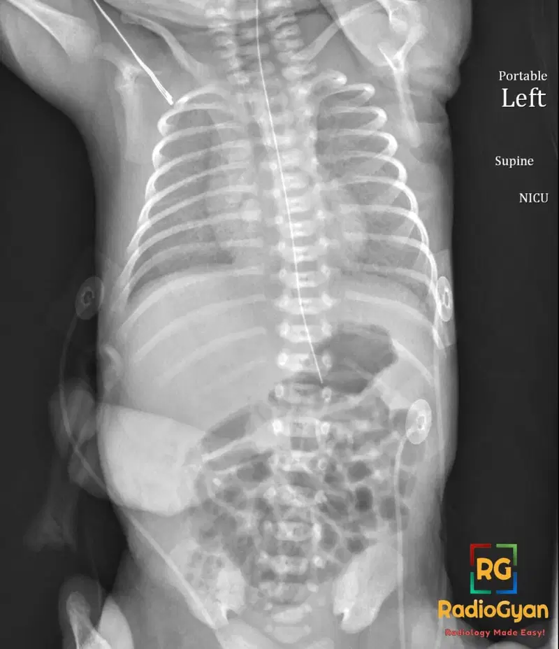
What causes an air bronchogram on chest radiography and CT?
Let me know in the comments.
Click to reveal the answer
Answer:
The air bronchogram is the appearance of an air-filled bronchus rendered visible as a branching lucency because the alveoli around it are opacified. In normal lung, an air-filled bronchus is invisible on a radiograph: it is surrounded by air-filled alveoli of identical attenuation. The sign therefore appears only when the surrounding airspaces are replaced by fluid, pus, blood, cells or tissue, or when the alveoli collapse around a still-patent airway, creating the density difference that outlines the bronchial lumen. It is the cardinal marker of an airspace (alveolar) process and simultaneously confirms that the proximal airway is patent rather than obstructed by a central mass. Classic causes are pneumonia and other consolidation, pulmonary oedema, pulmonary haemorrhage, non-obstructive atelectasis, and ARDS; a persistent air bronchogram within an opacity that fails to resolve on treatment should raise suspicion for a neoplasm, particularly lepidic-predominant adenocarcinoma or pulmonary lymphoma. The finding also localises disease to the lung itself, excluding a pleural or mediastinal origin.
Why is it called so?
The bronchus is filled with air (“air”) and is displayed along its branching course against the opacified lung like a contrast study of the airway (“bronchogram”), the appearance that was once produced deliberately by instilling contrast medium into the bronchial tree.
Pathophysiology
Opacification of the alveolar spaces by exudate, transudate, blood, or tumour cells, or their collapse from resorption of alveolar gas, raises the attenuation of the lung immediately surrounding an unobstructed bronchus. The bronchial lumen, still ventilated and therefore still of air attenuation, is thrown into relief against this high-attenuation background. Because the sign depends on airway patency, it is lost when the bronchus is itself obstructed, filled with secretions, or destroyed, which is why obstructive atelectasis and centrally obstructing tumours typically show no air bronchograms.
Alternative names: Air alveologram (used for the same phenomenon at acinar level)
Other associated named signs: Silhouette sign and CT angiogram sign (other markers of airspace disease), gloved finger sign (the mucoid-impaction opposite, in which bronchi are filled rather than aerated), and bulging fissure sign
References
Access all radiology signs posted so far: https://radiogyan.com/radiology-signs/

Improve Content And Help Your Colleagues!
Found an error in the post? Please let us know using our contact page and we will update it with due credits!
If you wish to contribute radiological images for this case (or any other article on the website), you can submit them here and you will be duly credited: Submit radiology case