


What causes the spine sign on the lateral chest radiograph?
Let me know in the comments.
Click to reveal the answer
Answer:
The spine sign is loss of the normal progressive increase in lucency of the thoracic vertebral bodies from above downwards on a lateral chest radiograph. Normally the upper thoracic vertebrae are relatively dense because the shoulder girdles and arm musculature overlie them, and each successive vertebra appears darker, more black, as that soft tissue is left behind and only aerated lower lobe overlies the caudal spine. When posterior lung is consolidated, collapsed or covered by pleural fluid, the added attenuation cancels this gradient, so the lower vertebrae appear as dense as or denser than those above; the change is most convincing when a prior normal lateral film is available for comparison. It indicates disease in a lower lobe or the posterior recesses and is a useful check whenever the frontal film looks normal but clinical suspicion persists, since retrocardiac and posterobasal disease is easily hidden on the PA view, where the only clue may be loss of the diaphragmatic outline. It is non-specific as to lobe and cause (pneumonia, lower lobe atelectasis, mass and free pleural effusion all erase the same gradient) with reported sensitivity of roughly 60-70% but a high positive predictive value of about 91-97%. A separate thoracic spine sign exists on lung ultrasound, where visibility of the vertebrae above the diaphragm indicates pleural effusion.
Why is it called so?
The sign is read off the appearance of the spine itself rather than of the lung: the thoracic spine is the only landmark whose density can be judged continuously from top to bottom on a lateral chest radiograph. The descriptive variant name more black sign names the normal appearance: each vertebra should look more black than the one above, and the moment they stop getting blacker the spine sign is positive.
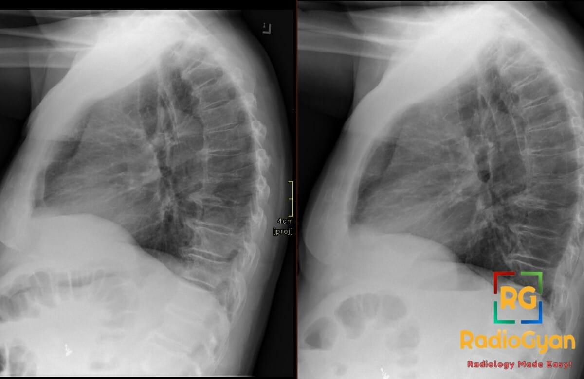
Pathophysiology
Radiographic density on a lateral projection is determined by the total attenuating tissue traversed by the beam. In the upper thorax the beam passes through both shoulders and the arm musculature, so the vertebrae there are relatively opaque; caudally, that soft tissue falls away and the beam meets only air-filled lung over each successive vertebra, producing the normal downward gradient of increasing lucency. Anything that replaces that basal air (alveolar exudate in pneumonia, resorbed air in collapse, tumour, or pleural fluid layering posteriorly) reinstates soft-tissue attenuation over the lower vertebrae and abolishes or reverses the gradient. Because air-filled lung contributes almost nothing to attenuation, only its replacement by fluid or tissue produces the change.
Alternative names: Spine sign of Felson, more black sign, vertebral fade-off sign
Other associated named signs: Silhouette sign (border effacement localises consolidation), air bronchogram, hilum overlay sign, cervicothoracic sign, flat waist sign, and the ultrasound thoracic spine sign of pleural effusion
References
Access all radiology signs posted so far: https://radiogyan.com/radiology-signs/

Improve Content And Help Your Colleagues!
Found an error in the post? Please let us know using our contact page and we will update it with due credits!
If you wish to contribute radiological images for this case (or any other article on the website), you can submit them here and you will be duly credited: Submit radiology case