


What causes biconcave vertebral bodies (codfish vertebrae) on sagittal radiographs of the spine?
Let me know in the comments.
Click to reveal the answer
Answer:
Codfish vertebra = both endplates concave, the body hourglass-shaped and the disc biconvex, because soft trabecular bone has yielded to the pressure of a normal nucleus pulposus. It is a shape, not a diagnosis. Three things decide the report: what softened the bone (osteoporosis in most adults), whether the endplate is depressed or merely low (fracture versus deformity), and whether the depression is a smooth arc (soft bone) or a squared central step (sickle cell, H-shaped vertebra).
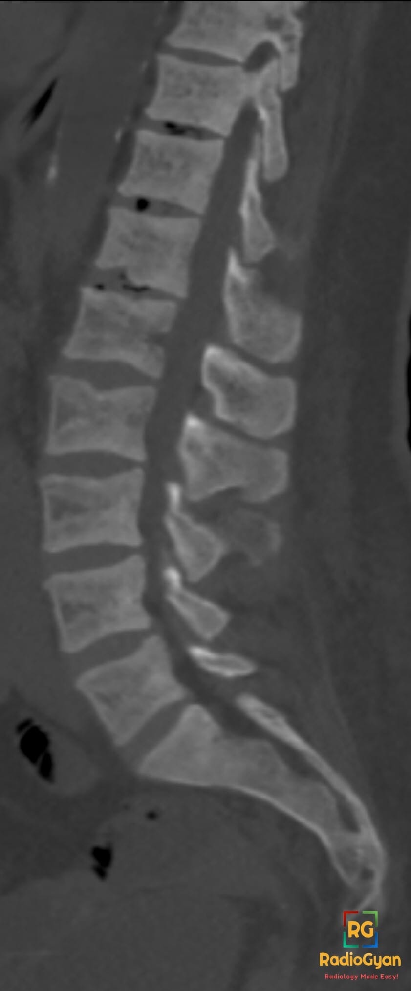
Why is it called so?
Fish vertebrae are amphicoelous, hollowed at both ends, and a softened human body comes to match. Useful pedantry: fish vertebra is the bone, fish mouth is the biconvex disc space between two of them [1].
Pathophysiology
The endplate sits between trabecular support below and nucleus pulposus pressure above. Weaken the trabeculae and the endplate bows inwards. The corollary is the useful bit: the sign needs a healthy turgid disc, so it appears at levels with preserved disc height. A flattened or sclerotic endplate at a degenerate, narrowed level is degenerative change, not this sign.
Alternative names: Fish vertebra, fish mouth vertebra, fishmouthing, biconcave vertebra, hourglass vertebra
Other associated named signs: H-shaped or Lincoln log vertebra of sickle cell disease, rugger jersey spine of renal osteodystrophy, hair-on-end skull of chronic haemolysis, and the picture frame vertebra of Paget disease
Causes
| Mechanism | Causes | Give-away elsewhere |
|---|---|---|
| Reduced bone quantity | Postmenopausal and senile osteoporosis, glucocorticoids, heparin, Cushing syndrome, hypogonadism, immobilisation, anorexia nervosa | Cortical thinning, coexisting wedge or crush deformities, normal biochemistry |
| Defective mineralisation | Osteomalacia, X-linked hypophosphataemia, hypophosphatasia | Looser zones, coarse indistinct trabeculae, blurred cortex |
| Hyperparathyroid and renal | Primary hyperparathyroidism, CKD mineral and bone disorder, dialysis | Rugger jersey spine, subperiosteal resorption of radial middle phalanges, brown tumours |
| Marrow expansion or replacement | Thalassaemia, chronic haemolysis, sickle cell disease, myeloma, leukaemia, metastases | Hair-on-end skull, widened diploe, lytic lesions, pedicle destruction if malignant |
| Inherited matrix disease | Osteogenesis imperfecta, homocystinuria, Marfan, Ehlers-Danlos | Blue sclerae, bowed long bones, ectopia lentis, dural ectasia, young patient |
Two corrections to the usual list. Sickle cell disease classically gives the squared H-shaped vertebra, not this shape; sickle patients do get smooth biconcavity, but from chronic-disease osteoporosis. Paget disease does not belong: it squares and enlarges the body (picture frame vertebra), the opposite morphology.
Codfish or H-shaped: read the step-off
| Feature | Codfish (fish) vertebra | H-shaped (Lincoln log) |
|---|---|---|
| Contour | Smooth curved arc | Squared step, sharp shoulders |
| Extent | Whole endplate, corner to corner | Central only; anterior and posterior thirds normal height |
| Mechanism | Global trabecular weakening | Infarction of the centrally supplied endplate |
| Cause | Osteoporosis, osteomalacia, renal osteodystrophy, marrow expansion | Sickle cell disease; less often Gaucher, thalassaemia |
| Specificity | Low | High for haemoglobinopathy |
Fracture or deformity?
Grade height loss with the Genant semiquantitative method on T4 to L4, typing each vertebra as wedge, biconcave or crush. The codfish vertebra is the biconcave type [2].
| Genant grade | Height loss | Area loss | Report as |
|---|---|---|---|
| 0 | None | None | Normal |
| 0.5 | Under 20% | Not applicable | Borderline deformity |
| 1 | 20% to 25% | 10% to 20% | Mild fracture |
| 2 | 25% to 40% | 20% to 40% | Moderate fracture |
| 3 | Over 40% | Over 40% | Severe fracture |
Height alone over-calls. The algorithm-based qualitative (ABQ) method calls a fracture only on endplate depression, buckling or cortical break; a merely short vertebra with an intact endplate is non-osteoporotic short vertebral height [3]. In MrOS (732 men aged 65+), fracture prevalence was 10% to 13% depending on method, but over 50% had short vertebral height without endplate depression, and their bone density matched men with normal vertebrae [4].
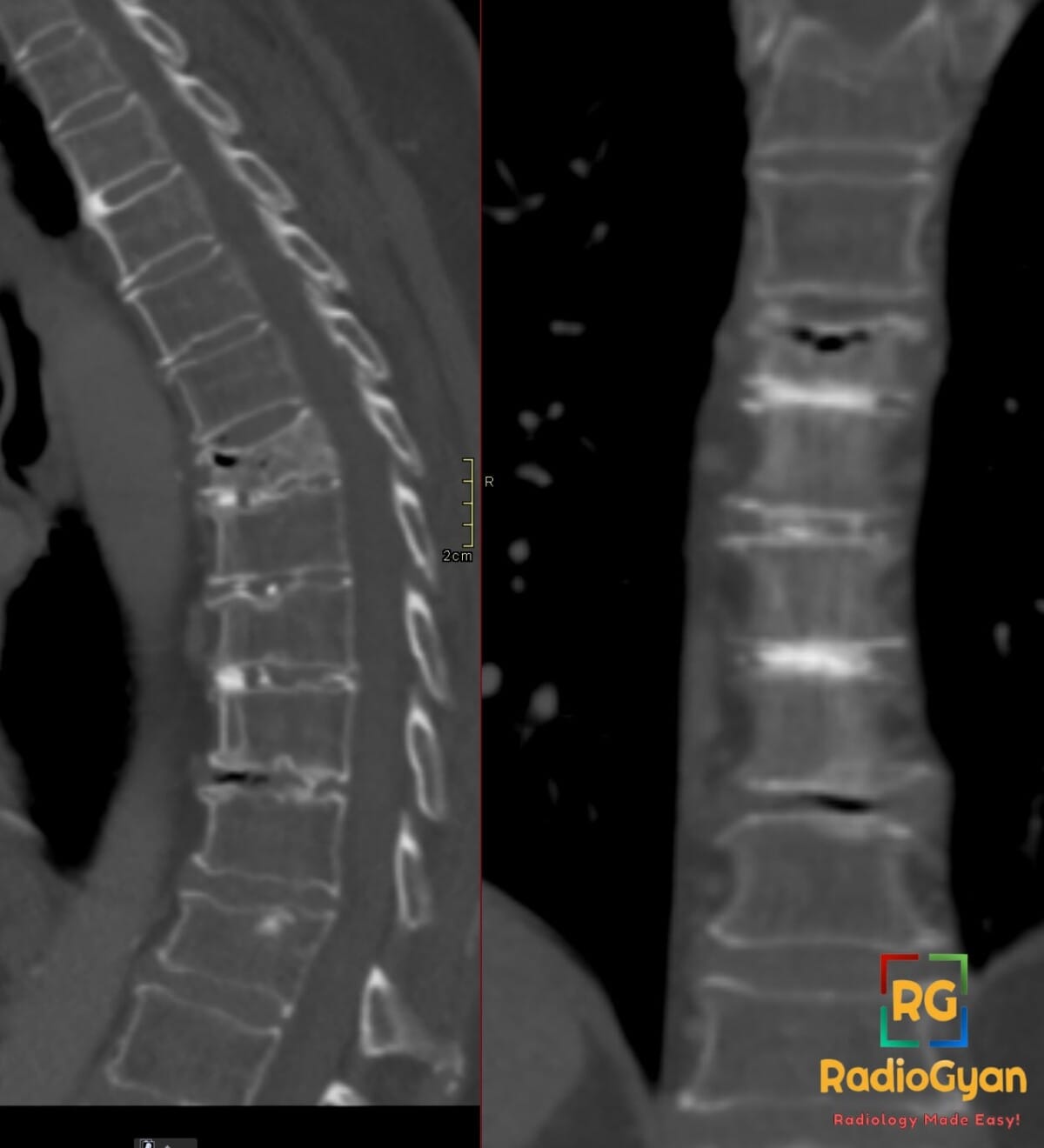
At the workstation: run the endplate, not the ruler. A smooth unbroken cortical line that sits low is a deformity. One that steps, buckles, condenses or disappears is a fracture, whatever the percentage says.
Mimics
| Mimic | How it differs |
|---|---|
| Cupid’s bow | Normal variant. Two parasagittal concavities of the inferior endplate in the lower lumbar spine; the midline endplate is normal [5] |
| Schmorl node | Focal rounded disc herniation with a sclerotic rim, not a global concavity [6] |
| Scheuermann disease | Anterior wedging of three or more contiguous thoracic bodies with irregular endplates and kyphosis |
| Limbus vertebra | Corticated triangular anterosuperior fragment with a matching corticated defect |
| Degenerative endplate change | Flat or sclerotic endplates at a level with reduced disc height |
| Malignant collapse | Convex posterior cortical bulge, pedicle involvement, soft tissue mass, diffuse T1 marrow replacement |
What to do next
- Say fracture when the endplate is depressed. The IMPACT study found a 34% false-negative rate in local reports, two thirds missed outright and one third hedged into uselessness [7].
- Give level, morphology, Genant grade and a count. Two or more changes risk category on its own.
- If it is a short vertebra with an intact endplate, say so, and say it is not a fracture.
- Recommend DXA with vertebral fracture assessment, plus calcium, phosphate, alkaline phosphatase, PTH, vitamin D and renal function. A vertebral fragility fracture is itself a treatment trigger under the 2024 UK NOGG guideline [8].
- MRI only for a specific question: acute or old, benign or malignant.
Frequently asked questions
What causes codfish vertebrae?
Any cause of generalised soft trabecular bone: osteoporosis (commonest by far), osteomalacia, hyperparathyroidism and renal osteodystrophy, marrow expansion in thalassaemia and chronic haemolysis, marrow replacement by myeloma or leukaemia, and osteogenesis imperfecta or homocystinuria in the young.
Is a codfish vertebra a fracture?
Only if the endplate is depressed, buckled or broken. A vertebra that is merely short with an intact endplate is a deformity, not a fracture. In the MrOS study over 50% of men aged 65 and over had short vertebral height of that kind, with bone density no lower than normal.
How do you tell codfish from H-shaped vertebrae?
By the shape of the depression. A smooth corner to corner arc means soft bone. A squared central step with preserved anterior and posterior thirds is the H-shaped or Lincoln log vertebra of sickle cell disease.
Why is it called a codfish vertebra?
Fish vertebrae are amphicoelous, concave at both ends, so a softened human body comes to look like one. Strictly, fish vertebra names the bone and fish mouth names the biconvex disc between two of them.
References
- Rexroad JT, Moser RP 3rd, Georgia JD. “Fish” or “fish mouth” vertebrae? AJR Am J Roentgenol. 2003;181(3):886-7. PMID: 12933500.
- Genant HK, Wu CY, van Kuijk C, Nevitt MC. Vertebral fracture assessment using a semiquantitative technique. J Bone Miner Res. 1993;8(9):1137-48. PMID: 8237484.
- Ferrar L, Jiang G, Adams J, Eastell R. Identification of vertebral fractures: an update. Osteoporos Int. 2005;16(7):717-28. PMID: 15868071.
- Ferrar L, Jiang G, Cawthon PM, et al. Identification of vertebral fracture and non-osteoporotic short vertebral height in men: the MrOS study. J Bone Miner Res. 2007;22(9):1434-41. PMID: 17563237.
- Ramirez H Jr, Navarro JE, Bennett WF. “Cupid’s bow” contour of the lumbar vertebral endplates detected by computed tomography. J Comput Assist Tomogr. 1984;8(1):121-4. PMID: 6690493.
- Pfirrmann CW, Resnick D. Schmorl nodes of the thoracic and lumbar spine. Radiology. 2001;219(2):368-74. PMID: 11323459.
- Delmas PD, van de Langerijt L, Watts NB, et al. Underdiagnosis of vertebral fractures is a worldwide problem: the IMPACT study. J Bone Miner Res. 2005;20(4):557-63. PMID: 15765173.
- Gregson CL, Armstrong DJ, Avgerinou C, et al. The 2024 UK clinical guideline for the prevention and treatment of osteoporosis. Arch Osteoporos. 2025;20(1):119. PMID: 40921943.
- Murphy WA Jr, DiVito DM. Fuller Albright, postmenopausal osteoporosis, and fish vertebrae. Radiology. 2013;268(2):323-6. PMID: 23882094.
Access all radiology signs posted so far: https://radiogyan.com/radiology-signs/

Improve Content And Help Your Colleagues!
Found an error in the post? Please let us know using our contact page and we will update it with due credits!
If you wish to contribute radiological images for this case (or any other article on the website), you can submit them here and you will be duly credited: Submit radiology case