


What causes sunburst periosteal reaction in bone on radiographic imaging?
Let me know in the comments.
Click to reveal the answer
Answer:
A sunburst periosteal reaction (sunray appearance) is caused by a rapidly growing lesion that breaks through the cortex and lifts the periosteum faster than it can lay down organised bone. Thin spicules of new bone and mineralised tumour radiate outward into the soft tissue mass, like rays from the sun. The classic cause is conventional osteosarcoma. It is also seen with Ewing sarcoma, osteoblastic and other metastases (including neuroblastoma in children) and, rarely, sclerotic myeloma.
The sign tells you how fast a lesion is growing, not what it is. Benign lesions can produce the same radiating pattern where bone is thin or the lesion is vascular: intraosseous hemangioma of the skull, intraosseous meningioma and odontogenic myxoma of the jaw. In a long bone of a young patient, treat a sunburst reaction as a primary bone sarcoma until proven otherwise and refer before biopsy.
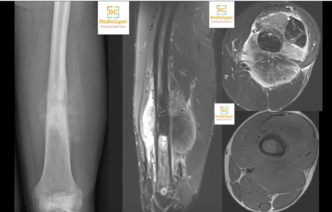
Why is it called so?
The name describes the look. On a radiograph or CT, fine linear bone densities fan out from the lesion at different angles and lengths, like rays of light spreading from the sun. Sunray and sunburst are used interchangeably. The term was in radiological use well before 1970; the earliest PubMed-indexed paper to carry it in its title is Lehrer, Maxfield and Nice’s 1970 description of “the periosteal sunburst pattern” in metastatic bone tumours. No primary source for the original coinage is indexed, so any single “first described by” claim should be treated with caution.
Pathophysiology
Periosteum responds to an insult by making new bone, and the shape of that bone records the speed of the insult. Ragsdale, Madewell and Sweet framed periosteal reaction as an index of the nature and intensity of the inciting process, not of its tissue of origin. A slow process gives the periosteum time to build thick, solid bone. A fast one does not.
In a sunburst reaction the lesion has already crossed the cortex and is expanding radially into the soft tissues. The periosteum is lifted and stretched, and bone forms along the fibrous and vascular strands that bridge the gap between the elevated periosteum and the cortex. Because the mass is pushing outward from a centre, those strands and the bone laid along them diverge. In osteosarcoma, malignant osteoid produced by the tumour itself mineralises along the same radiating framework, which is why the spicules are often coarse and irregular and extend into the soft tissue mass.
Hair-on-end reaction is the close relative: the spicules are fine, parallel and perpendicular to the cortex rather than divergent. Both are spiculated reactions and both are aggressive in a long bone.
Alternative names: sunray appearance, sun-ray spiculation, divergent spiculated periosteal reaction, radiating spiculated periosteal reaction.
Other associated named signs: Codman triangle (elevated periosteal cuff at the lesion margin, often seen alongside a sunburst reaction in osteosarcoma), hair-on-end (parallel perpendicular spicules), onion skin and laminated periosteal reaction.
Periosteal reaction patterns: where sunburst fits
| Pattern | What it looks like | What it implies | Typical causes |
|---|---|---|---|
| Solid (thick, smooth or undulating) | Dense new bone fused to the cortex | Slow process; periosteum has time to remodel | Osteoid osteoma, healing fracture, hypertrophic osteoarthropathy, venous stasis |
| Single lamellar | One thin shell separated from the cortex | Mild, early or slowly progressive process | Osteomyelitis, Langerhans cell histiocytosis, early healing fracture |
| Lamellated / onion skin | Several concentric layers | Intermittent or cyclical growth | Ewing sarcoma, osteomyelitis, osteosarcoma |
| Hair-on-end | Fine parallel spicules perpendicular to the cortex | Aggressive; uniform radial push | Ewing sarcoma; in the skull, marrow expansion (thalassaemia) |
| Sunburst (sunray) | Spicules of varying length diverging from a central point | Highly aggressive; rapidly expanding mass through the cortex | Osteosarcoma, metastases, Ewing sarcoma; skull hemangioma and meningioma as mimics |
| Codman triangle | Cuff of elevated periosteum at the lesion margin | Periosteum stripped faster than it can ossify centrally | Osteosarcoma, Ewing sarcoma, infection, subperiosteal haematoma |
| Disorganised / complex | Mixed, interrupted, amorphous new bone | Most aggressive; reaction breached by tumour | High-grade sarcoma |
Two practical points follow. First, the patterns overlap: a single osteosarcoma often shows sunburst spicules centrally, a Codman triangle at the margins and a disorganised reaction where the tumour has broken through its own new bone. Second, the pattern is not a diagnosis. Rana and colleagues and Wenaden and colleagues both stress that periosteal reaction aids characterisation of a bone lesion but is not specific for any one entity.
Causes of a sunburst periosteal reaction
| Group | Cause | What the evidence says |
|---|---|---|
| Primary malignant bone tumour | Conventional osteosarcoma | The classic cause. Sunburst reaction, Codman triangle and a mineralised soft tissue mass are frequently displayed (Crombé 2024) |
| Osteosarcoma outside long bones | Spiculated reaction in 21 of 24 (87.5%) head and neck tumours and 46 of 91 (50.5%) irregular and flat bone tumours; sunray reaction in 8 of 10 jaw tumours | |
| Periosteal (surface) osteosarcoma | Perpendicular reaction extending into a broad-based surface mass in 38 of 40 cases | |
| Ewing sarcoma | More often lamellated or hair-on-end, but a spiculated or sunburst component is well recognised | |
| Metastasis and marrow malignancy | Osteoblastic metastases | Described as a metastatic sunburst pattern since 1970 (Lehrer) and as spiculated reaction resembling osteosarcoma (Wyche 1978) |
| Neuroblastoma | Metastases to the mandible and femoral diaphysis with sunburst reaction; can mimic Ewing sarcoma in a child | |
| Other carcinomas | Case report: isolated skull metastasis from rectal adenocarcinoma with sunburst spicules resembling osteosarcoma | |
| Sclerotic myeloma / plasmacytoma | Rare; sternal sclerotic myeloma with peripheral sunburst reaction mimicking osteosarcoma | |
| Benign mimics | Intraosseous hemangioma (skull, face) | Radiating trabecular “sunburst” or honeycomb pattern; infants often lack it |
| Intraosseous meningioma | Mixed hyperostotic and lytic bone with radial spiculation, more frequent in WHO grade 2 and 3 tumours | |
| Odontogenic myxoma (jaw) | Sunburst reaction is uncommon but reported and mimics malignancy |
Conventional osteosarcoma remains the answer the exam wants, and the one that should drive urgent referral. But the spread of causes matters in practice. A sunburst reaction in the femur of a 70-year-old is far more likely to be a metastasis. A radiating pattern in a skull vault lesion of a middle-aged adult is more often a hemangioma than a sarcoma. And in a young child with a diaphyseal lesion, metastatic neuroblastoma can reproduce both the sunburst reaction and the clinical picture of Ewing sarcoma.
Age and site narrow the differential
| Where and in whom | Think first | Do not forget |
|---|---|---|
| Metaphysis of a long bone, age 10 to 25 | Conventional osteosarcoma | Ewing sarcoma, osteomyelitis with an atypical reaction |
| Diaphysis of a long bone, child | Ewing sarcoma | Metastatic neuroblastoma (under 5 years), osteosarcoma |
| Any bone, age over 40 | Metastasis | Secondary osteosarcoma (Paget disease, prior radiation), sclerotic myeloma |
| Surface of a long bone diaphysis | Periosteal osteosarcoma | Parosteal osteosarcoma, periosteal chondroma |
| Calvarium | Intraosseous hemangioma, intraosseous meningioma | Metastasis (including neuroblastoma in children), calvarial osteosarcoma |
| Mandible or maxilla | Osteosarcoma of the jaw | Odontogenic myxoma, neuroblastoma metastasis, hemangioma |
Not to be confused with the meningioma sunburst sign
The phrase “sunburst appearance” is also used for a completely different finding. In meningioma, the sunburst or spoke-wheel sign describes feeding vessels radiating from a central dural pedicle on catheter angiography, and sometimes the corresponding radiating enhancement or flow voids on MRI. It has nothing to do with periosteum.
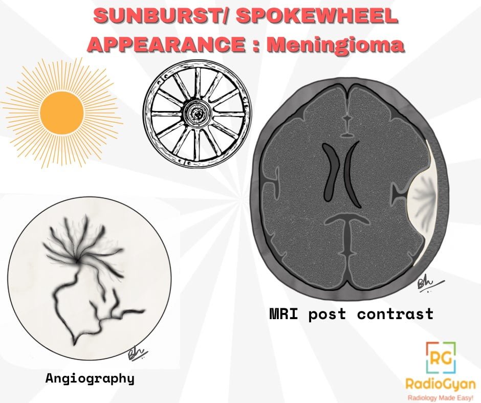
There is a genuine bone overlap, though. When a meningioma grows within the calvarium, it can produce hyperostosis with radial bony spiculation. In 65 intraosseous meningiomas, a mixed hyperostotic and lytic pattern with radial spiculations and a scalp mass were more frequent in higher-grade tumours, while plain hyperostosis was typical of WHO grade 1 disease. So a spiculated calvarial lesion is worth a close look at the adjacent dura.
How specific is the sign?
Not very, and it cannot be. A sunburst reaction tells you the periosteum has been overrun quickly. Any process that does that, malignant or benign, can produce it. The published reports make the point: metastatic sunburst patterns described since 1970, a rectal adenocarcinoma skull metastasis with spicules resembling osteosarcoma, a sclerotic sternal myeloma, benign odontogenic myxomas of the jaw, and calvarial hemangiomas that produce the pattern in adults but often not in infants.
The radiograph itself is also a limited judge. In 184 patients with confirmed bone tumours, agreement between two musculoskeletal radiologists on overall lesion aggressiveness was only kappa 0.22 on radiographs against 0.63 on CT, and about 10% of lesions were not seen on radiographs at all, half or more of them in the axial skeleton. That study measured aggressiveness overall rather than spicule pattern specifically, but the lesson carries over: in the pelvis, spine, skull and chest wall, confirm the reaction on CT before building a report around it.
What to do when you see one
- Read the whole lesion, not just the spicules. Zone of transition, cortical destruction, matrix (osteoid clouds versus chondroid rings and arcs), soft tissue mass and a Codman triangle together define aggressiveness.
- Get MRI of the whole involved bone. The ACR Appropriateness Criteria (Suspected Primary Bone Tumors, 2024 update) place radiographs first and MRI or CT as the usual next step for an aggressive or indeterminate lesion, to define extent and plan biopsy or surgery. Image the entire bone with adjacent joints to catch skip metastases.
- Use CT where radiographs are weak. CT shows cortical breach, matrix and spicule pattern better in flat and axial bones.
- Stage the patient. For a suspected bone sarcoma, the ESMO-EURACAN-GENTURIS guideline recommends chest CT for lung metastases, with bone scintigraphy or PET/CT for skeletal disease.
- Refer before biopsy. The 2025 UK bone sarcoma guideline states that patients with clinical or radiological findings suggestive of a primary bone tumour should be referred to a specialist centre and managed by an accredited bone sarcoma multidisciplinary team. The tract matters: tumour cells were found in 32% of open biopsy tracts versus 0.8% of percutaneous core biopsy tracts, and contaminated tracts were associated with local recurrence.
- In an older adult, look for a primary. A sunburst reaction after age 40 should prompt a search for metastatic disease and a check for Paget disease or prior radiation in the field.
After chemotherapy
Neoadjuvant chemotherapy often changes how an osteosarcoma looks on radiographs, and the change is easy to over-read. In 22 patients reviewed before and after chemotherapy, increased ossification or calcification and even an increase in tumour diameter were seen in more than half, and none of the radiographic features, periosteal reaction included, predicted histological response. A denser, better-defined sunburst after treatment is not proof of tumour kill. Response is graded on the resection specimen, with MRI used for surgical planning.
Reporting checklist
- Name the pattern precisely: divergent sunburst, parallel hair-on-end, lamellated, Codman triangle, or a mixture.
- Describe location in the bone (epiphysis, metaphysis, diaphysis; medullary or surface) and the patient’s age.
- Describe margin, cortical destruction, matrix mineralisation and the size of any soft tissue mass.
- State plainly that the pattern is aggressive, and give a short differential weighted by age and site.
- If the lesion is in the skull or jaw, say whether features of hemangioma, meningioma or odontogenic myxoma are present.
- Recommend MRI of the whole bone, staging if appropriate, and referral to a bone sarcoma centre before biopsy.
Common pitfalls
| Pitfall | Why it misleads | What to do instead |
|---|---|---|
| Calling sunburst diagnostic of osteosarcoma | The pattern reflects growth rate; metastases, Ewing sarcoma, myeloma and benign skull and jaw lesions can produce it | Report it as an aggressive pattern and give a differential weighted by age and site |
| Assuming absence of sunburst excludes sarcoma | Many sarcomas show lamellated, Codman or no visible reaction, especially in flat and axial bones | Judge aggressiveness on the whole lesion: margin, cortex, matrix, soft tissue mass |
| Relying on radiographs in the pelvis, spine or skull | About 10% of bone lesions were not seen on radiographs, half or more of them axial; agreement on aggressiveness was kappa 0.22 on radiographs vs 0.63 on CT | Use CT for axial and flat bones; MRI for extent |
| Confusing the meningioma “sunburst” vessel sign with bone spicules | Same name, different finding and modality | Say which one you mean: radiating vessels vs radiating bone spicules |
| Reading more mineralisation after chemotherapy as a good response | Increased ossification occurred in more than half of patients and did not predict histological response | Leave response grading to the resection specimen |
| Arranging a local biopsy before referral | Tract contamination 32% with open biopsy vs 0.8% with percutaneous biopsy | Refer to the bone sarcoma MDT first; let the surgeon plan the tract |
Frequently asked questions
What is a sunburst periosteal reaction?
A sunburst (sunray) periosteal reaction is a spiculated periosteal reaction in which thin bony spicules radiate outward from a lesion in a divergent, fan-like pattern, like rays from the sun. It means the periosteum has been breached and lifted faster than it can lay down organised bone, so it is one of the most aggressive periosteal reaction patterns.
Is a sunburst appearance always osteosarcoma?
No. Conventional osteosarcoma is the classic cause, but the pattern records how fast the lesion is growing, not what it is made of. It is also reported with Ewing sarcoma, osteoblastic and other metastases including neuroblastoma, sclerotic myeloma, and benign lesions such as intraosseous hemangioma of the skull, intraosseous meningioma and odontogenic myxoma of the jaw.
What is the difference between sunburst and hair-on-end periosteal reaction?
Both are spiculated reactions. In hair-on-end the spicules are fine, parallel and perpendicular to the cortex, reflecting a slightly less aggressive or more uniform process; it is classically linked with Ewing sarcoma and, in the skull, with marrow expansion in thalassaemia. In sunburst the spicules diverge from a central point and vary in length, reflecting a rapidly expanding mass. The distinction is descriptive, and both should be treated as aggressive in a long bone.
Is the sunburst appearance the same as the sunburst sign of meningioma?
No. The meningioma sunburst or spoke-wheel sign describes radiating feeding vessels on angiography or post-contrast imaging of a dural tumour. The sunburst periosteal reaction describes radiating bone spicules on radiographs or CT. A separate bone finding, radial spiculated hyperostosis, can occur in intraosseous meningioma and is associated with higher-grade tumours.
Which primary tumour most often shows a sunburst periosteal reaction?
Conventional osteosarcoma, typically in the metaphysis of the distal femur or proximal tibia in an adolescent. Spiculated periosteal reaction is also very frequent in osteosarcoma outside the long bones: 87.5 percent of head and neck osteosarcomas in one CT and MRI series, 50.5 percent of irregular and flat bone osteosarcomas in another, and perpendicular reaction in 38 of 40 periosteal osteosarcomas.
What imaging should follow a sunburst periosteal reaction on an X-ray?
Treat it as a suspected primary bone sarcoma. MRI of the whole involved bone with adjacent joints defines marrow and soft tissue extent and skip lesions; CT helps for axial and flat bones. Staging includes chest CT, with bone scintigraphy or PET/CT for skeletal disease. Refer to a bone sarcoma centre before any biopsy.
Why should biopsy wait for referral to a sarcoma centre?
Because the biopsy tract has to be excised with the tumour and a badly placed biopsy can compromise limb-salvage surgery. In one series, tumour cells were found in 32 percent of open biopsy tracts but only 0.8 percent of percutaneous core biopsy tracts, and contaminated tracts were associated with local recurrence. UK and ESMO guidelines both recommend that biopsy is planned by the treating sarcoma team.
Does more ossification after chemotherapy mean a good response in osteosarcoma?
Not reliably. In a study of 22 patients with osteogenic sarcoma, increased ossification or calcification and even tumour growth were seen in more than half of patients after neoadjuvant chemotherapy, and none of the radiographic features predicted histopathological response. Response is judged on the resection specimen, not on how mineralised the radiograph looks.
References
- Lehrer HZ, Maxfield WS, Nice CM. The periosteal “sunburst” pattern in metastatic bone tumors. Am J Roentgenol Radium Ther Nucl Med. 1970;108(1):154-61. PMID 5262633.
- Wyche LD, et al. Spiculated periosteal reaction in metastatic disease resembling osteosarcoma. Orthopedics. 1978;1(3):215-21. PMID 281665.
- Ragsdale BD, Madewell JE, Sweet DE. Radiologic and pathologic analysis of solitary bone lesions. Part II: periosteal reactions. Radiol Clin North Am. 1981;19(4):749-83. PMID 7323291.
- Holscher HC, Hermans J, Nooy MA, Taminiau AH, Hogendoorn PC, Bloem JL. Can conventional radiographs be used to monitor the effect of neoadjuvant chemotherapy in patients with osteogenic sarcoma? Skeletal Radiol. 1996;25(1):19-24. PMID 8717114.
- Murphey MD, Jelinek JS, Temple HT, Flemming DJ, Gannon FH. Imaging of periosteal osteosarcoma: radiologic-pathologic comparison. Radiology. 2004;233(1):129-38. PMID 15333772.
- Wenaden AE, Szyszko TA, Saifuddin A. Imaging of periosteal reactions associated with focal lesions of bone. Clin Radiol. 2005;60(4):439-56. PMID 15767101.
- Rana RS, Wu JS, Eisenberg RL. Periosteal reaction. AJR Am J Roentgenol. 2009;193(4):W259-72. PMID 19770293.
- Martínez-Lage JF, et al. Cavernous hemangiomas of the cranial vault in infants: a case-based update. Childs Nerv Syst. 2010;26(7):861-5. PMID 20405132.
- Ilica AT, Mossa-Basha M, Zan E, et al. Cranial intraosseous meningioma: spectrum of neuroimaging findings with respect to histopathological grades in 65 patients. Clin Imaging. 2014;38(5):599-604. PMID 24997535.
- Li Y, et al. Sclerotic multiple myeloma with an unusual sunburst periosteal reaction occurring in the sternum. Skeletal Radiol. 2015;44(5):749-54. PMID 25351419.
- Prasad GL, et al. Pediatric cranial intraosseous hemangiomas: a review. Neurosurg Rev. 2018;41(1):109-17. PMID 27558363.
- Barrientos-Ruiz I, Ortiz-Cruz EJ, Serrano-Montilla J, Bernabeu-Taboada D, Pozo-Kreilinger JJ. Are biopsy tracts a concern for seeding and local recurrence in sarcomas? Clin Orthop Relat Res. 2017;475(2):511-8. PMID 27655183.
- Luo Z, et al. Head and neck osteosarcoma: CT and MR imaging features. Dentomaxillofac Radiol. 2020;49(2):20190202. PMID 31642708.
- Strauss SJ, Frezza AM, Abecassis N, et al. Bone sarcomas: ESMO-EURACAN-GENTURIS-ERN PaedCan Clinical Practice Guideline for diagnosis, treatment and follow-up. Ann Oncol. 2021;32(12):1520-36. PMID 34500044.
- Luo Z, et al. Clinical and imaging features of 112 patients with irregular and flat bone osteosarcoma. Quant Imaging Med Surg. 2022;12(3):1988-2001. PMID 35284272.
- Gondim Teixeira PA, Lombard C, Moustache-Espinola P, et al. Initial characterization of focal bone lesions with conventional radiographs or computed tomography: diagnostic performance and interobserver agreement assessment. Can Assoc Radiol J. 2023;74(2):404-14. PMID 36207066.
- Liu M, et al. Isolated skull metastasis with unusual spiculated periosteal reaction from rectal cancer resembling osteosarcoma on FDG PET/CT. Clin Nucl Med. 2023;48(11):1006-8. PMID 37793159.
- Veloso MR, Soares MQ, Soares AB, Panzarella F, Nascimento M, Junqueira JL. Clinical and radiographic characteristics of osteosarcomas of the jaws: a retrospective study. J Clin Exp Dent. 2024;16(8):e998-e1003. PMID 39281792.
- Crombé A, Simonetti M, Longhi A, Hauger O, Fadli D, Spinnato P. Imaging of osteosarcoma: presenting findings, metastatic patterns, and features related to prognosis. J Clin Med. 2024;13(19):5710. PMID 39407770.
- Gerrand C, Amary F, Anwar HA, et al. UK guidelines for the management of bone sarcomas. Br J Cancer. 2025;132(1):32-48. PMID 39550489.
- Kumar M, et al. Metastatic neuroblastoma masquerading as Ewing’s sarcoma on X-ray: an imaging puzzle in pediatric oncology. J Orthop Case Rep. 2025;15(1):171-6. PMID 39801891.
- Expert Panel on Musculoskeletal Imaging; Ahlawat S, Lenchik L, et al. ACR Appropriateness Criteria Suspected Primary Bone Tumors: 2024 update. J Am Coll Radiol. 2025;22(5S):S440-54. PMID 40409893.
- Kalfarentzos E, et al. A rare and atypical manifestation of intraosseous hemangioma in the zygomatic bone. Diagnostics (Basel). 2025;15(15):1979. PMID 40804943.
- Moura AEO, et al. Odontogenic myxoma with rare sunburst periosteal reaction: report of two cases and review of the literature. Oral Radiol. 2026. Online ahead of print. PMID 42018241.
- Marquardt R, et al. Neuroblastoma metastasis to the mandible in children: a case report and focused narrative review. Children (Basel). 2026;13(8):989. PMID 42650329.
Access all radiology signs posted so far: https://radiogyan.com/radiology-signs/

Improve Content And Help Your Colleagues!
Found an error in the post? Please let us know using our contact page and we will update it with due credits!
If you wish to contribute radiological images for this case (or any other article on the website), you can submit them here and you will be duly credited: Submit radiology case