


What causes Thymic sail sign in thymus on chest X-ray?
Let me know in the comments.
Click to reveal the answer
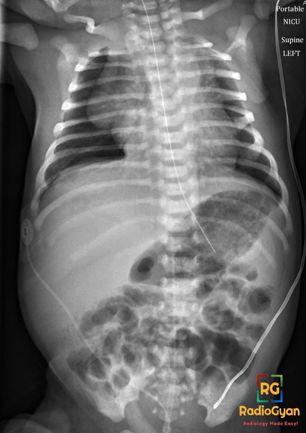
Normal prominent thymus in infants, appearing as a triangular soft tissue shadow (the Thymic sail sign) with convex lateral border along the right or left mediastinal border on chest radiograph. This is a physiological finding in approximately 8.8% of children, reflecting the normal size and position of the thymus in the anterior superior mediastinum, and is not associated with pathology.
Why is it called so?
Named for its resemblance to a sail due to the triangular shape of the thymic shadow with a convex lateral border projecting from the mediastinum, evoking the image of a ship’s sail billowing outward. The sign classically refers to the right thymic lobe as it abuts the minor (horizontal) fissure. This visual appearance is commonly referred to as the Thymic sail sign on pediatric chest X-ray.
Pathophysiology
In infants, the thymus is relatively large, occupying the anterior superior mediastinum behind the sternum and extending superiorly toward the neck and inferiorly over the heart. On frontal chest X-ray, the thymus forms a triangular soft tissue density with convex lateral margins due to its natural contour, bounded inferiorly by the minor fissure and continuous with the mediastinum without intervening air. This anatomy produces the characteristic Thymic sail sign on imaging.
Alternative names: None specific to this sign itself, though it is closely linked to the thymic wave sign (see below).
Other associated named signs: Thymic wave sign (undulating thymic border from adjacent rib impressions, indicating the thymus is soft and pliable, hence normal); spinnaker-sail sign (thymic lobes displaced and elevated by pneumomediastinum in neonates, resembling a ship’s spinnaker sail).
References
Access all radiology signs posted so far: https://radiogyan.com/radiology-signs/

Improve Content And Help Your Colleagues!
Found an error in the post? Please let us know using our contact page and we will update it with due credits!
If you wish to contribute radiological images for this case (or any other article on the website), you can submit them here and you will be duly credited: Submit radiology case