


What causes Pawnbroker’s sign in the thoracic lymph nodes on chest radiographs?
Let me know in the comments.
Click to reveal the answer
Pawnbroker’s sign is caused by bilateral hilar and right paratracheal lymphadenopathy, most commonly seen in sarcoidosis. This pattern of lymph node enlargement on chest radiographs reflects granulomatous inflammation within the lymphatic system of the mediastinum and hila.
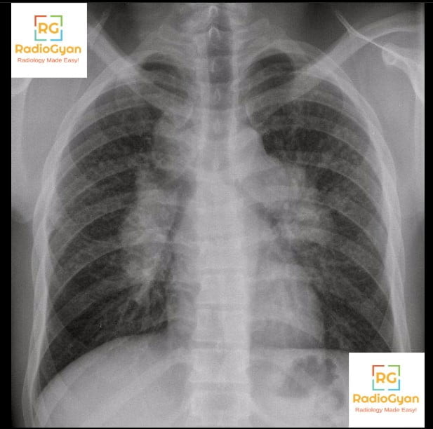
Sarcoidosis is a multisystem granulomatous disease of unknown etiology that frequently manifests with symmetrical enlargement of these lymph nodes. Bilateral hilar adenopathy is present in approximately 50% of sarcoidosis cases and may be accompanied by right paratracheal node enlargement.
Why is it called so?
It is named “Pawnbroker’s sign” because the lymph node enlargement pattern resembles the three suspended balls of the traditional pawnbroker’s symbol, historically linked to the Medici family crest and symbolizing monetary lending. The three nodal groups (right paratracheal, right hilar, and left hilar nodes) seen on chest X-ray correspond visually to the three balls hanging from a bar in the pawnbroker’s emblem.

Pathophysiology
The sign develops due to granulomatous infiltration and subsequent enlargement of mediastinal and hilar lymph nodes. Sarcoid granulomas accumulate within these nodes causing symmetrical swelling, which projects on chest radiographs as three distinct rounded opacities—right paratracheal, right hilar, and left hilar lymphadenopathy—that mimic the appearance of three balls suspended from a horizontal bar. This triad of lymphadenopathy arises from inflammation-driven lymph node hyperplasia and fibrosis typical of sarcoidosis.
Alternative names: 1-2-3 sign, Garland triad
Other associated named signs: Lambda sign (the gallium-67 scintigraphy equivalent pattern of the same lymph node distribution)
References
- Yoshimizu T, et al. The appearance of “lambda” and “panda” sign on Ga-67 scintigraphy in sarcoidosis. Kaku Igaku. 1991.
- Kita T, et al. Clinical significance of the serum IL-2R level and Ga-67 scan findings in making a differential diagnosis between sarcoidosis and non-Hodgkin’s lymphoma. Ann Nucl Med. 2007.
Access all radiology signs posted so far: https://radiogyan.com/radiology-signs/

Improve Content And Help Your Colleagues!
Found an error in the post? Please let us know using our contact page and we will update it with due credits!
If you wish to contribute radiological images for this case (or any other article on the website), you can submit them here and you will be duly credited: Submit radiology case