


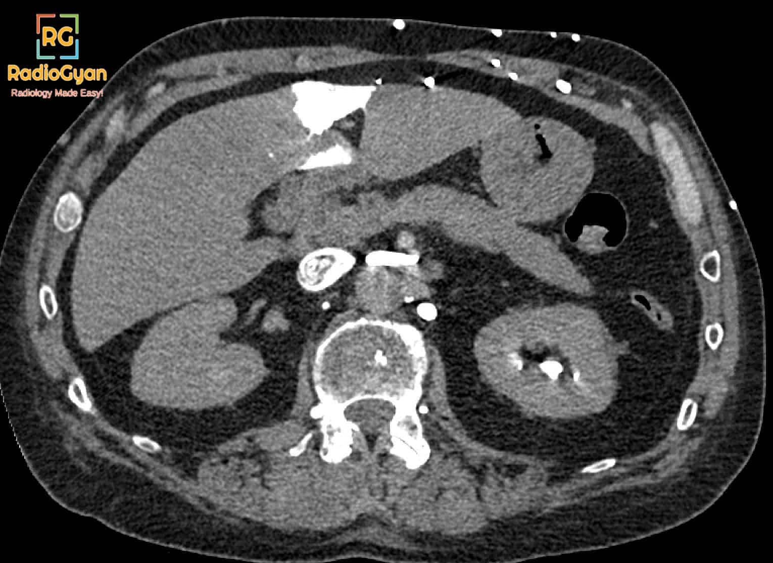
What causes the hot quadrate lobe sign on contrast-enhanced CT of the liver?
Let me know in the comments.
Click to reveal the answer
Answer:
The hot quadrate lobe sign is focal, intense, early enhancement of the quadrate lobe (segment IV) of the liver, just to the right of the falciform ligament and immediately beneath the anterior liver capsule, on the arterial phase of a contrast-enhanced CT or MRI performed through an upper limb vein. It is a marker of superior vena cava obstruction rather than of liver disease. When the SVC is narrowed or occluded, contrast injected into an arm vein is diverted through chest wall collaterals, the internal thoracic and superficial thoraco-abdominal veins, into the paraumbilical veins of Sappey, and so into the left portal vein. Segment IV therefore receives a bolus of undiluted contrast well before the rest of the liver opacifies through the normal route. The finding is confined to the arterial phase and equilibrates on the portal venous and delayed phases, which is the key to recognising it as a pseudo-lesion rather than a tumour: it is subcapsular and adjacent to the falciform ligament, has no mass effect, does not displace vessels, and becomes isodense to the rest of the parenchyma on later phases. On technetium-99m sulphur colloid scintigraphy the same physiology produces focal increased uptake in the same location, the focal hepatic hot spot sign. Its differentials on the arterial phase alone are focal nodular hyperplasia, hepatocellular adenoma, hepatocellular carcinoma, haemangioma and other transient hepatic attenuation differences. Its clinical significance is that it points to a long-standing, usually partial, SVC obstruction, and should prompt a search for the cause on the same study, most often a mediastinal or bronchogenic malignancy, fibrosing mediastinitis, or thrombosis around an indwelling central venous catheter or pacemaker lead.
Why is it called so?
“Hot” describes the focally increased signal of the region, the intense arterial enhancement on CT and MRI and the focal increase in radiotracer uptake on technetium-99m sulphur colloid scintigraphy, which makes it stand out against the rest of the liver. “Quadrate lobe” is the classical anatomical name for segment IV, the part of the liver in which that hot spot appears.
Pathophysiology
The paraumbilical veins of Sappey run in the falciform ligament and drain into the left portal vein, and they connect the systemic veins of the anterior abdominal and chest wall with the portal system. In a normal patient they carry negligible flow. When the SVC is obstructed above the level of the azygos vein, blood from the upper limbs and head is rerouted through the internal thoracic and superficial thoraco-abdominal veins, down the anterior chest and abdominal wall, and into these paraumbilical veins, forming a systemic-to-portal shunt. Contrast injected into an arm vein takes the same detour, so a concentrated bolus reaches the left portal vein and perfuses the small territory it supplies, segment IV, at a time when the remainder of the liver is still unopacified. The result is a sharply defined focus of dense enhancement in the quadrate lobe on the arterial phase only. Because it reflects flow rather than tissue, it fades as the systemic circulation equilibrates, and it disappears altogether if the study is performed through a lower limb vein. Chronicity matters: the collateral pathway takes time to open, so the sign implies established rather than acute obstruction.
Alternative names: Focal hepatic hot spot sign, hepatic hot spot sign
Other associated named signs: The hot caudate lobe sign of Budd-Chiari syndrome, in which the caudate lobe drains directly into the IVC and is spared while the congested remainder of the liver enhances poorly, and transient hepatic attenuation differences (THAD) more generally
References
Video
Detailed video with PACS based case images:

Access all radiology signs posted so far: https://radiogyan.com/radiology-signs/

Improve Content And Help Your Colleagues!
Found an error in the post? Please let us know using our contact page and we will update it with due credits!
If you wish to contribute radiological images for this case (or any other article on the website), you can submit them here and you will be duly credited: Submit radiology case