


What causes thumbprinting in the large bowel on plain radiography?
Let me know in the comments.
Click to reveal the answer
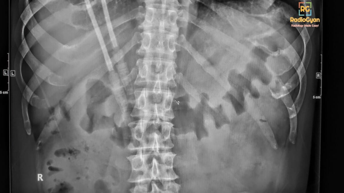
Thumbprinting in the large bowel on plain radiography is caused by thickening of the bowel wall, primarily due to submucosal edema or hemorrhage that results from inflammation or ischemia. This radiologic sign of bowel wall edema is commonly associated with conditions such as ischemic colitis, infectious colitis (including pseudomembranous colitis), inflammatory bowel disease, and other causes of bowel wall edema like diverticulitis or hemorrhage.
Why is it called so?
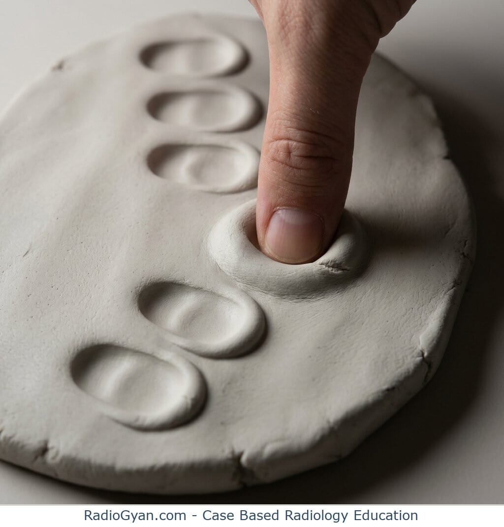
It is called “thumbprinting” because the thickened, inflamed haustral folds project inward into the gas-filled bowel lumen on radiographs, resembling rounded impressions similar to thumbprints. The appearance was described in 1963 by Schwartz, Boley and colleagues in Radiology, who illustrated these pseudotumor-like marginal indentations caused by hemorrhage or edema along the colon wall.
Pathophysiology
The pathological process involves edema and inflammation within the submucosa of the bowel wall, leading to localized swelling and thickening of the mucosal folds. This thickening creates nodular, thumb-shaped protrusions on imaging that indent the bowel lumen, giving the characteristic radiographic appearance known as thumbprinting. It reflects compromised bowel wall integrity often due to ischemia, infection, or inflammation.
Alternative names: Thumbprint sign (used interchangeably with thumbprinting).
Other associated named signs: Accordion sign and target (double halo) sign, both seen in colonic wall pathology such as colitis or ischemia.
Thumbprinting is a plain-radiograph sign of large-bowel wall thickening: smooth, rounded soft-tissue indentations, each roughly the width of a thumb, projecting into the gas-filled colonic lumen. They are edematous or hemorrhagic haustral folds seen in profile. The sign tells you the colonic submucosa is swollen. It does not tell you why, and it is not specific to any single disease.
The practical value of thumbprinting is that it converts a non-specific abdominal film into a positive finding: this patient has colitis of some kind, and needs a CT rather than reassurance. The commonest cause in an older patient is ischemic colitis; in a hospitalized patient on antibiotics it is Clostridioides difficile colitis. The distribution of the abnormal segment does most of the differential work, and that is what the sections below are organized around.
| At a glance | |
|---|---|
| What it is | Rounded soft-tissue indentations of the gas-filled colonic wall on an abdominal radiograph |
| What it means | Submucosal edema or hemorrhage thickening the haustral folds – i.e. colitis |
| Also called | Thumbprint sign, thumb-printing (of the colon) |
| Where it is seen | Most often the transverse and descending colon, where gas outlines the wall on a supine film |
| Commonest causes | Ischemic colitis; pseudomembranous (C. difficile) colitis; inflammatory bowel disease; infectious colitis |
| Specificity | Low. It localizes and confirms colitis but never establishes the cause on its own |
| Sensitivity | Low. A normal abdominal radiograph does not exclude colitis; radiographs often do not change management |
| Next step | Contrast-enhanced CT abdomen and pelvis, plus the clinical context (antibiotics, neutropenia, anticoagulation, travel) |
| Red flags on the same film | Pneumatosis, portal venous gas, free air, transverse colon > 6 cm, progression on serial films |
| Not to be confused with | The thumb sign of acute epiglottitis on a lateral neck radiograph |
Why the colon looks like that: the mechanism
Three things have to be true at once for thumbprinting to appear on a radiograph.
- The submucosa is expanded. Edema fluid or blood accumulates in the submucosal layer, which is the loosest and most distensible layer of the bowel wall. The mucosa and muscularis are pushed apart and the fold thickens.
- The thickening is fold-based, not diffuse. The haustral folds are already transverse shelves projecting into the lumen. When they swell they become rounded, polypoid mounds rather than thin lines, and they are spaced at haustral intervals – which is why the indentations look regular.
- There is gas next to them. The sign is a silhouette. Without a gas column outlining the wall the swollen folds have nothing to be seen against, which is why thumbprinting is usually reported in the transverse and descending colon on a supine film and is invisible in a fluid-filled or collapsed segment.
This is also why the sign is insensitive. A colon that is inflamed but spastic, fluid-filled or empty of gas will look normal, and the same wall thickening that produces obvious thumbprinting in one segment is undetectable in the next.
Why is it called thumbprinting?
Because each indentation is about the size and shape of a thumb pressed into soft clay. The description is deliberately literal: a row of smooth, convex soft-tissue impressions along the margin of the gas column, as if someone had pushed a thumb repeatedly into the bowel wall.
The classic radiographic account comes from Schwartz, Boley and colleagues in Radiology in 1963, describing the roentgenologic features of reversible vascular occlusion of the colon – the entity we now call ischemic colitis – and its overlap with ulcerative colitis. The pseudotumoral marginal indentations they illustrated, caused by submucosal hemorrhage, are the sign as radiologists still use it. The companion clinical paper by Boley and Schwartz appeared the same year. The word itself was already in radiological use for marginal filling defects, so the 1963 papers are best credited with tying the appearance to colonic ischemia rather than with coining the term.
A concise modern illustrated summary is Chawla et al., Abdominal Imaging 2015, in the “Classics in Abdominal Imaging” series.
What causes thumbprinting of the colon?
Anything that expands the colonic submucosa. The list below is grouped by mechanism, with the features that push you towards one cause rather than another.
| Cause | Typical distribution | What favors it |
|---|---|---|
| Ischemic colitis | Segmental, watershed – splenic flexure (Griffiths point) and rectosigmoid junction (Sudeck point); rectum usually spared | Age > 60, sudden cramping left-sided pain followed within 24 h by bloody diarrhea, hypotensive or post-operative episode, atrial fibrillation, vasoconstrictor drugs |
| Pseudomembranous (C. difficile) colitis | Pancolonic, often with the most marked wall thickening of any colitis | Recent antibiotics or hospitalization, watery diarrhea, marked leukocytosis; accordion sign on CT |
| Ulcerative colitis | Continuous from the rectum proximally, no skip areas | Younger patient, bloody diarrhea with urgency, known IBD; chronic disease gives the featureless lead-pipe colon rather than thumbprinting |
| Crohn colitis | Segmental with skip lesions, right colon and terminal ileum | Perianal disease, strictures, fistulae, comb sign on CT |
| Infectious colitis (Salmonella, Shigella, Campylobacter, E. coli O157:H7) | Right colon or pancolonic depending on organism | Acute febrile diarrheal illness, food or contact history, positive stool studies |
| Amebic colitis (Entamoeba histolytica) | Cecum and ascending colon | Residence in or travel from an endemic area, subacute course, may form an ameboma |
| Neutropenic enterocolitis (typhlitis) | Cecum and ascending colon | Neutropenia after chemotherapy, right iliac fossa pain and fever; a CT diagnosis, not a radiograph one |
| Intramural hemorrhage | Segmental, sharply demarcated | Anticoagulation, coagulopathy, blunt trauma; high-attenuation wall on unenhanced CT |
| Radiation colitis | Rectosigmoid, conforming to the radiation field | Pelvic radiation therapy, field-shaped rather than vascular-territory distribution |
| Angioedema (hereditary, or ACE-inhibitor related) | Small bowel more often than colon, long segment | Episodic self-limiting attacks, ascites, no fever or leukocytosis |
| Hypoalbuminemia and portal hypertensive colopathy | Diffuse, low-grade | Cirrhosis, nephrotic syndrome, anasarca; the patient is not acutely tender |
| Submucosal infiltration (lymphoma, rarely metastases) | Focal or segmental, mass-like | Known malignancy, bulky lymphadenopathy; the folds look nodular rather than smoothly edematous |
Reading the distribution: a practical approach
On the radiograph, the single most useful question is which segment. The colon has a predictable vascular and inflammatory geography, and the affected segment narrows a twelve-item list to two or three.
| Segment showing thumbprinting | Think first of | Reasoning |
|---|---|---|
| Splenic flexure and descending colon, rectum spared | Ischemic colitis | The splenic flexure is the watershed between superior and inferior mesenteric supply; the rectum has a dual systemic supply and is nearly always spared |
| Rectosigmoid junction, rectum spared | Ischemic colitis (Sudeck point) | The second watershed, between the last sigmoid branch and the superior rectal artery |
| Rectum continuously involved, extending proximally | Ulcerative colitis | UC starts distally and spreads continuously; rectal sparing argues strongly against it |
| Whole colon, marked thickening, out of proportion to a well-looking bowel gas pattern | Pseudomembranous colitis | The most severe wall thickening of any colitis, typically pancolonic |
| Cecum and ascending colon in a neutropenic patient | Neutropenic enterocolitis (typhlitis) | The cecum is the most distensible and least vascularized segment; a surgical-risk diagnosis |
| Cecum and ascending colon with a travel history | Amebic colitis | Ileocecal predilection; may coexist with a liver abscess |
| Isolated right colon | Isolated right colon ischemia | Uncommon but important – the ACG guideline flags it as carrying a worse prognosis and warranting assessment of the superior mesenteric artery |
| Segment inside a pelvic radiation therapy field | Radiation colitis | The abnormality respects the treatment field, not a vascular territory |
Mimics: when thumbprinting is over-called
Thumbprinting is easy to over-diagnose, particularly on a portable supine film. Before reporting it, work through the list below.
| Mimic | How to tell it apart |
|---|---|
| Normal haustral folds in a distended colon | Normal folds are thin, sharply defined lines that cross part of the lumen. Thumbprints are rounded, soft-tissue-dense mounds with a convex luminal margin and a wider base |
| Fecal residue and scybala | Stool contains mottled internal gas, is not attached to a single wall, and moves between films or on a decubitus view |
| Overlapping small-bowel loops | Follow each loop for its full length; valvulae conniventes cross the entire lumen and are more closely spaced than haustra |
| Under-penetrated or rotated projection | Reproduce the finding on a second view. A genuinely thickened wall persists; a projectional artefact does not |
| Diverticular muscular hypertrophy | Produces a saw-tooth, concertina sigmoid outline with visible diverticula, not smooth rounded impressions |
| Focal pericolic inflammation from diverticulitis | Causes localized ileus and pericolic soft tissue rather than the segmental, repeating haustral indentations of true thumbprinting |
| Extrinsic compression by a mass or fluid | Indentation is solitary and does not repeat at haustral intervals |
Thumbprinting versus the thumb sign: two different signs
These are searched interchangeably and mean entirely different things. The colonic sign is thumbprinting (plural impressions, abdomen). The airway sign is the thumb sign (a single thumb-shaped structure, lateral neck). Using the wrong one in a report or a viva is a recognizable error.
| Thumbprinting (thumbprint sign) | Thumb sign | |
|---|---|---|
| Region | Abdomen | Neck and airway |
| Radiograph | Supine or erect abdominal radiograph | Lateral soft-tissue neck radiograph |
| What is abnormal | Multiple edematous colonic haustral folds | A single swollen epiglottis |
| Underlying disease | Colitis – ischemic, infective, inflammatory or hemorrhagic | Acute epiglottitis (supraglottitis) |
| Associated findings | Loss of haustration, colonic dilatation, pneumatosis in advanced disease | Thickened aryepiglottic folds, obliterated vallecula (vallecula sign) |
| Urgency | Urgent CT and gastroenterology or surgical review | Airway emergency – do not distress the patient, involve anesthesia and ENT immediately |
| Modality of choice | Contrast-enhanced CT abdomen and pelvis | Direct laryngoscopy; imaging is adjunctive |
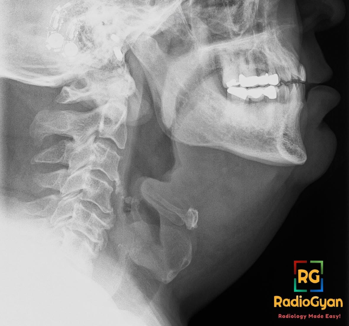
For the airway sign, lateral soft-tissue neck radiography has a reported sensitivity of roughly 88% in adult epiglottitis, but a normal film does not exclude it and imaging must never delay airway assessment.
The CT equivalent, and why CT is the real test
Thumbprinting is a legacy sign from an era of barium enemas and plain films. It remains worth recognizing because abdominal radiographs are still taken in large numbers, and because spotting it upgrades the urgency of the next study. But the diagnosis is made on CT. The ACR Appropriateness Criteria for acute nonlocalized abdominal pain are explicit that contrast-enhanced CT of the abdomen and pelvis is the first-line study, and that abdominal radiographs, although commonly performed, frequently do not alter management.
| Radiograph finding | CT correlate | Interpretation |
|---|---|---|
| Thumbprinting | Circumferential wall thickening with a low-attenuation, expanded submucosa | Submucosal edema – confirms colitis and defines its extent |
| (not visible on radiograph) | Target or double-halo sign: hyperenhancing mucosa, low-attenuation submucosa, enhancing muscularis | Preserved perfusion with edema; favors an inflammatory or reperfused ischemic segment |
| (not visible on radiograph) | Accordion sign: oral contrast trapped between grossly thickened haustral folds | Strongly suggests pseudomembranous colitis, though not pathognomonic |
| (not visible on radiograph) | Comb sign: engorged, splayed vasa recta | Active Crohn disease |
| Loss of haustration, featureless colon | Tubular colon with absent haustra | Chronic ulcerative colitis (lead-pipe colon) |
| Transverse colon > 6 cm with systemic toxicity | Colonic dilatation with abnormal haustral pattern and segmental mural thinning | Toxic megacolon – urgent surgical involvement |
| Curvilinear or bubbly lucency in the bowel wall | Pneumatosis intestinalis, with or without portal venous gas | Concern for transmural infarction; a surgical emergency in the right clinical setting |
| (not visible on radiograph) | Absent or reduced mural enhancement | Necrosis. In a 2026 validation of a CT severity score in 174 patients with ischemic colitis, decreased wall enhancement was an independent predictor of severe disease and improved the score to an AUC of 0.80 |
What to do when you see it
- Say where it is. Name the segment. The distribution carries more diagnostic weight than the sign itself.
- Look for the emergency findings on the same film. Pneumatosis, portal venous gas, free intraperitoneal gas, and colonic dilatation with a transverse colon diameter greater than 6 cm.
- Measure the transverse colon. Toxic megacolon by the Jalan criteria requires colonic dilatation greater than 6 cm without mechanical obstruction, plus systemic toxicity – at least three of fever, tachycardia above 120/min, neutrophilic leukocytosis and anemia, plus at least one of hypotension, hypovolemia, altered mental state or electrolyte derangement.
- Recommend contrast-enhanced CT. State it as a recommendation, not an observation. Where ischemia is suspected, arterial and portal venous phases allow assessment of mural enhancement and the mesenteric vessels.
- Ask for the context before you commit to a cause. Antibiotic exposure, neutrophil count, anticoagulation, travel, radiation therapy and known IBD each move the differential decisively.
- Flag the contraindication. Barium or water-soluble contrast enema is contraindicated in suspected toxic megacolon because of the perforation risk.
Reporting checklist
- Segment or segments involved, and whether the rectum is spared
- Length of the abnormal segment and whether involvement is continuous or skip
- Maximum transverse colonic diameter
- Haustral pattern: preserved, thickened or lost
- Pneumatosis intestinalis, portal venous gas, pneumoperitoneum
- Comparison with any previous film – progression on serial radiographs is itself an indication for surgical review
- An explicit recommendation for contrast-enhanced CT and the clinical correlation you need
The two causes you will meet most often
Ischemic colitis
The most common form of gastrointestinal ischemia, and the classic cause of thumbprinting. It is usually non-occlusive and transient, arising from a low-flow state rather than an embolus. The typical patient is elderly, presents with sudden cramping abdominal pain followed within a day by bloody diarrhea, and looks better than the imaging suggests. The ACG clinical guideline on colon ischemia recommends CT of the abdomen and pelvis with intravenous contrast as the initial imaging study and early colonoscopy for confirmation, and highlights isolated right colon ischemia as a subgroup with a worse prognosis that should prompt evaluation of the superior mesenteric artery. Most cases resolve with supportive management; surgery is reserved for gangrene, perforation or clinical deterioration.
Pseudomembranous (Clostridioides difficile) colitis
Suspect it in any inpatient with diarrhea, recent antibiotics and a strikingly thickened colon. Wall thickening is often the most marked of any colitis and is usually pancolonic, and the accordion sign on CT is a well-recognized though non-specific finding. Diagnosis is made on stool testing, not on imaging: the ACG clinical guideline bases diagnosis on testing patients with clinically significant diarrhea and sets out the current antibiotic and fecal microbiota transplantation pathways. The radiologist’s job is to recognize the pattern, exclude toxic megacolon and perforation, and say so.
Frequently asked questions
What is the thumbprinting sign on an abdominal X-ray?
Thumbprinting is the appearance of smooth, rounded soft-tissue indentations projecting into the gas-filled lumen of the large bowel on an abdominal radiograph. Each indentation is about the width of a thumb. They represent haustral folds thickened by submucosal edema or hemorrhage, and the sign therefore indicates colitis.
What causes thumbprinting of the colon?
Any process that expands the colonic submucosa. The commonest causes are ischemic colitis, pseudomembranous (Clostridioides difficile) colitis, inflammatory bowel disease and infectious colitis. Less common causes include amebic colitis, neutropenic enterocolitis, intramural hemorrhage in anticoagulated patients, radiation colitis, angioedema and submucosal infiltration by lymphoma.
Is thumbprinting the same as the thumb sign?
No. Thumbprinting is a colonic sign on an abdominal radiograph, caused by multiple edematous haustral folds. The thumb sign is an airway sign on a lateral soft-tissue neck radiograph, caused by a single swollen epiglottis in acute epiglottitis. The names are similar, the diseases are unrelated, and only the epiglottitis sign is an airway emergency. Note that “thumbprint sign” is used as a synonym for colonic thumbprinting, which is the main source of the confusion.
Which part of the colon is most often affected?
The transverse and descending colon are where thumbprinting is most often seen, partly because those segments hold gas on a supine film. In ischemic colitis the abnormality favors the watershed zones, the splenic flexure and the rectosigmoid junction, and the rectum is nearly always spared because of its dual blood supply.
Can thumbprinting be seen on CT?
The equivalent CT finding is circumferential bowel wall thickening with a low-attenuation, edematous submucosa, often with a target or double-halo appearance. CT is far more sensitive than the radiograph, shows the full extent of involvement, and adds findings the radiograph cannot show, such as reduced mural enhancement, pericolic stranding, engorged vasa recta and mesenteric vascular disease.
Does thumbprinting mean the bowel is dead?
No. Thumbprinting reflects a swollen submucosa, which in most cases is reversible – the majority of ischemic colitis is transient and settles with supportive care. The findings that do suggest transmural infarction are pneumatosis intestinalis, portal venous gas, free intraperitoneal gas and, on CT, absent mural enhancement. Those require urgent surgical involvement.
How do I tell thumbprinting from normal haustral folds?
Normal haustra are thin, sharply defined lines that partially cross the lumen at regular intervals. Thumbprints are broad-based, rounded, soft-tissue-dense mounds with a convex margin bulging into the gas column, and the affected segment usually shows other abnormalities such as loss of the normal fold pattern or dilatation. If you are unsure, look at a second projection and compare with the unaffected colon on the same film.
Related radiology signs
- Accordion sign – the CT counterpart most associated with pseudomembranous colitis
- Lead-pipe colon – the featureless, haustra-free colon of chronic ulcerative colitis
- Apple-core sign – the annular colonic lesion of carcinoma
- Small bowel feces sign – a small-bowel obstruction sign on CT
References
- Schwartz S, Boley S, Lash J, Sternhill V. Roentgenologic aspects of reversible vascular occlusion of the colon and its relationship to ulcerative colitis. Radiology. 1963;80:625-635. PMID: 13987392
- Chawla A, Tim WL, Lim TC. The colonic thumbprinting sign. Abdominal Imaging. 2015;40(7):2918-2920. PMID: 26088343
- Brandt LJ, Feuerstadt P, Longstreth GF, Boley SJ. ACG clinical guideline: epidemiology, risk factors, patterns of presentation, diagnosis, and management of colon ischemia (CI). American Journal of Gastroenterology. 2015;110(1):18-44. PMID: 25559486
- Kelly CR, Fischer M, Allegretti JR, et al. ACG clinical guidelines: prevention, diagnosis, and treatment of Clostridioides difficile infections. American Journal of Gastroenterology. 2021;116(6):1124-1147. PMID: 34003176
- Scheirey CD, Fowler KJ, Therrien JA, et al; Expert Panel on Gastrointestinal Imaging. ACR Appropriateness Criteria® acute nonlocalized abdominal pain. Journal of the American College of Radiology. 2018;15(11S):S217-S231. PMID: 30392591
- Kawamoto S, Horton KM, Fishman EK. Pseudomembranous colitis: spectrum of imaging findings with clinical and pathologic correlation. RadioGraphics. 1999;19(4):887-897. PMID: 10464797
- Valtchev L, Vannier M, Dacher JN, et al. Assessing the severity of ischemic colitis: validation of a CT scan severity score in 174 consecutive patients. European Radiology. 2026;36(7):5775-5783. PMID: 41813909
- Ong SCL, Mohaidin N. Imaging features of toxic megacolon. BMJ Case Reports. 2018;2018:bcr2018227121. PMID: 30275028
- Chew FY, Shen TC, Chuang BK. Acute epiglottitis and the thumb sign. QJM. 2024;117(8):597. PMID: 38598428
- Solomon P, Weisbrod M, Irish JC, Gullane PJ. Adult epiglottitis: the Toronto Hospital experience. Journal of Otolaryngology. 1998;27(6):332-336. PMID: 9857318
Access all radiology signs posted so far: https://radiogyan.com/radiology-signs/

Improve Content And Help Your Colleagues!
Found an error in the post? Please let us know using our contact page and we will update it with due credits!
If you wish to contribute radiological images for this case (or any other article on the website), you can submit them here and you will be duly credited: Submit radiology case