


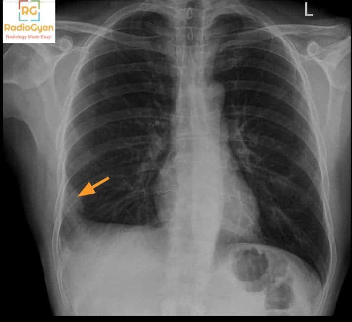
What causes Hampton hump on chest radiography?
Let me know in the comments.
Click to reveal the answer
Answer:
A peripheral wedge-shaped, pleural-based opacity in the lung, known as Hampton’s hump, is caused by pulmonary infarction resulting from occlusion of a pulmonary artery branch, most commonly due to acute pulmonary embolism. This radiographic finding represents hemorrhage and necrosis of lung parenchyma distal to the embolus. While highly suggestive of pulmonary embolism, it is not pathognomonic and can also occur with other causes of pulmonary infarction. The presence of Hampton’s hump correlates with increased risk of severe pulmonary embolism and is associated with a higher likelihood of pleural effusion and in-hospital mortality.
Why is it called so?
The sign is named after American radiologist Aubrey Otis Hampton, who together with pathologist Benjamin Castleman first described this radiographic appearance in 1940 by correlating postmortem chest radiographs with autopsy findings in cases of pulmonary embolism and infarction. The term “hump” refers to the characteristic convex medial margin of the opacity, which curves toward the hilum.
Pathophysiology
Hampton’s hump develops when a pulmonary artery branch is occluded by an embolus, leading to ischemia and subsequent infarction of the dependent lung parenchyma. Within 24–72 hours, alveolar wall necrosis and hemorrhage result in consolidation visible as a wedge-shaped opacity with its base against the pleura. If blood supply is not restored, this area may progress to scarring. The convex medial margin of the opacity reflects the interface between infarcted and normal lung tissue.
Alternative names: None
Other associated named signs: Westermark sign (regional oligemia), Palla sign (enlarged right descending pulmonary artery)
References
Access all radiology signs posted so far: https://radiogyan.com/radiology-signs/

Improve Content And Help Your Colleagues!
Found an error in the post? Please let us know using our contact page and we will update it with due credits!
If you wish to contribute radiological images for this case (or any other article on the website), you can submit them here and you will be duly credited: Submit radiology case