


What causes Double density sign in heart on frontal chest radiograph?
Let me know in the comments.
Click to reveal the answer
Answer:
An enlarged left atrium. As the left atrium dilates it extends posteriorly and to the right, so its right margin is projected through the right cardiac silhouette on the frontal chest radiograph as a second curved opacity. Mitral valve disease is the classic cause, but anything that raises left atrial pressure or volume will produce it.
Why is it called so?
It is named for the two overlapping densities seen along the right heart border: the usual right atrial border laterally, and a second, more medial curved edge formed by the enlarged left atrium lying behind it. Two edges where there should be one.
Pathophysiology
Chronic pressure or volume overload of the left atrium, from mitral stenosis, mitral regurgitation, raised left ventricular filling pressure or atrial fibrillation, drives atrial remodelling and dilatation. Once the atrium is large enough, its right margin reaches beyond the interatrial septum, meets aerated right lower lobe and acquires a visible edge of its own. The sign is specific to the frontal projection: on the lateral view the same enlargement shows as posterior bulging of the atrial wall and backward displacement of the oesophagus, not as a double density.
Alternative names: Double atrial shadow, double atrial contour, double right heart border sign
Other associated named signs: Widening of the carinal angle, third mogul sign (prominent left atrial appendage bulge along the left heart border), and the walking man sign, all further radiographic indicators of left atrial enlargement.
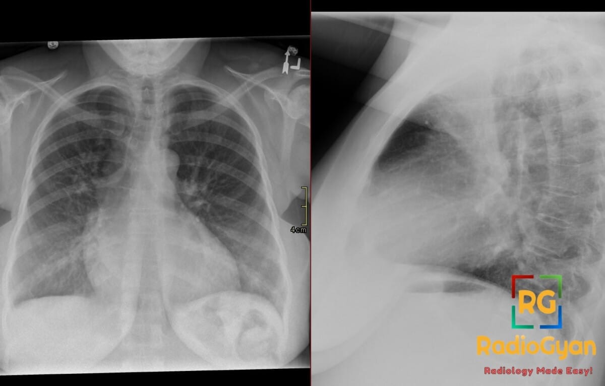
How to measure left atrial size on the frontal radiograph
Higgins and colleagues measured from the middle of the double density to the left main bronchus on adult PA films. The distance was under 7 cm in 96 per cent of 148 normal patients and over 7 cm in 90 per cent of 48 patients with left atrial enlargement on echocardiography. Carinal angles have been proposed since and discriminate poorly.
| Measurement on the PA film | Threshold | How well it performs |
|---|---|---|
| Middle of the double density to the left main bronchus | over 7 cm | The original Higgins criterion: 90 per cent of enlarged atria exceeded it, 96 per cent of normal ones did not. A less reliable gauge of atrial size in children |
| Interbronchial angle | over 76.4 degrees | Best angle cut-off in 122 reviewed patients, but only 61 per cent sensitive and 63 per cent specific |
| Subcarinal angle | over 65.4 degrees | Same series: 51 per cent sensitive, 66 per cent specific. The authors called tracheal bifurcation widening insensitive, nonspecific and of little value |
| Subcarinal angle and sub-angle distance against cardiac ultrasound | no useful cut-off | Area under the ROC curve only 0.62 to 0.63 in 159 adults; the authors judged chest radiographs unreliable for predicting left atrial enlargement |
The sign names the diagnosis, it does not measure the atrium
The one radiographic measurement that worked is the one nobody performs. Everything layered on top of it tests badly: carinal angles correlate weakly with atrial size, and when a deep learning study benchmarked human readers, radiologists reached an area under the curve of only 0.651 for left atrial enlargement, against 0.858 for the algorithm on the same films. Read the double density as a pointer to a diagnosis, not a measure of chamber size.
- Say what else on the film supports it: a widened carinal angle, a third mogul, pulmonary venous congestion, mitral annular calcification.
- Look at the left ventricle. A normal or small left ventricle with a big atrium points to mitral stenosis; a big atrium with a big ventricle points to regurgitation.
- Measure if you are going to claim enlargement: middle of the double density to the left main bronchus, over 7 cm in an adult.
- Do not grade severity from the radiograph. Echocardiography, with left atrial volume indexed to body surface area, is the reference standard.
- In a patient with no known valve disease, a new double density is often the first clue to atrial fibrillation or chronically raised filling pressures.
Causes of left atrial enlargement
Mitral valve disease is the classic cause and still dominates where rheumatic heart disease is common. Elsewhere the usual culprits are raised left ventricular filling pressure and atrial fibrillation, in which the atrium remodels and dilates with no valve lesion at all.
| Cause | Mechanism | Other clues on the same film |
|---|---|---|
| Rheumatic mitral stenosis | Fixed inflow obstruction raising left atrial pressure | Normal or small left ventricle, upper lobe venous diversion, Kerley B lines, mitral annular or valvular calcification |
| Mitral regurgitation | Volume overload of atrium and ventricle | Left atrium and left ventricle both enlarged |
| Left ventricular failure, hypertension, aortic valve disease | Raised filling pressure transmitted back to the atrium | Cardiomegaly, pulmonary oedema, aortic calcification |
| Atrial fibrillation | Atrial myopathy and electrical remodelling | Often the only abnormality on the film |
| Left atrial myxoma or thrombus | Mass effect and functional inflow obstruction | Focal contour change, calcification within a myxoma |
| Left to right shunt (ventricular septal defect, patent ductus arteriosus) | Increased pulmonary venous return | Plethoric lungs, enlarged central pulmonary arteries |
The other double density sign: osteoid osteoma
The same name is used for an unrelated finding in musculoskeletal nuclear medicine. On bone scintigraphy or SPECT, an osteoid osteoma shows a central focus of intense tracer uptake in the nidus surrounded by a wider zone of less intense but still increased uptake, also called the hotter spot within hot area sign. It has nothing to do with the heart, so check whether a report means the chest film or the bone scan.
Frequently asked questions
What does the double density sign mean on a chest x-ray?
It means the left atrium is enlarged. The dilated atrium extends to the right behind the right atrium and its margin becomes visible as a second curved line inside the right heart border on the frontal film.
What measurement confirms left atrial enlargement on a frontal radiograph?
More than 7 cm from the middle of the double density to the left main bronchus on an adult PA film. In the original series 90 per cent of echocardiographically enlarged atria exceeded 7 cm and 96 per cent of normal ones did not. It is a less reliable gauge in children.
Does the double density sign always mean mitral stenosis?
No. Mitral stenosis is the classic teaching association, but mitral regurgitation, left ventricular failure, hypertension, atrial fibrillation, a left atrial mass and left to right shunts all enlarge the atrium. Judging the left ventricle on the same film narrows it quickly.
What is the double density sign in osteoid osteoma?
A different sign with the same name. On bone scintigraphy, an osteoid osteoma produces intense uptake in the central nidus with a surrounding rim of lower but still increased uptake. It is also called the hotter spot within hot area sign.
References
- Higgins CB, Reinke RT, Jones NE, et al. Left atrial dimension on the frontal thoracic radiograph: a method for assessing left atrial enlargement. AJR Am J Roentgenol. 1978;130(2):251-5.
- Murray JG, Brown AL, Anagnostou EA, et al. Widening of the tracheal bifurcation on chest radiographs: value as a sign of left atrial enlargement. AJR Am J Roentgenol. 1995;164(5):1089-92.
- Quinton SJ, Ker JA, Rheeder P, et al. The reliability of chest radiographs in predicting left atrial enlargement. Cardiovasc J Afr. 2010;21(5):274-9.
- Nam JG, Kim J, Noh K, et al. Automatic prediction of left cardiac chamber enlargement from chest radiographs using convolutional neural network. Eur Radiol. 2021;31(11):8130-40.
- Lang RM, Badano LP, Mor-Avi V, et al. Recommendations for cardiac chamber quantification by echocardiography in adults: an update from the American Society of Echocardiography and the European Association of Cardiovascular Imaging. J Am Soc Echocardiogr. 2015;28(1):1-39.e14.
- Otto CM, Nishimura RA, Bonow RO, et al. 2020 ACC/AHA Guideline for the Management of Patients With Valvular Heart Disease. Circulation. 2021;143(5):e72-e227.
- Floria M, Radu S, Gosav EM, et al. Left atrial structural remodelling in non-valvular atrial fibrillation: what have we learnt from CMR? Diagnostics (Basel). 2020;10(3).
- Helms CA. Osteoid osteoma. The double density sign. Clin Orthop Relat Res. 1987;(222):167-73.
Access all radiology signs posted so far: https://radiogyan.com/radiology-signs/

Improve Content And Help Your Colleagues!
Found an error in the post? Please let us know using our contact page and we will update it with due credits!
If you wish to contribute radiological images for this case (or any other article on the website), you can submit them here and you will be duly credited: Submit radiology case