


What causes Bat-wing appearance on chest xray?
Let me know in the comments.
Click to reveal the answer
Answer:
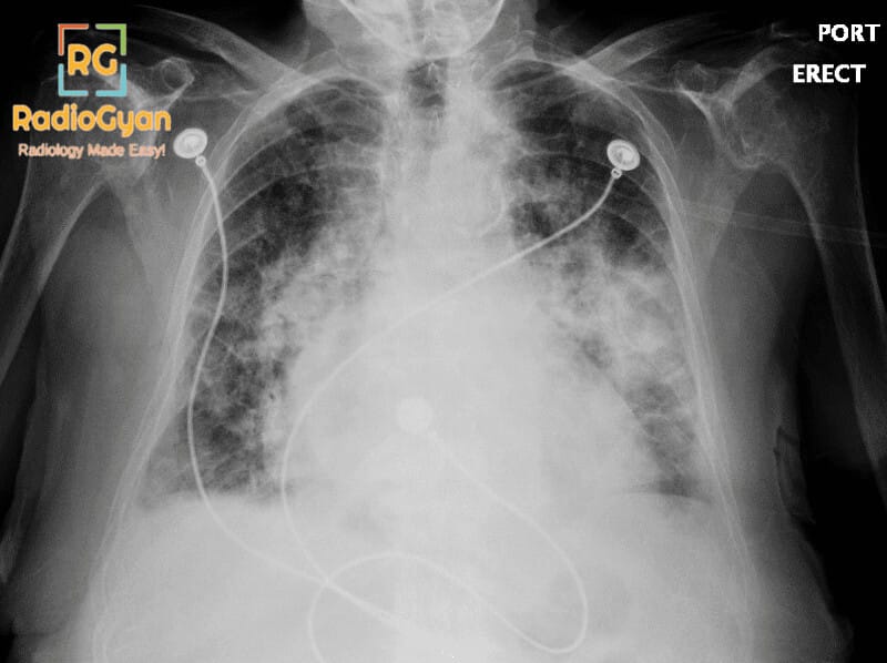
The bat-wing appearance represents bilateral symmetrical perihilar opacities that typically indicate accumulation of fluid or cellular material in the alveoli and interstitium of the lungs. The most common cause is pulmonary edema from cardiac sources such as heart failure, characterized by fluid accumulation due to elevated pulmonary venous pressure and increased hydrostatic pressure in the pulmonary capillaries. However, this sign is not exclusive to cardiac causes and can also be seen in pneumonia, pulmonary hemorrhage, inhalation injuries, sarcoidosis, bronchoalveolar carcinoma, and pulmonary alveolar proteinosis. The appearance typically demonstrates a ground-glass pattern and characteristically spares the lung cortices, preserving the peripheral lung zones while concentrating in the central perihilar regions. The sign is symmetrical and may be associated with cardiomegaly when the underlying cause is cardiac in nature. Air bronchograms may be visible, representing patent airways surrounded by consolidated or edematous lung tissue.
Why is it called so?
The sign is named “bat-wing appearance” because the bilateral symmetrical perihilar opacities that radiate outward from the hilum resemble the wings of a bat in extension, creating a characteristic butterfly or wing-like silhouette on frontal radiographic views. The symmetrical distribution and shape mimicking a bat’s outstretched wings provide an intuitive visual descriptor for this radiological pattern.
Pathophysiology
The bat-wing appearance develops through the accumulation of fluid in the pulmonary alveoli and interstitial spaces. In cardiac pulmonary edema, elevated left atrial pressure (from left ventricular dysfunction, mitral valve disease, or fluid overload) increases hydrostatic pressure in the pulmonary capillaries, forcing fluid across the capillary membrane into the interstitium and alveolar spaces. This fluid-filled tissue appears opaque on radiographs. The perihilar concentration occurs because pulmonary venous pressure is highest centrally at the hilum and decreases peripherally. As edema progresses, it advances from the perihilar regions toward the periphery. The bilateral symmetry reflects the uniform distribution of pulmonary venous pressure elevation across both lungs. In non-cardiac causes such as pneumonia or alveolar proteinosis, similar central consolidation patterns develop through different mechanisms—either inflammatory cellular infiltration or protein deposition—but produce the same radiographic appearance.
Alternative names:
Butterfly appearance, butterfly pattern, batwing sign, perihilar shadowing
Other associated named signs:
Kerley B lines (horizontal septal lines indicating pulmonary edema), upper zone vessel enlargement (reflecting pulmonary venous hypertension), Cardiomegaly (enlarged cardiac silhouette in heart failure), Reverse bat-wing or reversed bat-wing appearance (peripheral or subpleural consolidation representing the opposite distribution pattern)
The bat-wing appearance (also called the butterfly pattern) is bilateral, symmetrical airspace opacification centred on the perihilar and central lung, with sparing of the outer one-third of each lung (the “cortex”). The shape of the central opacity, fanning out from both hila, resembles the outstretched wings of a bat. It is a classic sign of severe, rapidly developing alveolar pulmonary edema, most often cardiogenic, but the same distribution can be produced by any process that floods the central alveoli. It is an uncommon presentation of pulmonary edema and, when present, usually signals acute and severe disease.
What is the bat-wing appearance?
The bat-wing appearance describes confluent alveolar (airspace) opacity that is concentrated in the central, perihilar lung and fades before it reaches the outer lung, so the peripheral cortex remains relatively lucent. The opacity is bilateral and broadly symmetrical, and often contains air bronchograms. It is the alveolar-filling counterpart of the more common, evenly graded pattern of pulmonary edema, and it tends to appear when fluid accumulates quickly and heavily.
- Modality: most often recognised on the frontal chest radiograph; the correlate on CT is central ground-glass or consolidation with peripheral and subpleural sparing.
- Distribution: central and perihilar, bilateral and symmetrical, sparing the outer third of the lungs.
- Finding: confluent airspace opacity, frequently with air bronchograms, that resolves rapidly when the underlying cause (typically fluid overload) is treated.
Why the central lung? Pathophysiology
In cardiogenic edema, a rise in left atrial and pulmonary venous pressure increases hydrostatic pressure in the pulmonary capillaries and drives fluid across the capillary membrane into the interstitium and then the alveoli. Several factors are thought to concentrate the fluid centrally in the bat-wing variant: the central lung carries a greater blood volume and is more distensible than the periphery; the peripheral cortex has more efficient lymphatic clearance and greater respiratory motion, which helps clear fluid; and when edema forms very rapidly, clearance mechanisms are overwhelmed centrally first. The result is dense central alveolar flooding with a relatively dry peripheral cortex. The exact mechanism remains debated, and the pattern is not specific to cardiac disease—any cause of rapid, heavy alveolar filling can reproduce it.
How to recognise it on the chest radiograph
- Check symmetry and centrality. The opacity should be bilateral, symmetrical and hug both hila, with a clear zone in the outer lung.
- Look for air bronchograms. These confirm an alveolar-filling process rather than a purely interstitial or pleural abnormality.
- Judge the heart and vascular pedicle. Cardiomegaly and a widened vascular pedicle push strongly towards a cardiogenic or overload cause.
- Correlate with time and treatment. Cardiogenic edema typically changes over hours and clears quickly with diuresis; opacity that persists for days or weeks argues for a non-edematous cause.
Causes of a bat-wing pattern
Although the bat-wing pattern is most strongly associated with cardiogenic pulmonary edema, any process that rapidly fills the central alveoli with fluid, blood, pus or other material can reproduce it. The clinical context and supporting radiographic features usually narrow the list.
| Cause | Why it produces a central pattern | Clue that favours it |
|---|---|---|
| Cardiogenic pulmonary edema (most common) | Rapid, severe rise in pulmonary capillary hydrostatic pressure with central alveolar flooding | Cardiomegaly, widened vascular pedicle, upper-lobe blood diversion, Kerley B lines, pleural effusions; clears with diuresis |
| Renal failure / fluid overload (uraemic edema) | Combined hydrostatic overload and increased capillary permeability | Known renal failure or fluid overload; heart size may be normal |
| Non-cardiogenic edema / ARDS | Diffuse alveolar damage with permeability edema; usually patchy and peripheral but can be central | Normal heart size, prominent air bronchograms, a precipitating insult (sepsis, aspiration, pancreatitis) |
| Diffuse alveolar haemorrhage | Blood fills the central alveolar spaces | Haemoptysis, falling haemoglobin, vasculitis, SLE or anticoagulation |
| Pneumocystis (PJP) pneumonia | Perihilar alveolar and ground-glass filling | Immunocompromise or HIV, subacute dyspnoea, raised LDH |
| Pulmonary alveolar proteinosis | Lipoproteinaceous material fills the alveoli in a perihilar distribution | Disproportionately mild symptoms, no cardiomegaly or effusion, crazy-paving on CT |
| Inhalation injury or near-drowning | Toxic or aspirated fluid causes permeability alveolar flooding | Clear exposure history |
| Invasive mucinous adenocarcinoma (formerly bronchioloalveolar carcinoma) | Tumour cells and mucin fill the alveoli, mimicking edema | Chronic and non-resolving; does not clear with diuresis; may cause bronchorrhoea |
Cardiogenic versus non-cardiogenic edema
When a bat-wing pattern is due to edema, the chest radiograph can often suggest whether the cause is cardiac, renal (overload) or a capillary-permeability injury. Milne and colleagues showed that a small set of features distinguishes these groups with high accuracy.
| Feature | Cardiogenic / overload edema | Permeability edema (ARDS) |
|---|---|---|
| Heart size | Usually enlarged | Usually normal |
| Vascular pedicle width | Widened | Normal or narrow |
| Distribution of blood flow | Balanced or inverted (upper-lobe diversion) | Normal or balanced |
| Distribution of edema | Even, central / perihilar | Patchy, often peripheral |
| Septal (Kerley B) lines | Common | Uncommon |
| Peribronchial cuffing | Common | Uncommon |
| Pleural effusions | Common | Uncommon |
| Air bronchograms | Uncommon | Common |
Reverse bat-wing appearance
The reverse (reversed) bat-wing pattern is the mirror image: peripheral, subpleural airspace opacity with relative sparing of the central perihilar lung. It is characteristically seen in chronic eosinophilic pneumonia (“photographic negative of pulmonary edema”) and cryptogenic organising pneumonia, and peripheral, reverse-bat-wing distributions have also been reported in COVID-19 pneumonia. Recognising the reversed pattern steers the differential away from hydrostatic edema and towards these organising and eosinophilic processes.
Clinical significance and pitfalls
- Signals severe, acute disease. A true bat-wing pattern usually reflects rapid, heavy alveolar flooding and demands prompt clinical correlation and treatment, not a wait-and-watch approach.
- Not synonymous with heart failure. Cardiogenic edema is the commonest cause, but haemorrhage, infection, proteinosis and even malignancy can look identical; always weigh the clinical context.
- Use the tempo. Edema changes over hours and clears with diuresis; a “bat-wing” opacity that persists for days to weeks should raise pneumonia, alveolar proteinosis, haemorrhage or mucinous adenocarcinoma.
- Correlate with the heart and pleura. Cardiomegaly, a widened vascular pedicle, septal lines and effusions support a cardiac or overload cause; their absence should prompt a non-cardiogenic differential.
Frequently asked questions
What is the bat-wing appearance on a chest X-ray?
The bat-wing or butterfly appearance is bilateral, symmetrical airspace opacity concentrated in the central perihilar lung with sparing of the peripheral cortex. It is a classic pattern of severe, rapidly developing alveolar pulmonary edema, most often cardiogenic.
What causes a bat-wing pattern?
The most common cause is cardiogenic pulmonary edema. Other causes include renal failure or fluid overload, non-cardiogenic edema and ARDS, diffuse alveolar haemorrhage, Pneumocystis pneumonia, pulmonary alveolar proteinosis, inhalation injury, and invasive mucinous adenocarcinoma.
Why does the edema spare the periphery of the lung?
The central lung has a greater blood volume and is more distensible, while the peripheral cortex has more efficient lymphatic clearance and greater respiratory motion. When edema forms rapidly, central clearance is overwhelmed first, concentrating fluid centrally and leaving a relatively dry peripheral cortex. The exact mechanism is still debated.
How do you tell cardiogenic from non-cardiogenic edema?
Cardiogenic and overload edema tend to show an enlarged heart, a widened vascular pedicle, Kerley B lines, peribronchial cuffing and pleural effusions. Permeability edema (ARDS) usually shows a normal heart size, a patchy peripheral distribution and prominent air bronchograms.
What is the reverse bat-wing appearance?
The reverse bat-wing pattern is peripheral, subpleural airspace opacity that spares the central lung, the photographic negative of pulmonary edema. It is typical of chronic eosinophilic pneumonia and cryptogenic organising pneumonia and has also been reported in COVID-19 pneumonia.
References
- Milne EN, Pistolesi M, Miniati M, Giuntini C. The radiologic distinction of cardiogenic and noncardiogenic edema. AJR Am J Roentgenol. 1985;144(5):879-894. PMID: 3872571.
- Gluecker T, Capasso P, Schnyder P, et al. Clinical and radiologic features of pulmonary edema. RadioGraphics. 1999;19(6):1507-1531. PMID: 10555672.
- Han J, Xiang H, Ridley WE, Ridley LJ. Bat wing or butterfly sign: pulmonary oedema. J Med Imaging Radiat Oncol. 2018;62(Suppl 1):18. PMID: 30309074.
- Kumar A, Kumar S, Katiyar V, et al. “Crazy-paving” pattern: a characteristic presentation of pulmonary alveolar proteinosis and a review of the literature from India. Lung India. 2016;33(3):335-342. PMID: 27186004.
- Ghosh S, Nandolia KK, Tale S, et al. Reverse Batwing sign in COVID-19 pneumonia. QJM. 2020;113(9):659-660. PMID: 32649770.
Access all radiology signs posted so far: https://radiogyan.com/radiology-signs/

Improve Content And Help Your Colleagues!
Found an error in the post? Please let us know using our contact page and we will update it with due credits!
If you wish to contribute radiological images for this case (or any other article on the website), you can submit them here and you will be duly credited: Submit radiology case