


What causes the double duct sign in the hepatopancreatobiliary system on imaging?
Let me know in the comments.
Click to reveal the answer
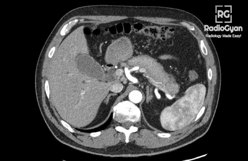
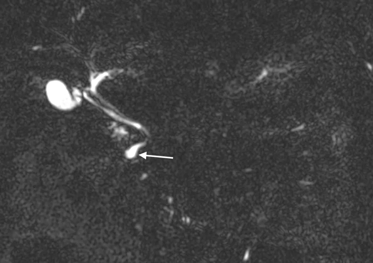
The double duct sign is simultaneous dilatation of the distal common bile duct and the main pancreatic duct, produced by a single obstructing lesion where the two ducts converge at the ampulla of Vater. It is best appreciated on CT or MRCP as two dilated parallel tubular structures pointing at the pancreatic head.
The sign matters because the lesion sitting at that convergence is malignant more often than not: pancreatic head adenocarcinoma, ampullary carcinoma and distal cholangiocarcinoma head the list. But it is not a cancer test. Across fifty years of series the positive predictive value is about 65%, with sensitivity 72 to 77% and specificity 73 to 80%, and the single strongest modifier is not duct calibre but jaundice: malignancy was found in 85.5% of patients with a double duct sign and obstructive jaundice, against 5.9% of those without it.
So the practical reading is two-sided. In a jaundiced patient, a double duct sign is periampullary cancer until proven otherwise. In an anicteric patient with normal bloods and no mass, the commonest answers are stones, chronic pancreatitis and plain age-related duct capacitance, and the job is to exclude a small tumour efficiently rather than to scan indefinitely.

Why is it called so?
It is named the double duct sign because imaging shows two distinct ducts dilated at once, the common bile duct and the main pancreatic duct, reflecting their anatomical convergence near the ampulla of Vater. The term comes from Freeny, Bilbao and Katon in 1976, who reviewed 40 deliberately similar ERCP studies blinded to the diagnosis. Irregular, nodular or rat-tailed pancreatic duct encasement occurred exclusively in carcinoma, and when the adjacent bile duct was involved in the same way, diagnostic certainty rose. That is the sign in its original form: paired strictures on contrast injection, not simply two wide ducts on a CT.
Six years later the same group reassessed it. Of 1180 ERCPs, 52 had a double duct sign; 30 were malignant and 22 benign. Their conclusion, still the most quoted sentence about this sign, was that the double duct sign per se is not disease specific, and that the surrounding ductal detail is what separates benign from malignant.
Pathophysiology
The common bile duct and the main pancreatic duct drain through a shared channel at the ampulla of Vater, surrounded by the sphincter of Oddi. Any lesion at or immediately adjacent to that channel obstructs both outflows simultaneously, so bile and pancreatic juice back up together and both ducts dilate upstream. Anything that obstructs one duct alone, such as a mid bile duct stricture or a pancreatic body tumour, cannot produce the sign.
That is also why the sign is anatomically non-specific: intraluminal (a stone), mural (ampullary adenoma or carcinoma), intramural-inflammatory (paraduodenal pancreatitis), extrinsic (a debris-filled duodenal diverticulum) and functional (sphincter spasm) causes all act at the same point.
Alternative names: double duct sign of Freeny; biductal dilatation. Some authors restrict “double duct sign” to paired strictures on ERCP and use “biductal dilatation” for the cross-sectional appearance, which is worth keeping in mind when comparing older papers with modern series.
Other associated named signs: the duct-penetrating sign and the duct-interrupted sign on MRCP, the interrupted duct sign of isoattenuating carcinoma, Courvoisier’s sign clinically, and the bulging papilla of main-duct IPMN.
How dilated is dilated?
Most false-positive double duct signs are a measurement problem. Duct calibre rises with age, and the conventional cut-offs were never population-derived. In a population-based MRCP study of healthy adults from the Study of Health in Pomerania, the 95th percentile for the common bile duct was 8 mm under 65 years and 11 mm at 65 or older, and for the pancreatic duct 3 mm and 4 mm respectively. Judged against conventional limits, 10.9% of healthy bile ducts and 18.2% of healthy pancreatic ducts would have triggered a workup.
| Duct | Age under 65 years | Age 65 years or older | Practical note |
|---|---|---|---|
| Common bile duct (MRCP, 95th percentile) | Up to 8 mm | Up to 11 mm | 10.9% of healthy adults exceed the conventional 6 to 7 mm cut-off |
| Main pancreatic duct (MRCP, 95th percentile) | Up to 3 mm | Up to 4 mm | 18.2% of healthy adults exceed the conventional 3 mm cut-off |
| Post-cholecystectomy common bile duct | Commonly 10 mm or more without obstruction | Compare with any prior study before calling it new | |
| Main pancreatic duct in a cystic lesion (Kyoto 2024) | 5 to 9.9 mm = worrisome feature; 10 mm or more = high-risk stigma | Think main-duct IPMN, not a periampullary mass | |
Two corollaries for reporting. First, a mildly wide bile duct and a 3.5 mm pancreatic duct in a 78-year-old with no jaundice is usually physiology, particularly after cholecystectomy. Second, the shape of the abnormality matters more than the number: proportionate smooth dilatation of both ducts with no stricture and no mass is a different finding from abrupt truncation. If you write about this in reports, our note on dilatation versus dilation covers the terminology, and the thresholds used here sit alongside the rest of our normal radiology measurements.
What the double duct sign actually predicts
| Study | Population | What the double duct sign meant |
|---|---|---|
| Freeny 1976 (ERCP, n = 40 blinded studies) | Look-alike ERCPs | Coined the term. Irregular pancreatic duct encasement occurred only in carcinoma; adding a matching bile duct lesion raised diagnostic certainty |
| Plumley 1982 (ERCP, 52 of 1180) | Unselected ERCP series | 30 malignant, 22 benign (58% malignant). Concluded the sign alone is not disease-specific |
| Menges 2000 (ERCP, 43 of 1209) | Malignant and benign pancreatic lesions | 15% had no pancreatic carcinoma; stenosis length did not discriminate |
| Kalady 2004 (ERCP, 355 duct strictures) | Pancreatic duct strictures | Sensitivity 77%, specificity 80%, PPV 65%. Isolated pancreatic duct stricture malignant in 12% vs 79% when a bile duct stricture coexisted |
| Krishna 2012 (CT or MRI, n = 234) | Referred for EUS | Malignancy in 85.5% with obstructive jaundice vs 5.9% without |
| Cohen 2014 (EUS, n = 82) | No jaundice, no mass on CT or MRI | Mass on EUS in 6 of 68 (9%): 4 pancreatic, 1 ampullary carcinoma, 1 ampullary adenoma. No new cancers at median 13 months |
| Sinha 2015 (MRCP, 4-year database) | Jaundiced and anicteric | Commonest cause choledocholithiasis. Malignancy in 48% of jaundiced and 0% of anicteric patients |
| Ten Berge 2015 (n = 241) | Periampullary lesions | Sensitivity 72%, specificity 73%, adjusted odds ratio 6.37 for malignancy |
| Kanchustambam 2020 (meta-analysis, n = 177) | Non-jaundiced, incidental, normal cross-sectional imaging | Periampullary tumour in 5% (95% CI 0 to 10), benign causal pathology in 22% (95% CI 10 to 34) |
| Yao 2024 (n = 103, 20 incidental) | Low-risk incidental double duct sign | No pancreatic head or periampullary tumour over a median 7.3 years of follow-up |
| Askar 2024 (n = 97, 64 non-jaundiced) | Tertiary HPB centre, normal bilirubin | Malignancy in 10.9%, including 4.3% with entirely normal serology. 53% never characterised, at a workup cost of 38,927 pounds |
Read together, the series say something simple. The double duct sign is a good alerting sign and a poor discriminating one. It roughly sextuples the odds of malignancy in a periampullary lesion (odds ratio 6.37), and that is genuinely useful. It cannot be used to make or exclude the diagnosis on its own, because one in four to one in three cases is benign even in surgical cohorts, and because 15% of classic ERCP double duct stenoses in one series had no cancer at all, with chronic pancreatitis confirmed on the resected specimen.
Jaundice changes everything
| Clinical context | Approximate risk of malignancy | Reasonable next step |
|---|---|---|
| Obstructive jaundice plus double duct sign | 85% (48% in a community MRCP series) | Pancreas-protocol CT if not already done, urgent HPB multidisciplinary referral, tissue only if it changes management |
| Mass on CT or MRI, no jaundice | High | Staging CT plus EUS-guided tissue acquisition |
| No jaundice, abnormal liver enzymes or raised CA 19-9 | About 11% | EUS. Deranged enzymes and raised CA 19-9 both tracked with malignancy |
| No jaundice, normal bloods, no mass | About 5%, and 4.3% of malignancies had normal serology | One good EUS. If negative, stop scanning rather than enter open-ended surveillance |
| Abdominal pain as the dominant symptom | Low; pain tracked with benign disease | Look for stones, chronic pancreatitis, paraduodenal pancreatitis |
The asymmetry is striking. Among jaundiced patients the sign behaves as the textbooks say. Among anicteric patients the same appearance carries roughly a 5 to 11% cancer risk, and in the MRCP series of Sinha and colleagues no anicteric patient had malignancy at all. The one caveat is that normal blood tests do not make the risk zero: in a 2024 tertiary-centre cohort, 2 of 64 non-jaundiced patients (4.3%) had a malignant periampullary tumour despite entirely normal serology.
Causes of the double duct sign
| Malignant cause | Imaging pointers |
|---|---|
| Pancreatic head or uncinate ductal adenocarcinoma | Hypoenhancing mass on pancreatic phase, abrupt duct truncation, upstream parenchymal atrophy, vascular encasement. The commonest cause overall |
| Ampullary carcinoma | Small polypoid lesion bulging into the duodenum, dual duct dilatation out of proportion to mass size, earlier jaundice |
| Distal (intrapancreatic) cholangiocarcinoma | Periductal thickening with an abrupt shouldered stricture, delayed enhancement, relative sparing of pancreatic parenchyma |
| Duodenal or periampullary adenocarcinoma | Eccentric duodenal wall mass, luminal narrowing, secondary ampullary involvement |
| Main-duct or mixed-type IPMN with high-grade change | Diffuse duct dilatation, enhancing mural nodule, bulging papilla, mucin extrusion at ERCP |
| Pancreatic neuroendocrine tumour or carcinoma | Usually hyperenhancing; duct obstruction implies larger or higher-grade disease |
| Metastasis or lymphoma in the pancreatic head | Known primary (renal cell, melanoma, lung), bulky nodes, preserved duct calibre relative to mass size in lymphoma |
| Ampullary adenoma with focal carcinoma | Villous papillary lesion; a benign biopsy does not exclude invasion |
| Benign cause | Clue that it is not cancer |
|---|---|
| Choledocholithiasis, impacted or recently passed distal stone | Colicky pain, fluctuating enzymes, stone on MRCP or EUS, gallbladder stones. The commonest cause in an MRCP series |
| Chronic pancreatitis | Parenchymal and intraductal calcification, side-branch ectasia, multiple strictures, pseudocysts, long smooth biliary stricture |
| Mass-forming autoimmune (IgG4-related) pancreatitis | Sausage-shaped gland, capsule-like rim, homogeneous delayed enhancement, duct-penetrating sign, other organ involvement, raised IgG4 |
| Paraduodenal (groove) pancreatitis | Duodenal wall thickening in 88.8%, intramural cysts in 82.6%, patchy delayed enhancement of the groove, restricted diffusion in only 3.6% |
| Benign post-inflammatory or post-instrumentation stricture | Long, smooth, symmetrical narrowing; previous ERCP, stent or surgery |
| Ampullary adenoma or adenomyoma | Small papillary lesion, no parenchymal invasion, slow or no change over time |
| Lemmel syndrome (periampullary duodenal diverticulum) | Debris-filled diverticulum indenting the intrapancreatic duct; recognise it before calling a mass |
| Sphincter of Oddi dysfunction, including opioid-related spasm | Episodic pain, normal ducts distally, opioid or chronic opium use in the history |
| Age-related capacitance and post-cholecystectomy dilatation | Both ducts mildly and proportionally dilated, no stricture, no jaundice, stable over years |
| Choledochocele (type III choledochal cyst) | Cystic dilatation of the intraduodenal bile duct at the ampulla, dilating both ducts without a solid mass |
Benign mimics worth knowing
Three benign entities account for most of the diagnostic trouble.
Choledocholithiasis. A stone impacted at, or recently passed through, the ampulla dilates both ducts and inflames the papilla. It was the commonest cause of a double duct sign in an MRCP database review, and it is the reason a colicky pain history pulls the probability away from cancer. Missing it means an ERCP with stone extraction is delayed while a cancer workup runs.
Mass-forming autoimmune pancreatitis. IgG4-related disease in the pancreatic head reproduces the whole malignant phenotype, including the double duct sign, and it responds to steroids rather than surgery. The discriminators are reproducible: homogeneous enhancement, the duct-penetrating sign and an ADC below 0.94 x 10⁻³ mm2/s. Any two of those three gave 98.7% specificity for autoimmune pancreatitis over adenocarcinoma, and all three gave 100%. Diagnosis should follow the International Consensus Diagnostic Criteria, which combine imaging of parenchyma and duct, serology, other organ involvement, histology and steroid response.
Paraduodenal (groove) pancreatitis. The signature is in the duodenal wall, not the pancreas. A systematic review of 292 CT, 231 MRI and 115 EUS examinations found duodenal wall thickening in 88.8%, intramural cysts in 82.6% and increased second-part enhancement in 76.3%, with a solid groove mass in only 40.9%, which typically enhances patchily in the portal venous phase, becomes iso- or hyperintense on delayed images in 100% of cases, and restricts diffusion in only 3.6%.
Two less common causes are worth naming because they are easy to miss and easy to fix. Lemmel syndrome is obstruction from a periampullary duodenal diverticulum, often debris-filled, compressing the intrapancreatic ducts. Sphincter of Oddi dysfunction, including opioid or opium-related spasm, produces episodic dual dilatation with no lesion at all. And a choledochocele (type III choledochal cyst) can dilate both ducts from within the duodenal wall.
Telling benign from malignant on imaging
| Finding | Favours | Reported performance |
|---|---|---|
| Duct-penetrating sign on MRCP (duct traverses the lesion, unobstructed) | Inflammatory mass | Sensitivity 85%, specificity 96%, accuracy 94%. MRCP area under the curve 0.98 vs 0.84 for arterial-phase CT |
| Abrupt duct truncation or irregular stenosis | Carcinoma | Present in 96% of carcinomas in the same series |
| Homogeneous enhancement plus duct penetration plus ADC below 0.94 x 10⁻³ mm2/s | Mass-forming autoimmune pancreatitis | Any two of three: 80% sensitivity, 98.7% specificity. All three: 100% specificity |
| Short bile duct stricture sited away from the papilla, abrupt transition, the two duct lesions close together | Carcinoma | Discriminating ERCP features since 1982 |
| Long biliary stricture, calcification, pseudocyst, side-branch ectasia | Chronic pancreatitis | Malignancy under 1% when pancreas divisum, duct stones or a pseudocyst is present |
| Stricture in the head or neck | Carcinoma | Odds ratio 42 on multivariate analysis |
| History of pancreatitis, irregular side branches | Benign disease | Both strongly protective in the same model |
| Duodenal wall thickening with intramural cysts, no restricted diffusion | Paraduodenal pancreatitis | Wall thickening in 88.8%, cysts in 82.6%, restricted diffusion in 3.6% |
The single most useful cross-sectional discriminator remains the duct-penetrating sign: on MRCP, a main pancreatic duct that runs through the abnormal segment, narrowed but patent, argues for an inflammatory mass. In the original description the main duct was unobstructed in 85% of inflammatory masses and obstructed or irregularly stenosed in 96% of carcinomas, giving 85% sensitivity, 96% specificity and 94% accuracy, with MRCP outperforming arterial-phase CT and MRI (area under the curve 0.98 vs 0.84 and 0.76). Restricting the sign to a completely normal-calibre duct makes it 100% specific but only 36% sensitive.
For a mass-forming lesion where the differential is cholangiocarcinoma versus adenocarcinoma versus inflammation, the enhancement timing usually separates them: adenocarcinoma stays hypoenhancing, cholangiocarcinoma enhances late and progressively, and autoimmune pancreatitis enhances homogeneously with a capsule-like rim.
When there is no mass on CT
A double duct sign with no visible tumour is not reassurance, it is an indication for endoscopic ultrasound. Two numbers explain why. About 11% of pancreatic adenocarcinomas are isoattenuating to normal parenchyma on multiphasic CT, and in those cases the only evidence of tumour is indirect: an interrupted pancreatic duct, distal parenchymal atrophy, a convex contour deformity, or dual duct dilatation itself. Separately, 14% of resected adenocarcinomas showed no secondary signs at all, and those tumours clustered in the uncinate process (50% versus 5%) and were at an earlier stage, which is exactly the group worth finding.
Endoscopic ultrasound answers the question well. For bile duct dilatation of unknown cause on cross-sectional imaging, EUS achieved an area under the curve of 0.98 for identifying a pathological obstruction, 0.91 for malignancy and 1.00 for choledocholithiasis; male sex, ALT or ALP three times the upper limit of normal, and intrahepatic duct dilatation predicted a true obstruction. In patients with a double duct sign, EUS-guided fine-needle aspiration was 92.8 to 98.5% accurate for malignancy.
The mirror-image finding is just as useful: when EUS shows no mass, later cancer is rare. In 62 such patients followed for a median of 13 months none developed malignancy, and in an incidental cohort followed for a median of 7.3 years none developed a pancreatic head or periampullary tumour. That is the evidence base for stopping, instead of committing a well patient to years of scans. The cost of not stopping is measurable: one centre recorded 80 multidisciplinary discussions, 103 CT and 65 MRI examinations and 29 EUS procedures in 64 non-jaundiced patients.
How to image it properly
- Pancreas-protocol CT. Thin-section dual-phase acquisition (late arterial or pancreatic phase plus portal venous phase) with multiplanar reformats. Report using the Society of Abdominal Radiology and American Pancreatic Association template, which standardises tumour, duct, arterial and venous descriptors and resectability terminology.
- MRCP. Best for duct morphology: level and length of the stricture, duct penetration, stones, side branches, anomalous junction and cystic lesions. Add diffusion-weighted imaging, which contributes an ADC threshold for the autoimmune pancreatitis differential.
- Endoscopic ultrasound. The test of choice when CT and MRI show no cause, and the route to tissue when tissue is needed.
- ERCP. Now therapeutic rather than diagnostic: stone extraction, stenting, sphincterotomy, papillectomy. The original double duct sign lives here, but nobody should be scoped purely to look for it.
Do not call a main-duct IPMN a double duct sign
Diffuse main pancreatic duct dilatation with a patulous papilla and mucin extrusion is main-duct IPMN, a different disease with its own algorithm. The 2024 international evidence-based Kyoto guidelines treat a main pancreatic duct of 10 mm or more as a high-risk stigma, along with obstructive jaundice in a patient with a cystic lesion of the pancreatic head and an enhancing mural nodule of 5 mm or more; a duct of 5 to 9.9 mm is a worrisome feature, as are acute pancreatitis, raised CA 19-9 and new-onset diabetes. Getting this label right decides whether the patient is referred for resection, for surveillance, or for a periampullary cancer workup.
The ampullary biopsy trap
If the lesion is at the papilla, resist the temptation to let a benign forceps biopsy close the case. In 241 patients with periampullary lesions, endoscopic biopsies were 96% specific but only 71% sensitive with a negative predictive value of 51%, while the double duct sign itself carried an adjusted odds ratio of 6.37 for malignancy. ESGE accordingly recommends endoscopic ultrasound plus MRCP for staging ampullary tumours and advises against diagnostic or therapeutic papillectomy when adenoma has not been proven. In practice, a double duct sign with a benign ampullary biopsy means repeat sampling or resection, decided on the imaging phenotype.
Reporting checklist
- State both measurements and the level at which each duct changes calibre, and compare with any prior study.
- Say whether the transition is abrupt or gradual, and whether the pancreatic duct penetrates the abnormal segment.
- Name a mass if you see one, and describe upstream parenchymal atrophy, contour deformity and an interrupted duct if you do not.
- Look specifically at the duodenal wall and the periampullary region for groove pancreatitis, a diverticulum or a polypoid ampullary lesion.
- Record the benign pointers you can see: calcification, duct stones, pseudocyst, side-branch ectasia, multiple strictures.
- Assess vessels and nodes if malignancy is plausible, using the standard resectability descriptors.
- Close with the clinical modifier: whether the patient is jaundiced, and recommend MRCP or EUS rather than a repeat CT when no cause is visible.
Common pitfalls
| Pitfall | Why it misleads | Fix |
|---|---|---|
| Applying textbook duct cut-offs to an 80-year-old | Almost one in five healthy pancreatic ducts exceeds 3 mm | Use age-adjusted limits: 11 mm bile duct and 4 mm pancreatic duct at 65 years or older |
| Calling a main-duct IPMN a double duct sign | Diffuse duct dilatation with a bulging papilla is a different disease and a different algorithm | Look for mucin, mural nodules and a patulous ampulla; classify with the Kyoto 2024 criteria |
| Stopping at a negative CT | About 11% of adenocarcinomas are isoattenuating and 14% show no secondary signs at all | Report the duct findings as the abnormality and recommend EUS |
| Treating a benign ampullary biopsy as the answer | Negative predictive value only 51% | Repeat sampling or resect based on the imaging phenotype |
| Missing the duodenal diverticulum | Debris in a periampullary diverticulum mimics an ampullary mass | Scroll the duodenum on coronal images before reporting a periampullary mass |
| Open-ended surveillance of an unexplained double duct sign | Half of non-jaundiced cases are never characterised, at real cost and anxiety | One good EUS plus normal bloods is a reasonable endpoint in the non-jaundiced patient |
Frequently asked questions
What is the double duct sign?
The double duct sign is simultaneous dilatation of the common bile duct and the main pancreatic duct, caused by a single obstructing lesion at or near the ampulla of Vater. It was originally described on ERCP as paired strictures, and is now most often reported on CT or MRCP as two dilated parallel tubular structures converging on the pancreatic head.
Does the double duct sign always mean pancreatic cancer?
No. Malignancy is the single commonest group of causes, but pooled series put the positive predictive value at roughly 65 percent, with sensitivity 72 to 77 percent and specificity 73 to 80 percent. In an unselected ERCP population only 58 percent of double duct signs were due to pancreatic cancer, and choledocholithiasis was the commonest cause in one MRCP series.
How does jaundice change the risk?
It is the strongest single modifier. In 234 patients with a double duct sign on CT or MRI, malignancy was found in 85.5 percent of those with obstructive jaundice but only 5.9 percent of those without. In an MRCP series, 48 percent of jaundiced patients had malignancy and none of the anicteric patients did. Painless jaundice plus a double duct sign should be treated as periampullary cancer until proven otherwise.
What should be done for an incidental double duct sign in a patient who is not jaundiced?
Correlate with bilirubin, liver enzymes and CA 19-9, then discuss for endoscopic ultrasound. A meta-analysis of 177 non-jaundiced patients with incidental double duct sign and otherwise normal cross-sectional imaging found a periampullary tumour in 5 percent and benign causal pathology in 22 percent. If EUS shows no mass, the subsequent risk is low: one cohort of 20 incidental cases had no pancreatic head or periampullary tumour over a median 7.3 years.
How dilated does a duct have to be?
Use age-adjusted limits. In a population-based MRCP study of healthy adults, the 95th percentile for the common bile duct was 8 mm under 65 years and 11 mm at 65 years or older, and for the pancreatic duct 3 mm under 65 years and 4 mm at 65 years or older. Judged by conventional cut-offs, 10.9 percent of healthy common bile ducts and 18.2 percent of healthy pancreatic ducts would have been called abnormal.
Which benign conditions cause a double duct sign?
Choledocholithiasis with an impacted or recently passed distal stone, chronic pancreatitis, mass-forming autoimmune (IgG4-related) pancreatitis, paraduodenal or groove pancreatitis, benign post-inflammatory or post-instrumentation strictures, ampullary adenoma, periampullary duodenal diverticulum compressing the ampulla (Lemmel syndrome), sphincter of Oddi dysfunction including opioid-related spasm, and age-related or post-cholecystectomy ductal capacitance.
Which imaging findings favour a benign cause?
A duct that runs through the abnormal segment rather than stopping at it. The duct-penetrating sign on MRCP had 85 percent sensitivity, 96 percent specificity and 94 percent accuracy for an inflammatory mass rather than carcinoma. Other benign pointers are a long smooth biliary stricture, parenchymal calcification, pancreatic duct stones, pseudocysts, side-branch ectasia and multiple strictures. Abrupt duct truncation, a short stricture away from the papilla, upstream parenchymal atrophy and restricted diffusion favour cancer.
Can there be a double duct sign with no visible mass?
Yes, and it is a recognised trap. About 11 percent of pancreatic adenocarcinomas are isoattenuating on multiphasic CT, where an interrupted duct, distal parenchymal atrophy and contour deformity may be the only clues, and 14 percent show no secondary signs at all, disproportionately in the uncinate process. Endoscopic ultrasound is the next test: for bile duct dilatation of unknown cause it gave an area under the curve of 0.98 for detecting a pathological obstruction.
Is a negative endoscopic biopsy of the ampulla reassuring?
Not reliably. In 241 patients with periampullary lesions, endoscopic biopsies had 96 percent specificity but only 71 percent sensitivity and a negative predictive value of 51 percent, while the double duct sign itself carried an odds ratio of 6.37 for malignancy. A benign biopsy in the presence of a double duct sign does not close the case.
References
- Freeny PC, Bilbao MK, Katon RM. “Blind” evaluation of ERCP in the diagnosis of pancreatic carcinoma: the “double duct” and other signs. Radiology. 1976;119(2):271-4. PMID 1265253.
- Plumley TF, Rohrmann CA, Freeny PC, Silverstein FE, Ball TJ. Double duct sign: reassessed significance in ERCP. AJR Am J Roentgenol. 1982;138(1):31-5. PMID 6976709.
- Menges M, Lerch MM, Zeitz M. The double duct sign in patients with malignant and benign pancreatic lesions. Gastrointest Endosc. 2000;52(1):74-7. PMID 10882966.
- Ichikawa T, Sou H, Araki T, et al. Duct-penetrating sign at MRCP: usefulness for differentiating inflammatory pancreatic mass from pancreatic carcinomas. Radiology. 2001;221(1):107-16. PMID 11568327.
- Prokesch RW, Chow LC, Beaulieu CF, Bammer R, Jeffrey RB Jr. Isoattenuating pancreatic adenocarcinoma at multi-detector row CT: secondary signs. Radiology. 2002;224(3):764-8. PMID 12202711.
- Kalady MF, Peterson B, Baillie J, et al. Pancreatic duct strictures: identifying risk of malignancy. Ann Surg Oncol. 2004;11(6):581-8. PMID 15150064.
- Shimosegawa T, Chari ST, Frulloni L, et al. International consensus diagnostic criteria for autoimmune pancreatitis: guidelines of the International Association of Pancreatology. Pancreas. 2011;40(3):352-8. PMID 21412117.
- Krishna N, Tummala P, Reddy AV, Mehra M, Agarwal B. Dilation of both pancreatic duct and the common bile duct on CT and MRI scans in patients with or without obstructive jaundice. Pancreas. 2012;41(5):767-72. PMID 22450366.
- Oterdoom LH, van Weyenberg SJ, de Boer NK. Double-duct sign: do not forget the gallstones. J Gastrointestin Liver Dis. 2013;22(4):447-50. PMID 24369328.
- Al-Hawary MM, Francis IR, Chari ST, et al. Pancreatic ductal adenocarcinoma radiology reporting template: consensus statement of the Society of Abdominal Radiology and the American Pancreatic Association. Radiology. 2014;270(1):248-60. PMID 24354378.
- Cohen J, Sawhney MS, Pleskow DK, et al. Double-duct sign in the era of endoscopic ultrasound: the prevalence of occult pancreaticobiliary malignancy. Dig Dis Sci. 2014;59(9):2280-5. PMID 24705640.
- Sinha R, Gardner T, Padala K, Greenaway JR, Joy D. Double-duct sign in the clinical context. Pancreas. 2015;44(6):967-70. PMID 26087354.
- Ten Berge JC, Suker M, Bruno MJ, et al. Are a double duct sign or endoscopic biopsies reliable predictors of malignancy in periampullary lesions. Dig Surg. 2015;32(4):306-11. PMID 26138752.
- Tamada T, Ito K, Kanomata N, et al. Pancreatic adenocarcinomas without secondary signs on multiphasic multidetector CT: association with clinical and histopathologic features. Eur Radiol. 2016;26(3):646-55. PMID 26084602.
- Choi SY, Kim SH, Kang TW, Song KD, Park HJ, Choi YH. Differentiating mass-forming autoimmune pancreatitis from pancreatic ductal adenocarcinoma on the basis of contrast-enhanced MRI and DWI findings. AJR Am J Roentgenol. 2016;206(2):291-300. PMID 26797355.
- Kanchustambam SRV, Sharma A, Perkins Z, Patel A. Diagnostic performance of EUS in non-jaundiced patients with an incidental finding of double duct sign on cross-sectional imaging: a systematic review and meta-analysis. Pancreatology. 2020;20(5):992-6. PMID 32631792.
- Vanbiervliet G, Strijker M, Arvanitakis M, et al. Endoscopic management of ampullary tumors: European Society of Gastrointestinal Endoscopy (ESGE) guideline. Endoscopy. 2021;53(4):429-48. PMID 33728632.
- Pausawasdi N, Hongsrisuwan P, Kamani L, Maipang K, Charatcharoenwitthaya P. Diagnostic value of endoscopic ultrasonography for common bile duct dilatation without identifiable etiology detected from cross-sectional imaging. Clin Endosc. 2022;55(1):122-7. PMID 34974680.
- Beyer G, Kasprowicz F, Hannemann A, et al. Definition of age-dependent reference values for the diameter of the common bile duct and pancreatic duct on MRCP: a population-based, cross-sectional cohort study. Gut. 2023;72(9):1738-44. PMID 36828626.
- Bonatti M, De Pretis N, Zamboni GA, et al. Imaging of paraduodenal pancreatitis: a systematic review. World J Radiol. 2023;15(2):42-55. PMID 36874260.
- Yao L, Amar H, Aroori S. Incidental double duct sign: should we be worried? Results from a long-term follow-up study. Ann Hepatobiliary Pancreat Surg. 2024;28(1):53-8. PMID 37914668.
- Ohtsuka T, Fernandez-del Castillo C, Furukawa T, et al. International evidence-based Kyoto guidelines for the management of intraductal papillary mucinous neoplasm of the pancreas. Pancreatology. 2024;24(2):255-70. PMID 38182527.
- Mahmoud Askar A, Zahir Hussain WH, Chavda R, et al. Clinical significance and resource burden of double duct sign in non-jaundiced patients. Cureus. 2024;16(3):e56252. PMID 38623128.
- Maino C, Castagnoli F, Vernuccio F, et al. CT and MR evaluation of main pancreatic duct dilation: a radiologist’s guide to differentiating benign from malignant causes. Eur J Radiol. 2026;199:112803. PMID 41932105.
Access all radiology signs posted so far: https://radiogyan.com/radiology-signs/

Improve Content And Help Your Colleagues!
Found an error in the post? Please let us know using our contact page and we will update it with due credits!
If you wish to contribute radiological images for this case (or any other article on the website), you can submit them here and you will be duly credited: Submit radiology case