


What causes tram-tracking in the lungs on chest X-ray?
Let me know in the comments.
Click to reveal the answer
Answer:
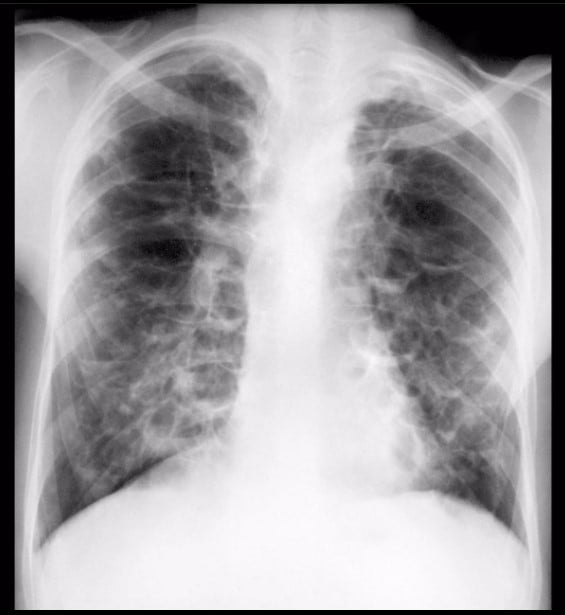
Bronchiectasis. The tram-track sign is a pair of parallel linear opacities produced by the thickened walls of an abnormally dilated bronchus projected along its length. A normal bronchus tapers steadily and its wall is too thin to see beyond the hila; when the airway is dilated and its wall is thickened, both walls become visible and, because the airway no longer tapers, they run parallel like tram rails.
| At a glance | |
|---|---|
| What it is | Two parallel thin linear opacities on a chest radiograph, usually 1–3 mm apart, running towards the periphery |
| What it means | A dilated, thick-walled bronchus seen along its long axis – the radiographic face of bronchiectasis |
| Also called | Tram-track sign, tram-line sign, tram-lines, tramline shadowing |
| End-on equivalent | Ring shadow on radiography; signet-ring sign on CT |
| Best test | Volumetric thin-section CT, not the radiograph |
| Careful | The same name is used for four unrelated findings outside the chest |
Why is it called so?
Named for the resemblance to the parallel rails of a tramway track. The two rails are the two walls of a single dilated bronchus seen in longitudinal or tangential projection. The term is one of the descriptive radiographic patterns catalogued in the Fleischner Society glossary, which lists tram-track as a synonym for parallel line opacities arising from bronchial wall thickening.
Pathophysiology
Bronchiectasis is the end point of the vicious vortex: impaired mucociliary clearance allows bacterial colonisation, which drives neutrophilic airway inflammation, which in turn damages the bronchial wall and worsens clearance. Neutrophil serine proteases released from activated neutrophils digest elastin and the cartilage-containing wall, while chronic inflammation causes peribronchial fibrosis and smooth muscle hypertrophy.
The mechanical consequence is the radiographic appearance. The wall thickens, so it becomes visible against aerated lung. The lumen dilates and loses the normal centrifugal tapering, so the two wall shadows stay a constant distance apart instead of converging. Where the same airway is caught end-on rather than along its length, the pair of lines becomes a ring shadow. The damage is structurally irreversible, which is why the sign persists between infections.
Four other tram-tracks: the same name, four unrelated diagnoses
This is the single most common source of confusion with this sign. “Tram-track” is a shape, not a disease, and it was coined independently in several organ systems. If you meet the phrase without context, none of these can be assumed.
| Where | What the two lines are | Diagnosis |
|---|---|---|
| Chest radiograph or CT | Parallel walls of a dilated, thick-walled bronchus | Bronchiectasis |
| Brain CT (or SWI) | Parallel gyriform cortical calcification following the crests of adjacent gyri | Sturge-Weber syndrome. A late finding: the leptomeningeal vascular malformation is visible on contrast-enhanced MRI long before calcification appears, so a normal CT does not exclude it |
| Orbital CT or MRI, axial plane | Enhancing thickened optic nerve sheath on either side of a non-enhancing central optic nerve | Optic nerve sheath meningioma – but the sign is not specific and is also seen in optic perineuritis, sarcoidosis, lymphoma, metastasis and idiopathic orbital inflammation |
| Neonatal brain imaging | Paired linear periventricular or cortical calcification | Reported as an unusual pattern of congenital toxoplasmosis |
| Hip ultrasound after arthroplasty | Two parallel echogenic lines in the joint | Polyethylene liner dissociation after total hip arthroplasty |
Only the first is a chest sign. The rest share the noun and nothing else.
How much can the chest radiograph actually tell you?
Less than the classic teaching implies. In the reference comparative study, chest radiography was 87.8% sensitive and only 74.4% specific for bronchiectasis against high-resolution CT, and radiographic severity correlated with CT severity (r = 0.62). Of 47 patients with an abnormal radiograph, 11 had a completely normal HRCT – roughly one in four abnormal films was a false alarm. Of 37 normal radiographs, only 5 had CT disease and all of it was mild cylindrical bronchiectasis.
Two practical conclusions follow. A normal radiograph makes clinically relevant bronchiectasis unlikely, which is useful. But tram-tracking on a film is a prompt for CT, not a diagnosis, because a quarter of the time the airways turn out to be normal. The things that most often masquerade as tram-tracking are listed below.
| Mimic | Typical setting | How to separate it |
|---|---|---|
| Bronchial wall thickening without dilatation | Asthma, chronic bronchitis, acute viral bronchitis | Walls are thickened but the airway still tapers; on CT the broncho-arterial ratio is normal |
| Crowded vessels and bronchi | Lobar collapse or any volume loss | Lines converge towards the collapsed segment; look for fissural displacement |
| Interstitial oedema | Heart failure | Peribronchial cuffing is usually perihilar, symmetrical and accompanied by septal lines and effusions; it resolves |
| Overlapping bony or soft-tissue structures | Costal cartilage, companion shadows of the ribs, scapular edge, skin folds | Follow the line beyond the lung edge – it will cross the chest wall |
| Traction bronchiectasis | Pulmonary fibrosis | Surrounding reticulation, volume loss and honeycombing; the airway is irregular rather than smoothly dilated |
The CT criteria that actually define bronchiectasis
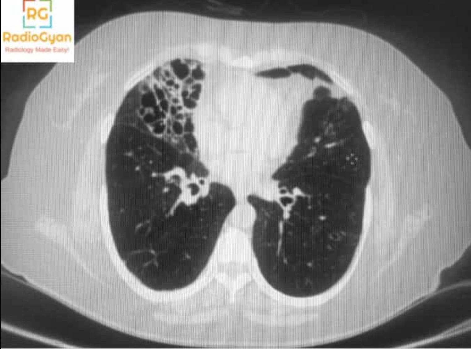
Volumetric thin-section CT is the diagnostic standard. Bronchiectasis is defined morphologically, by dilatation of the airway relative to its companion pulmonary artery, supported by secondary signs.
| Criterion | Type | Notes |
|---|---|---|
| Broncho-arterial ratio greater than 1 | Direct | The classic threshold. Produces the signet-ring sign on axial images: the dilated bronchus is the ring, the artery the gemstone |
| Lack of tapering | Direct | Constant bronchial calibre over at least 2 cm distal to a branch point. This is the tram-track in cross-sectional form, and the most reproducible criterion |
| Visibility of airways in the periphery | Direct | Bronchi seen within 1 cm of the costal pleura, or touching the mediastinal pleura |
| Bronchial wall thickening | Indirect | Subjective; correlates with disease activity rather than presence |
| Mucus plugging, tree-in-bud | Indirect | Indicates infected or retained secretions and small-airway involvement |
| Mosaic attenuation and air trapping on expiration | Indirect | Small-airway disease, often the earliest change |
The broncho-arterial ratio is softer than it looks. In never-smoking adults with normal lungs the mean ratio was 0.79 and it exceeded 1 in 8.5% of healthy subjects – driven by small arteries, not large bronchi, which means hypoxic conditions and altitude push the ratio up without any airway disease. The ratio also increases with normal ageing, so an isolated ratio just above 1 in an elderly patient is weak evidence. In children the opposite correction applies: without lung disease the ratio averages well below the adult figure, and a paediatric threshold nearer 0.8 is used. Use the ratio with the supporting criteria, never alone.
Traction bronchiectasis is a different finding
Dilated airways inside fibrotic lung are being pulled open from the outside. Calling this “bronchiectasis” in a report sends the patient down an airway-disease pathway when the disease is interstitial. The extent of traction bronchiectasis on CT is an independent predictor of survival across fibrotic interstitial lung diseases, so the label is not cosmetic.
| Feature | True bronchiectasis | Traction bronchiectasis |
|---|---|---|
| Surrounding lung | Normal, hyperinflated or consolidated | Reticulation, architectural distortion, honeycombing, volume loss |
| Airway contour | Smoothly dilated, cylindrical, varicose or cystic | Irregular, corkscrew, angulated |
| Wall | Thickened | Thin, stretched |
| Secretions | Mucus plugging and tree-in-bud common | Usually absent |
| Distribution | Follows the underlying cause (see below) | Follows the fibrosis – subpleural and basal in usual interstitial pneumonia |
| Implication | Airway clearance, sputum culture, cause workup | Interstitial lung disease workup, antifibrotic pathway |
Morphology: what the tram-track becomes as disease advances
Reid classified bronchiectasis by the shape of the dilated airway, and the three types have distinct radiographic faces. Tram-tracking belongs to the mildest of the three, which is why it is the pattern most often missed.
| Type | Airway shape | Radiographic appearance |
|---|---|---|
| Cylindrical (tubular) | Uniform dilatation, wall roughly parallel | Tram-tracking along the airway; ring shadows end-on |
| Varicose | Alternating dilatation and constriction | Beaded, irregular parallel lines; string-of-pearls contour |
| Cystic (saccular) | Ballooned airways ending in sacs | Clustered ring shadows and cysts, often with air-fluid levels; the “bunch of grapes” appearance |
Reading the distribution: what the pattern points to
Bronchiectasis is a pattern, not a diagnosis. The 2025 European Respiratory Society guideline makes a strong recommendation that every patient undergoes standardised testing to identify the underlying cause, and the imaging distribution is the first step in narrowing that search. In the European EMBARC registry the commonest identified aetiologies were post-infective disease, chronic obstructive pulmonary disease, connective tissue disease and immunodeficiency, with a large idiopathic group.
| Distribution | Think of |
|---|---|
| Upper lobes | Cystic fibrosis, previous tuberculosis, chronic sarcoidosis, radiation |
| Central or perihilar, upper zone predominant, with high-attenuation mucus | Allergic bronchopulmonary aspergillosis – look for the finger-in-glove sign |
| Right middle lobe and lingula, with tree-in-bud nodules | Non-tuberculous mycobacterial infection (nodular bronchiectatic form, the “Lady Windermere” phenotype) |
| Lower lobes, bilateral | Post-infective, aspiration, antibody deficiency, rheumatoid arthritis, alpha-1 antitrypsin deficiency |
| Lower lobes with situs abnormality or dextrocardia | Primary ciliary dyskinesia and Kartagener syndrome |
| Focal, confined to one lobe or segment | Obstruction until proven otherwise – endobronchial tumour, foreign body, broncholith, extrinsic node |
| Unilateral hyperlucent lung with bronchiectasis | Swyer-James-MacLeod syndrome (post-infectious constrictive bronchiolitis) |
| Central, with a tracheal diameter above 3 cm | Mounier-Kuhn syndrome (tracheobronchomegaly) |
What to do when you see it
Reporting checklist
- Say bronchiectasis, not “tram-tracking” alone – name the finding the clinician acts on.
- State whether it is true or traction bronchiectasis, and give the surrounding lung as the reason.
- Give distribution by lobe and morphology (cylindrical, varicose, cystic).
- Report activity markers: mucus plugging, tree-in-bud, consolidation, air-fluid levels. These change management more than extent does.
- If disease is focal, explicitly comment on the proximal airway and say whether an obstructing lesion is excluded.
- Flag features that point to a specific cause: high-attenuation mucus, situs inversus, tracheomegaly, upper-lobe predominance, oesophageal dilatation.
How it is managed in 2026
Bronchiectasis on imaging alone is not a disease. The guidelines define clinically significant bronchiectasis as compatible radiology plus a compatible clinical syndrome, typically chronic cough with sputum and recurrent infections. The 2025 European Respiratory Society clinical practice guideline updates the 2017 version and the 2019 British Thoracic Society guideline, and upgraded several recommendations from conditional to strong.
| Intervention | 2025 ERS recommendation | Note |
|---|---|---|
| Airway clearance techniques taught to all patients | Strong for | Extended beyond productive cough – patients with a dry cough and mucus plugging on CT may also benefit |
| Standardised testing to identify the underlying cause | Strong for | Also to assess severity, activity and treatable traits |
| Pulmonary rehabilitation where exercise capacity is impaired | Strong for | |
| Long-term macrolides in patients at high risk of exacerbations | Strong for | Exclude non-tuberculous mycobacteria first; never as monotherapy in NTM disease |
| Long-term inhaled antibiotics with chronic Pseudomonas aeruginosa | Strong for | Conditional for other chronic pathogens |
| Mucoactive drugs where airway clearance alone fails | Conditional for | Recombinant DNase is recommended against |
| Long-term non-macrolide oral antibiotics | Conditional against | Reserve for specific situations |
| Inhaled corticosteroids | Conditional against | Unless there is coexisting asthma or COPD |
The definition of “high risk of exacerbations” also changed. The old threshold of three or more exacerbations a year was replaced by individualised risk: two or more exacerbations in the previous year, or one severe exacerbation, or one exacerbation plus severe daily symptoms.
The therapeutic landscape changed at the same time. Brensocatib, an oral reversible dipeptidyl peptidase 1 inhibitor that blocks activation of neutrophil serine proteases, reduced the annualised rate of pulmonary exacerbations over 52 weeks in the phase 3 ASPEN trial, and the 25 mg dose also reduced the decline in post-bronchodilator FEV1. It was approved by the FDA in August 2025 for non-cystic fibrosis bronchiectasis at age 12 and above – the first drug ever approved for the condition. A finding that was once reported and forgotten now has a treatment attached to it, which is the strongest argument for describing it properly.
Alternative names: Tram track sign; tram-line sign; tramline shadowing; parallel line opacities.
Other associated named signs: Signet-ring sign (dilated bronchus and smaller adjacent artery on CT), ring shadow (the same airway end-on), tree-in-bud (small-airway secretions), finger-in-glove sign (mucoid impaction, classically in allergic bronchopulmonary aspergillosis), string of pearls (varicose bronchiectasis).
Frequently asked questions
What does the tram-track sign mean on a chest X-ray?
It means bronchiectasis. Two parallel linear opacities are the thickened walls of a dilated bronchus seen along its length, instead of the single thin line a normal bronchus makes. The bronchus has lost its normal tapering, so the two walls run in parallel like tram rails.
Is the tram-track sign specific to bronchiectasis?
No. On a chest radiograph, parallel lines are also produced by bronchial wall thickening without dilatation in asthma and chronic bronchitis, by crowded vessels in volume loss, and by overlapping ribs or costal cartilage. On a radiograph alone the sign is suggestive, not diagnostic.
Why does the same name appear in neuroimaging and orbital imaging?
Tram-track is a purely descriptive term for any pair of parallel lines, so it was coined independently in several organs. The three common ones are bronchiectasis on chest imaging, gyriform cortical calcification in Sturge-Weber syndrome on brain CT, and the enhancing sheath around a non-enhancing optic nerve in optic nerve sheath meningioma on orbital imaging. They share nothing but the shape.
Can a normal chest X-ray exclude bronchiectasis?
Largely, but not completely. In a comparative study against high-resolution CT the chest radiograph was about 88 percent sensitive and only 74 percent specific, and radiographic abnormality tracked with CT severity. A normal film makes clinically relevant bronchiectasis unlikely, but mild cylindrical disease is missed, so CT is still needed when the clinical suspicion is high.
What are the CT criteria for bronchiectasis?
The main criterion is bronchial dilatation relative to the accompanying pulmonary artery, the broncho-arterial ratio, classically greater than 1 and producing the signet-ring sign. Supporting criteria are lack of normal tapering over at least 2 cm and visibility of bronchi within 1 cm of the costal pleura or touching the mediastinal pleura.
Is a broncho-arterial ratio above 1 always abnormal?
No. In never-smokers with normal lungs the mean ratio was 0.79 and 8.5 percent of healthy subjects exceeded 1, usually because the artery was small rather than the bronchus large. The ratio also rises with age and at altitude, and in children a threshold of about 0.8 is more appropriate. Treat a borderline ratio as a soft finding and look for the supporting features.
How do I tell traction bronchiectasis from true bronchiectasis?
Traction bronchiectasis sits inside fibrotic lung. Look for reticulation, architectural distortion, volume loss and honeycombing around the dilated airway, and for an irregular, corkscrew contour. True bronchiectasis occurs in otherwise normal or hyperinflated lung, with smooth dilatation, wall thickening, mucus plugging and tree-in-bud. The distinction matters because the treatment pathways are completely different.
What does the distribution of bronchiectasis tell you?
Upper lobe disease suggests cystic fibrosis, allergic bronchopulmonary aspergillosis or previous tuberculosis. Right middle lobe and lingular disease with tree-in-bud suggests non-tuberculous mycobacterial infection. Lower lobe disease suggests post-infective disease, aspiration, primary ciliary dyskinesia or antibody deficiency. Focal disease in one segment should prompt a search for an obstructing lesion.
How is bronchiectasis treated now?
The 2025 European Respiratory Society guideline makes strong recommendations for teaching airway clearance techniques, for testing every patient to find the underlying cause, for long-term macrolides in patients at high risk of exacerbations, and for long-term inhaled antibiotics when there is chronic Pseudomonas aeruginosa infection. Inhaled corticosteroids are not recommended unless there is coexisting asthma or COPD.
Is there a drug approved specifically for bronchiectasis?
Yes, since 2025. Brensocatib, an oral dipeptidyl peptidase 1 inhibitor, reduced the annualised exacerbation rate in the phase 3 ASPEN trial and was approved by the FDA in August 2025 for non-cystic fibrosis bronchiectasis in patients aged 12 and over. It is the first approved therapy for the condition.
Related radiology signs
- Signet-ring sign – the same dilated bronchus seen end-on on CT
- Gloved-finger sign – mucoid impaction in dilated bronchi
- Williams-Campbell syndrome – congenital cartilage deficiency causing bronchiectasis
- Bat-wing appearance – the perihilar pulmonary oedema pattern
- Silhouette sign – localising disease on the chest radiograph
- Steeple sign – subglottic narrowing in croup
References
- Hansell DM, Bankier AA, MacMahon H, McLoud TC, Müller NL, Remy J. Fleischner Society: glossary of terms for thoracic imaging. Radiology. 2008;246(3):697-722. PMID: 18195376
- Cantin L, Bankier AA, Eisenberg RL. Bronchiectasis. AJR Am J Roentgenol. 2009;193(3):W158-171. PMID: 19696251
- van der Bruggen-Bogaarts BA, van der Bruggen HM, van Waes PF, Lammers JW. Screening for bronchiectasis. A comparative study between chest radiography and high-resolution CT. Chest. 1996;109(3):608-611. PMID: 8617064
- Diaz AA, Young TP, Maselli DJ, et al. Bronchoarterial ratio in never-smokers adults: implications for bronchial dilation definition. Respirology. 2017;22(1):108-113. PMID: 27538197
- Kapur N, Masel JP, Watson D, Masters IB, Chang AB. Bronchoarterial ratio on high-resolution CT scan of the chest in children without pulmonary pathology: need to redefine bronchial dilatation. Chest. 2011;139(6):1445-1450. PMID: 20864608
- Chalmers JD, Haworth CS, Flume P, et al. European Respiratory Society clinical practice guideline for the management of adult bronchiectasis. Eur Respir J. 2025;66(6):2501126. PMID: 41016738
- Hill AT, Sullivan AL, Chalmers JD, et al. British Thoracic Society guideline for bronchiectasis in adults. Thorax. 2019;74(Suppl 1):1-69. PMID: 30545985
- Chalmers JD, Burgel PR, Daley CL, et al. Phase 3 trial of the DPP-1 inhibitor brensocatib in bronchiectasis. N Engl J Med. 2025;392(16):1569-1581. PMID: 40267423
- Chalmers JD, Polverino E, Crichton ML, et al. Bronchiectasis in Europe: data on disease characteristics from the European Bronchiectasis registry (EMBARC). Lancet Respir Med. 2023;11(7):637-649. PMID: 37105206
- Marinescu DC, Hague CJ, Muller NL, et al. CT honeycombing and traction bronchiectasis extent independently predict survival across fibrotic interstitial lung disease subtypes. Radiology. 2025;314(2):e240232. PMID: 39903073
- Akpinar E. The tram-track sign: cortical calcifications. Radiology. 2004;231(2):515-516. PMID: 15128996
- Johns TT, Citrin CM, Black J, Sherman JL. CT evaluation of perineural orbital lesions: evaluation of the “tram-track” sign. AJNR Am J Neuroradiol. 1984;5(5):587-590. PMID: 6435424
- Kanamalla US. The optic nerve tram-track sign. Radiology. 2003;227(3):718-719. PMID: 12773677
- Nair AG, Iyer VR, Gopinathan I. Revisiting the “tram-track” sign. Indian J Ophthalmol. 2017;65(11):1217-1218. PMID: 29133657
- Surendrababu NR, Kuruvilla KA, Jana AK. Unusual pattern of calcification in congenital toxoplasmosis: the tram-track sign. Pediatr Radiol. 2006;36(6):569. PMID: 16520973
- Jang JH, Jin W, Lee GY, et al. The tram track sign: a characteristic sonographic feature of polyethylene liner dissociation after total hip arthroplasty. J Ultrasound Med. 2014;33(11):2027-2032. PMID: 25336480
Access all radiology signs posted so far: https://radiogyan.com/radiology-signs/

Improve Content And Help Your Colleagues!
Found an error in the post? Please let us know using our contact page and we will update it with due credits!
If you wish to contribute radiological images for this case (or any other article on the website), you can submit them here and you will be duly credited: Submit radiology case