


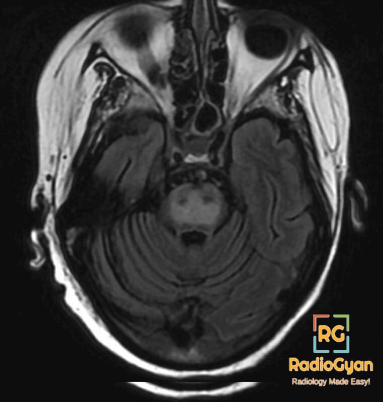
What causes the trident sign in the pons on MRI?
Let me know in the comments.
Click to reveal the answer
Answer:
The trident sign of the pons is a symmetrical, trident- or bat-wing-shaped area of T2 and FLAIR hyperintensity, with restricted diffusion in the acute phase, occupying the central basis pontis, principally the transverse pontocerebellar fibres, while sparing the peripheral rim of the pons, the tegmentum and the paired corticospinal tracts. The two spared corticospinal tracts notch the confluent central hyperintensity into a middle prong flanked by two lateral prongs, so the abnormal signal itself is trident-shaped on the axial image. It is the classic appearance of osmotic demyelination syndrome, formerly called central pontine myelinolysis, and follows rapid correction of hyponatraemia, most often in chronic alcoholism, malnutrition, liver transplantation, severe burns or prolonged diuretic use. Two caveats matter clinically. MRI is frequently normal in the first 24 to 48 hours even in a severely affected patient, so a negative early study does not exclude the diagnosis and repeat imaging at one to two weeks is indicated; and diffusion-weighted imaging is the most sensitive early sequence, showing restricted diffusion before the T2 change is conspicuous. Roughly half of patients also have extrapontine myelinolysis involving the basal ganglia, thalami, cerebellum, lateral geniculate bodies or subcortical white matter, which should be sought on the same study. The piglet sign is the related appearance in which the ventral pons is involved and the spared regions outline a piglet’s snout, and the two patterns evolve into one another over time. Differentials for central pontine T2 hyperintensity are pontine infarction, which is unilateral and respects an arterial territory, chronic microangiopathic change, which is patchy and asymmetric, glioma, which is expansile and mass-forming, and CLIPPERS, which produces punctate perivascular enhancement peppering the pons and brachium pontis. Correcting serum sodium by no more than 8 to 10 mmol/L in 24 hours, or 8 mmol/L in a high-risk patient, is the accepted prevention, and recovery is often better than the imaging appearance suggests.
Why is it called so?
The two spared corticospinal tracts notch the confluent hyperintense central pontine signal into three prongs, a middle prong and two lateral prongs, so the abnormal signal itself resembles a trident on the axial image.
Pathophysiology
Chronic hyponatraemia allows brain cells to extrude organic osmolytes to stay in osmotic balance with the dilute plasma. When plasma sodium is raised faster than those osmolytes can be regenerated, water is drawn abruptly out of the cells and the oligodendrocytes, which have the highest energy demand and the least capacity to recover osmolytes, are injured. The result is non-inflammatory demyelination with preserved axons and neuronal cell bodies. The central pons is affected preferentially because the grey and white matter there are intimately intermingled in a grid of transverse pontocerebellar fibres and descending tracts, which makes the tissue especially vulnerable to osmotic shift; the peripheral pons and the compact corticospinal tracts are relatively resistant and remain of normal signal, producing the trident outline. Restricted diffusion in the first days reflects intramyelinic oedema and myelin splitting, which then gives way to established T2 hyperintensity and, in survivors, to atrophy or complete resolution.
Alternative names: Trident-shaped pontine lesion, bat-wing sign, central pontine myelinolysis sign
Other associated named signs: The piglet sign of osmotic demyelination, the hot cross bun sign of multiple system atrophy, and the trident sign of the spinal cord in neurosarcoidosis, a different sign of the same name
References
Access all radiology signs posted so far: https://radiogyan.com/radiology-signs/

Improve Content And Help Your Colleagues!
Found an error in the post? Please let us know using our contact page and we will update it with due credits!
If you wish to contribute radiological images for this case (or any other article on the website), you can submit them here and you will be duly credited: Submit radiology case