
What causes Dromedary hump in kidney on imaging?
Let me know in the comments.
Click to reveal the answer
Answer:
Dromedary hump is a prominent focal bulge on the lateral border of the left kidney due to impression from the adjacent spleen, representing a normal anatomic variant that may mimic a renal mass or pseudotumor; it exhibits identical imaging characteristics to adjacent renal cortex across modalities including ultrasound, CT, MRI, and nuclear medicine, with normal calyceal extension into the bulge and normal radiotracer uptake on Tc-99m DMSA scan.
Why is it called so?:
Named after the prominent single hump on the back of a dromedary camel, reflecting the similar focal bulge on the lateral contour of the left kidney.
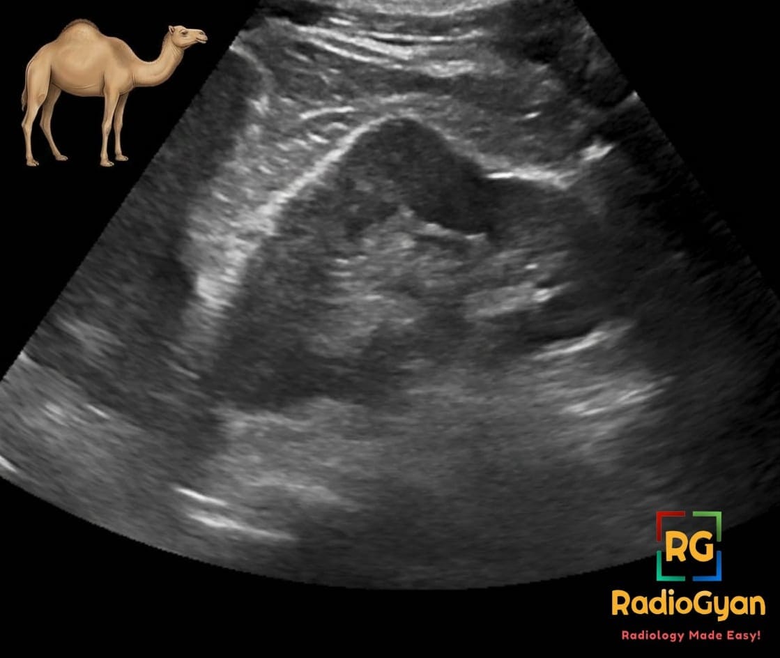
Pathophysiology:
The bulge develops embryologically from molding and adaptation of the superolateral left renal cortex by chronic pressure from the adjacent spleen during renal development and growth.
Alternative names: Splenic hump
Other associated named signs: Columns of Bertin
Access all radiology signs posted so far: https://radiogyan.com/radiology-signs/

Improve Content And Help Your Colleagues!
Found an error in the post? Please let us know using our contact page and we will update it with due credits!
If you wish to contribute radiological images for this case (or any other article on the website), you can submit them here and you will be duly credited: Submit radiology case