


What causes an apple core sign in the colon on barium enema or CT imaging?
Let me know in the comments.
Click to reveal the answer
Answer: The apple core sign, also known as the napkin ring sign, is most frequently caused by a stenosing annular colorectal carcinoma; however, other etiologies include lymphoma, chlamydia infection, and post-radiation therapy from radiosurgery. The classic appearance is that of a short, irregular, circumferential colonic stricture with overhanging margins and ulceration. Clinically, patients may present with abdominal pain, weight loss, diarrhea (which may be bloody), and iron deficiency anemia. Confirmation and differentiation usually require colonoscopy and histological assessment.
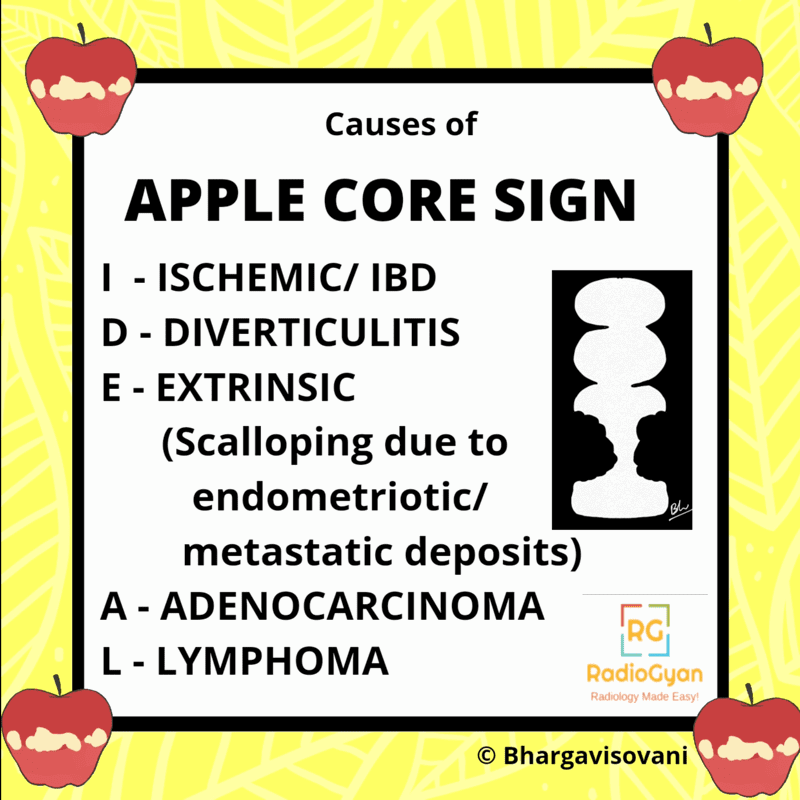
Why is it called so?
The sign is named for its visual resemblance to the core of an apple after the flesh has been eaten—a short segment of marked luminal stenosis with abrupt, “shouldered” margins mimics the appearance of the remaining central core, hence the term apple core sign. The term napkin ring sign was coined by Russell Carman to describe a similar circumferential narrowing. Both terms highlight the concentric narrowing with overhanging edges.
Pathophysiology
The apple core sign arises due to eccentric thickening of the colonic wall—typically by an infiltrating neoplasm—leading to severe luminal stenosis. The tumor grows circumferentially, destroying the mucosa and creating ulceration, with the remaining intact margins appearing as “shoulders” on imaging. This results in a narrowed, eccentric, irregular lumen often measuring 3–6 cm in length. Inflammatory and infectious processes may produce similar morphology by causing transmural inflammation, fibrosis, and stricturing.

Alternative names: Napkin ring sign (bowel).
Other associated named signs: None commonly cited in the literature for this specific pathology.
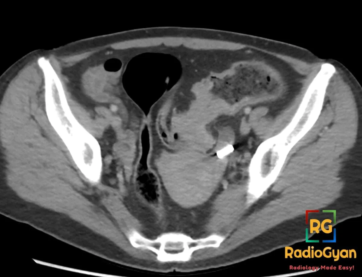
The apple core sign (also called the napkin ring sign) is a short, circumferential, irregular narrowing of the bowel lumen with abrupt, overhanging (“shouldered”) margins and destroyed mucosa. On a barium study or CT the residual stenotic lumen flanked by the two rounded shoulders resembles the core of an apple after the flesh has been eaten. It is the classic imaging appearance of an annular constricting colorectal carcinoma, but any process that produces a short circumferential stricture can reproduce it, so the sign always needs clinical and endoscopic correlation.
What is the apple core sign?
The apple core sign describes a short segment of bowel, usually only a few centimetres long, that is narrowed all the way around its circumference. The narrowed lumen is irregular because the mucosa is ulcerated and destroyed, and it ends abruptly at each side against rounded, overhanging edges (the “shoulders”). Those shoulders are the intact bowel wall being pushed aside by the tumour, and their sudden, cliff-like transition from normal calibre to tight stenosis is what gives the lesion its characteristic look. The same appearance is called the napkin ring lesion by pathologists and endoscopists, describing the encircling ring of tumour.
- Modality: historically seen on the double-contrast barium enema; today it is most often recognised on contrast-enhanced CT and CT colonography, where 3D endoluminal reconstructions reproduce the barium appearance.
- Morphology: short (classically 3–6 cm), circumferential, irregular luminal narrowing with abrupt shouldered margins.
- Common site: the sigmoid and descending (left) colon, where the lumen is narrower and stool is more solid, so annular tumours declare themselves earlier with obstruction.
Why is it called the apple core sign?
The name comes from the everyday image of an eaten apple: once the flesh is gone, what remains is a thin central core flanked by two rounded ends. On imaging, the tight residual lumen represents the core, and the two shouldered margins of preserved bowel wall represent the rounded ends of the apple. The alternative term, napkin ring, captures the same idea from a different angle: the tumour forms a complete ring around the bowel like a ring slipped around a rolled napkin. Both terms describe a concentric, encircling lesion with a strangled central lumen.
Pathophysiology
Most annular colonic cancers begin as a flat or polypoid mucosal lesion that grows circumferentially, spreading around the bowel wall rather than only along its length. As the tumour encircles the lumen it invades and destroys the mucosa (producing the irregular, ulcerated inner surface) and infiltrates the wall, which becomes rigid and cannot distend. The result is a fixed, short, concentric stenosis with abrupt margins where tumour meets normal wall. Benign processes reach a similar end point by a different route: transmural inflammation, fibrosis and scarring (as in diverticulitis, Crohn disease, ischaemia or tuberculosis) can also stiffen and narrow a short segment of colon, which is why the sign is suggestive of, but not specific for, malignancy.
How to recognise it and confirm the diagnosis
- Measure the segment. A short lesion (a few centimetres) with abrupt shoulders favours carcinoma; a longer, tapering narrowing favours an inflammatory or ischaemic stricture.
- Inspect the margins and mucosa. Overhanging, shouldered edges and a destroyed, irregular mucosal surface point to malignancy; smooth, symmetric, tapering margins point to a benign cause.
- Look beyond the lumen on CT. An eccentric soft-tissue mass, pericolic tumour extension, regional lymphadenopathy and liver lesions strongly favour cancer; symmetric wall thickening with prominent fat stranding and a diverticulum favours diverticulitis.
- Always correlate and confirm. Colonoscopy with biopsy is required for tissue diagnosis; CT of the chest, abdomen and pelvis is used for staging, with pelvic MRI added for rectal tumours.
Causes of an apple core sign
An annular constricting colorectal carcinoma is by far the commonest cause, and it must be excluded first. However, a range of malignant and benign processes can produce an identical short circumferential stricture. The clinical context and the surrounding imaging findings usually narrow the differential.
| Cause | Why it produces an apple core | Clue that favours it |
|---|---|---|
| Annular colorectal adenocarcinoma (most common) | Circumferential tumour growth destroys mucosa and rigidly narrows a short segment | Short lesion, shouldered margins, soft-tissue mass, nodes, liver metastases; older patient with anaemia or weight loss |
| Serosal / peritoneal metastases (ovary, stomach, breast, pancreas) | Serosal tumour deposits desmoplastically tether and constrict the wall | Known primary malignancy, peritoneal disease, ascites |
| Lymphoma | Usually causes aneurysmal dilatation, but occasionally infiltrates and strictures a segment | Bulky nodes, homogeneous wall thickening, less mucosal destruction |
| Diverticulitis (with stricture) | Repeated transmural inflammation and fibrosis narrow the sigmoid | Diverticula, prominent pericolic fat stranding, longer segment, preserved mucosa |
| Ischaemic colitis stricture | Healing ischaemic injury leaves a fibrotic narrowing, often at the splenic flexure | Watershed location, smooth tapering margins, vascular risk factors |
| Crohn disease | Transmural inflammation and fibrosis produce a strictured segment | Skip lesions, mural stratification, mesenteric changes, younger patient |
| Intestinal tuberculosis / amoebiasis | Granulomatous or chronic infection scars and narrows the bowel, classically ileocaecal | Endemic exposure, ileocaecal location, an amoeboma mass with amoebiasis |
| Endometriosis | Serosal implants on the rectosigmoid cause fibrosis and extrinsic narrowing | Woman of reproductive age, cyclical symptoms, intact overlying mucosa |
| Radiation or lymphogranuloma venereum stricture | Chronic fibrosis after pelvic radiotherapy or chlamydial infection narrows the rectosigmoid | Relevant history, long smooth stricture, rectal involvement |
Malignant versus benign apple core
The single most useful task is to decide whether a short circumferential stricture is malignant or benign, because it changes management from staging and resection to conservative treatment. A few imaging features shift the balance, though tissue is always needed for a final answer.
| Feature | Favours malignant (carcinoma) | Favours benign (inflammatory / ischaemic) |
|---|---|---|
| Length of segment | Short (a few centimetres) | Often longer |
| Margins / transition | Abrupt, overhanging, shouldered | Smooth, gradual, tapering |
| Mucosa | Destroyed, irregular, ulcerated | Relatively preserved |
| Wall thickening | Eccentric, mass-like, non-stratified | Symmetric, mural stratification often preserved |
| Surrounding tissues | Nodes, pericolic tumour deposits, liver metastases | Inflammatory fat stranding, diverticula, mesenteric changes |
| Clinical context | Older age, weight loss, iron-deficiency anaemia, PR bleeding | Acute pain and fever, known IBD, ischaemic risk factors |
Clinical significance and pitfalls
- Treat as cancer until proven otherwise. A short apple-core stricture, especially in the left colon of an older patient, is a colorectal carcinoma until histology says otherwise.
- It is a sign, not a diagnosis. Benign strictures from diverticulitis, ischaemia, Crohn disease and tuberculosis can look identical; the diagnosis rests on biopsy plus the clinical picture.
- Watch for obstruction. A tight annular lesion can present as large-bowel obstruction; look for proximal dilatation and assess the competence of the ileocaecal valve.
- Do not forget staging. Once carcinoma is suspected, complete CT chest, abdomen and pelvis, add pelvic MRI for rectal tumours, and search for a synchronous second cancer at colonoscopy.
Frequently asked questions
What is the apple core sign in radiology?
The apple core sign is a short, circumferential, irregular narrowing of the bowel lumen with abrupt shouldered margins and destroyed mucosa. The strangled central lumen flanked by two rounded shoulders resembles the core of an eaten apple. It is the classic appearance of an annular constricting colorectal carcinoma.
What is the most common cause of the apple core sign?
A stenosing annular colorectal adenocarcinoma is by far the most common cause. Other causes include peritoneal or serosal metastases, lymphoma, diverticulitis with stricture, ischaemic colitis, Crohn disease, intestinal tuberculosis, amoebiasis, endometriosis and radiation stricture.
How do you tell a malignant apple core from a benign one?
Malignant lesions tend to be short with abrupt shouldered margins, destroyed mucosa, eccentric mass-like wall thickening and features such as lymph nodes or liver metastases. Benign strictures tend to be longer, with smooth tapering margins, relatively preserved mucosa and surrounding inflammatory or ischaemic changes. Colonoscopy and biopsy are required for a definitive diagnosis.
Is the apple core sign the same as the napkin ring sign?
Yes, in the bowel the two terms describe the same annular constricting lesion. Apple core is the radiographic description of the strangled lumen and shouldered margins, while napkin ring describes the encircling ring of tumour around the bowel wall. Note that napkin ring sign has a separate, unrelated meaning in coronary CT, where it describes a high-risk atherosclerotic plaque.
Which imaging test shows the apple core sign best?
It was classically demonstrated on the double-contrast barium enema. Today contrast-enhanced CT and CT colonography are used, because they show the lesion, the bowel wall and the extent of spread and allow staging in one examination. Colonoscopy provides direct visualisation and tissue for biopsy.
References
- Fonseca EKUN, Tridente CF, Ogawa RE, Yamauchi FI, Baroni RH. Apple core sign in colorectal cancer. Abdom Radiol (NY). 2017;42(7):2001-2002. PMID: 28214962.
- Freyschmidt J. The apple core sign. Eur Radiol. 2002;12(1):245-247. PMID: 11868104.
- Ganguly A, Meredith S, Probert C, et al. Colorectal cancer mimics: a review of the usual suspects with pathology correlation. Abdom Radiol (NY). 2016;41(9):1851-1866. PMID: 27178338.
- Cain BT, Huang LC. Benign Colonic Strictures. Dis Colon Rectum. 2021;64(9):1041-1044. PMID: 34108366.
Access all radiology signs posted so far: https://radiogyan.com/radiology-signs/

Improve Content And Help Your Colleagues!
Found an error in the post? Please let us know using our contact page and we will update it with due credits!
If you wish to contribute radiological images for this case (or any other article on the website), you can submit them here and you will be duly credited: Submit radiology case