


What causes Codman triangle in bone on radiography?
Let me know in the comments.
Click to reveal the answer
Codman triangle is caused by a rapidly growing lesion—most often a tumor such as osteosarcoma or Ewing sarcoma, but also possibly infection, hemorrhage, or abscess—that elevates the periosteum away from the underlying cortex faster than the periosteum can respond with new bone formation. This results in a characteristic radiologic appearance where the periosteum, partially ossified at its margins, forms a triangular or “V”-shaped reaction visible at the margin of the lesion on imaging modalities. The sign is strongly associated with aggressive bone lesions, including primary bone malignancies (osteosarcoma, Ewing sarcoma, chondrosarcoma), metastatic disease, aggressive benign tumors (aneurysmal bone cyst, giant cell tumor), and osteomyelitis.
Why is it called so?
The term “Codman triangle” honors Ernest Amory Codman, an American surgeon who described the radiographic finding in the context of Ewing sarcoma in the early 20th century, though the phenomenon was first described by Ribbert in 1914. The name reflects both the triangular radiographic appearance and Codman’s contribution to its clinical recognition.
Pathophysiology
A Codman triangle forms because an aggressive lesion grows so rapidly that it outstrips the ability of the periosteum to lay down new bone in organized layers. Instead of producing a smooth, continuous periosteal reaction (such as seen in benign, slow-growing processes), the lesion abruptly lifts the periosteum, which then forms a thin shell of new bone at its edges. The central portion of the elevated periosteum does not ossify, creating a two-sided, V-shaped or triangular area of new bone at the junction of the elevated periosteum and the cortex—often with a third side left open, hence the “triangle.”
Alternative names: None.
Other associated named signs: None directly associated; however, other aggressive periosteal reactions (e.g., spiculated or “sunburst,” laminated or “onion-skin”) may also be present in the same aggressive bone lesions that cause Codman triangle.
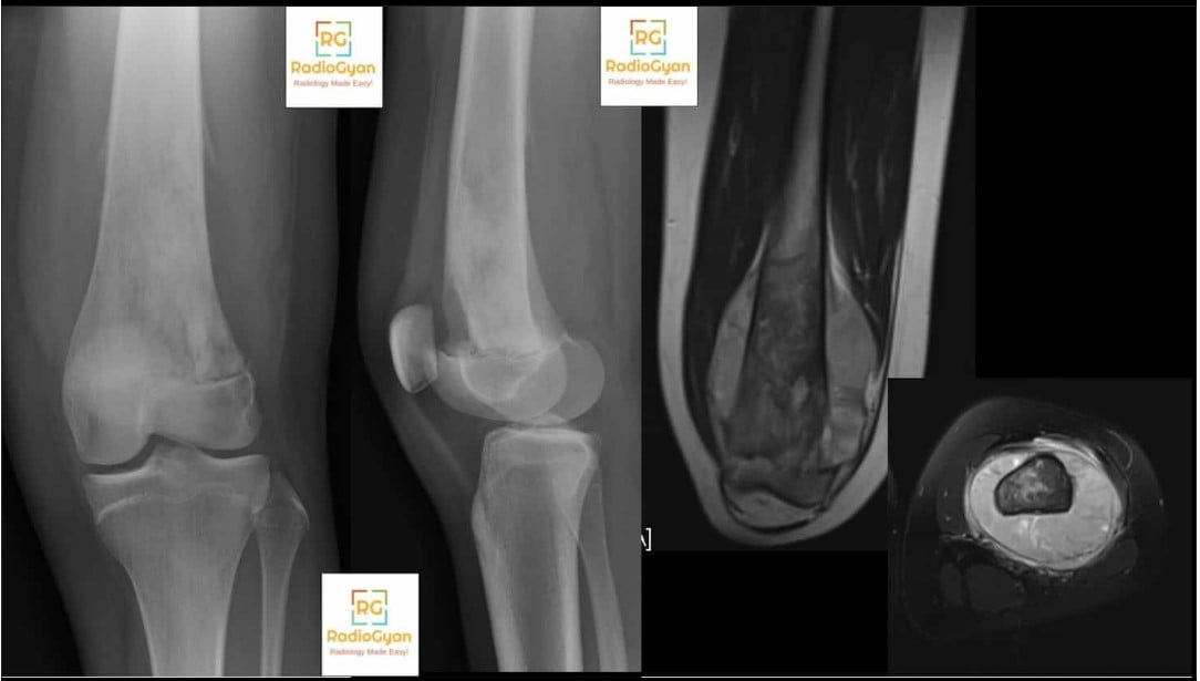
The Codman triangle is an interrupted periosteal reaction: a small, triangular cuff of new bone formed at the edge of a lesion where the periosteum has been lifted off the cortex faster than it can ossify. It is one of the classic radiographic markers of an aggressive bone lesion, and it is most often seen in osteosarcoma, followed by Ewing sarcoma. Two points matter more than the pattern itself. First, a Codman triangle tells you how fast something is growing, not what it is – pus and blood produce it as readily as tumour. Second, its absence is meaningless: most aggressive bone lesions do not show one, so a lesion without a Codman triangle is not thereby reassuring.
What is a Codman triangle?
The periosteum has two layers: an outer fibrous layer and an inner cambium layer packed with osteoprogenitor cells. When anything lifts the periosteum away from the cortex, the cambium responds by laying down new bone. How that new bone looks on the radiograph depends almost entirely on how fast the periosteum is being lifted, not on what is doing the lifting. A slow process gives the cambium time to build a solid, continuous shell. A fast one outruns it.
In a Codman triangle, the lesion strips the periosteum so quickly that only the leading edge – the acute angle where the elevated periosteum is still anchored to intact cortex – manages to ossify. The stripped central portion is destroyed or outpaced and never mineralises. What remains on the radiograph is a two-sided figure: the cortex forms one side, the ossified cuff of periosteum forms the second, and the third side is open toward the lesion. This is why the Codman triangle is really a pseudotriangle, and why it usually appears in pairs, one at each end of the lesion.
Periosteal reaction patterns: what each one means
Ragsdale, Madewell and Sweet’s classic analysis established the principle that still governs how these patterns are read: periosteal reaction encodes the rate of a process, and rate correlates with aggressiveness but not with histology. Patterns divide into continuous (the periosteum keeps up) and interrupted (it does not).
| Pattern | Appearance | What it implies | Typical causes |
|---|---|---|---|
| Solid / thick continuous | Dense, uniform shell fused to cortex | Slow, long-standing, benign | Osteoid osteoma, chronic osteomyelitis, stress fracture, healed trauma, hypertrophic osteoarthropathy |
| Single lamella | One thin line parallel to cortex | Recent but not necessarily aggressive | Early infection, early stress response, trauma |
| Lamellated (onion-skin) | Multiple concentric layers | Intermittent or phasic growth; indeterminate | Ewing sarcoma, osteomyelitis, osteosarcoma, LCH |
| Spiculated, perpendicular (hair-on-end) | Fine spicules at right angles to cortex | Fast; aggressive | Ewing sarcoma, osteosarcoma, thalassaemia (skull), metastases |
| Spiculated, divergent (sunburst) | Coarse spicules radiating from a point | Fast; aggressive | Osteosarcoma, high-grade metastases, meningioma (skull) |
| Interrupted (Codman triangle) | Triangular cuff of new bone at the lesion edge, unossified centrally | Very fast; aggressive | Osteosarcoma, Ewing sarcoma, subperiosteal abscess, subperiosteal haematoma, metastases, aggressive ABC |
Two practical consequences follow. Periosteal reaction takes time to mineralise – typically one to three weeks – so an early radiograph in acute osteomyelitis or an early stress injury can be entirely normal. And children make periosteal bone faster and more floridly than adults, because the paediatric periosteum is thicker, more cellular and only loosely attached to the cortex. The same insult produces a dramatic reaction in a child and almost nothing in an elderly patient.
Related patterns are covered separately: sunburst periosteal reaction, onion-skin periosteal reaction and hair-on-end appearance.
Why is it called the Codman triangle?
The sign is named for Ernest Amory Codman (1869-1940), the Boston surgeon better known for founding the end-result idea in hospital outcomes auditing and for the shoulder eponyms that bear his name. Codman was one of the drivers of the American College of Surgeons Registry of Bone Sarcoma, and it was in that work in the 1920s that he described the little trumpet-shaped cuff of reactive bone at the upper limit of a bone sarcoma that appears on the radiograph as a triangular space beneath the uplifted periosteal edge – an observation he included among his diagnostic criteria for bone sarcoma.
Secondary sources widely credit Ribbert with the first description in 1914 and describe Codman’s account as being made in the setting of Ewing sarcoma. Both claims are repeated far more often than they are sourced, and neither traces cleanly to an accessible primary paper; treat the priority question as unsettled. What is not in doubt is that the eponym has outlived the classification it belonged to. Note also the modern convention: the possessive form has been dropped, so Codman triangle rather than Codman’s triangle.
Causes of a Codman triangle
Anything that lifts periosteum quickly will do it. The list is dominated by malignancy, but the non-neoplastic causes are the ones that get missed.
| Category | Entities | Features that point to it |
|---|---|---|
| Primary bone malignancy | Osteosarcoma (commonest), Ewing sarcoma (second commonest), chondrosarcoma, undifferentiated pleomorphic sarcoma of bone | Osteoid or chondroid matrix, wide zone of transition, cortical destruction, soft-tissue mass; age 10-25 for osteosarcoma and Ewing |
| Metastasis and marrow malignancy | Skeletal metastases (including prostate and neuroblastoma), lymphoma of bone, leukaemia, myeloma | Age over 40 (or under 5 for neuroblastoma), multiple lesions, known primary, permeative pattern out of proportion to symptoms |
| Infection | Acute osteomyelitis with subperiosteal abscess, tuberculous osteomyelitis, syphilis, eumycetoma | Fever, raised inflammatory markers, sinus tract, sequestrum or involucrum, marrow oedema with rim-enhancing subperiosteal collection on MRI |
| Haemorrhage | Subperiosteal haematoma from trauma, haemophilia, scurvy, or non-accidental injury | History or coagulopathy; fluid-fluid or blood-degradation signal on MRI; no soft-tissue mass; resolves on follow-up |
| Rapidly expanding benign or locally aggressive lesion | Aneurysmal bone cyst, Langerhans cell histiocytosis, giant cell tumour, osteoblastoma | Fluid-fluid levels (ABC), lytic skull or long-bone lesion in a child (LCH), epiphyseal subarticular location (giant cell tumour) |
How specific is the Codman triangle?
Less specific and far less sensitive than its reputation suggests, and this is the single most useful thing to know about it.
It is not diagnostic of osteosarcoma
Osteosarcoma is the commonest cause, but a subperiosteal abscess in acute osteomyelitis, a subperiosteal haematoma in a haemophiliac or a child with scurvy, a sclerotic prostate metastasis and a rapidly expanding aneurysmal bone cyst all lift periosteum in exactly the same way and produce exactly the same appearance. The mechanism is mechanical, so the pattern cannot distinguish tumour from pus from blood. The clinical context, the patient’s age, the matrix and the rest of the aggressiveness checklist do the work.
Its absence does not downgrade a lesion
Across published radiographic series of osteosarcoma, some form of periosteal reaction is present in roughly two thirds of cases, but a Codman triangle specifically is reported in a minority – quoted figures vary widely between series and readers, and the sign is detected less often on CT and MRI than on radiographs because cross-sectional imaging shows the soft-tissue mass rather than the thin mineralised cuff. Any assessment that leans on the presence of a Codman triangle to call a lesion aggressive will miss most aggressive lesions.
Even the judgement ‘this looks aggressive’ is poorly reproducible
In a study of 184 patients with confirmed bone tumours, two musculoskeletal radiologists reading radiographs achieved only fair agreement on overall lesion aggressiveness (kappa 0.22), rising to 0.63 on CT, with benign-malignant accuracy of 72.8 to 76.5 per cent for both modalities. Around 10 per cent of the confirmed lesions were not visible on radiographs at all, and half to two thirds of those were in the axial skeleton. Radiographs remain the correct first test for a peripheral lesion, but they are a triage tool, not an arbiter.
Reading the radiograph: the aggressiveness checklist
A Codman triangle is one line in a checklist, not a verdict. Work through all of it before committing.
| Feature | Suggests non-aggressive | Suggests aggressive |
|---|---|---|
| Zone of transition | Narrow, sharply defined, sclerotic rim (Lodwick IA) | Wide, imperceptible, moth-eaten or permeative (Lodwick III) |
| Cortex | Intact, expanded and remodelled, thinned but continuous | Destroyed, breached, permeated |
| Periosteal reaction | Absent, or solid and continuous | Interrupted (Codman), lamellated, spiculated |
| Soft-tissue component | Absent or minimal | Discrete mass, often with mineralised matrix |
| Matrix | Rings and arcs (chondroid, in a benign context), ground glass (fibrous dysplasia) | Cloud-like or amorphous osteoid; disorganised chondroid with destruction |
| Growth over time | Static on comparison films | Measurable change over weeks |
Then anchor the differential with age and site, which narrow it faster than any single sign:
- Under 5 years: metastatic neuroblastoma, osteomyelitis, Langerhans cell histiocytosis, leukaemia.
- 5 to 25 years: osteosarcoma (metaphysis of long bones, especially about the knee), Ewing sarcoma (diaphysis and flat bones), osteomyelitis, LCH.
- 25 to 40 years: giant cell tumour, chondrosarcoma, lymphoma of bone.
- Over 40 years: metastasis, myeloma, chondrosarcoma, lymphoma, secondary osteosarcoma in Paget disease or a radiation field.
Osteosarcoma: the commonest cause
Conventional high-grade intramedullary osteosarcoma is the most frequent primary bone sarcoma of childhood and adolescence and, after myeloma, the second most frequent primary bone tumour overall. It arises in the metaphysis of a long bone – distal femur, proximal tibia and proximal humerus account for most cases – and typically produces a mixed lytic and sclerotic lesion with cloud-like osteoid matrix, cortical destruction, a soft-tissue mass and an aggressive periosteal reaction that may be a sunburst, a Codman triangle or an irregular hybrid. Subtypes behave differently on imaging: telangiectatic osteosarcoma is purely lytic with fluid-fluid levels and often has little or no periosteal reaction, while surface subtypes such as parosteal osteosarcoma sit on the cortex without medullary involvement.
Treatment is neoadjuvant chemotherapy, wide local excision and adjuvant chemotherapy. The backbone remains MAP – high-dose methotrexate, doxorubicin and cisplatin. EURAMOS-1, the largest randomised trial in the disease, tested adding maintenance pegylated interferon alfa-2b for good histological responders and did not show a statistically significant event-free survival benefit, so MAP alone remains standard for that group.
Ewing sarcoma: the second commonest cause
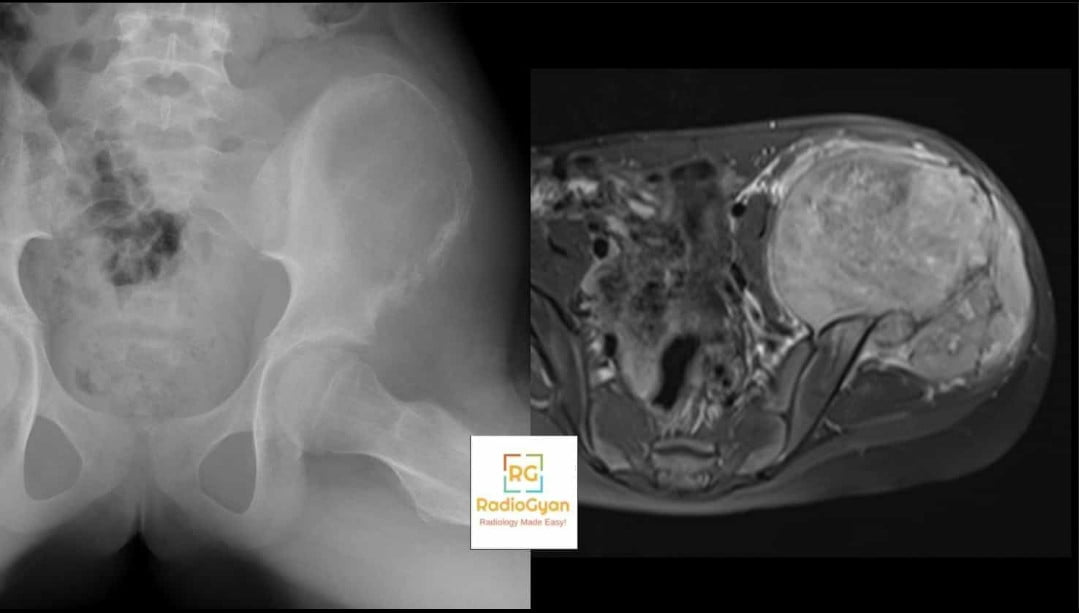
Ewing sarcoma is a small round cell sarcoma defined by a FET::ETS fusion, most commonly EWSR1::FLI1, and the 2020 WHO classification separates it from the round cell sarcomas carrying other fusions, which behave differently. It favours the diaphysis of long bones and the flat bones of the pelvis, ribs and scapula, and produces permeative or moth-eaten destruction with a lamellated or spiculated periosteal reaction; a Codman triangle is common at the margins.
Two features cause real diagnostic trouble. The soft-tissue mass is typically far larger than the bone destruction would predict, and in flat bones the bone changes can be almost invisible on radiographs while the mass is enormous. And the clinical presentation – fever, raised ESR and CRP, leucocytosis, local pain and swelling – mimics osteomyelitis closely enough that patients are commonly treated for infection first. When a presumed osteomyelitis fails to settle, image it properly rather than extending antibiotics.
Mimics and false Codman triangles
Before acting on a Codman triangle, confirm that it is one.
| Mimic | How to tell it apart |
|---|---|
| Physiological periosteal reaction of infancy | Symmetrical, bilateral, diaphyseal, smooth and continuous in an infant roughly 1 to 6 months old. It is a continuous pattern, not an interrupted one, and it is never focal or triangular. |
| Healing fracture callus | Bridging, continuous, matures over serial films, and centred on a fracture line rather than on a marrow lesion. |
| Distal femoral cortical irregularity (cortical desmos) | Posteromedial distal femoral metaphysis in an adolescent, at the adductor magnus insertion. Characteristic site, no marrow lesion, no soft-tissue mass. |
| Nutrient foramen or overlapping skin fold | Follows an oblique lucent channel or crosses bone margins; resolves on a repeat or orthogonal view. |
| Osteochondroma seen edge-on | Cortex and medulla are continuous with the parent bone – the defining feature – and there is no interrupted periosteum. |
| A genuine Codman triangle from infection or haemorrhage | Not a false sign but a false diagnosis. Fever and inflammatory markers, a rim-enhancing subperiosteal collection, or a coagulopathy point away from tumour; MRI usually resolves it. |
What to do when you see one
The value of the sign is that it starts a pathway. The current ACR Appropriateness Criteria for suspected primary bone tumours are explicit that radiographs are the most appropriate initial study, and that for a lesion that looks indeterminate or aggressive on radiographs, MRI or CT is the appropriate next step for anatomical extent, viability and biopsy or surgical planning.
- Complete the radiographic assessment. Two orthogonal views of the whole bone, and check for a second lesion.
- MRI of the entire involved bone, with contrast, before biopsy. Whole-bone coverage is not optional: skip metastases are found in a meaningful minority of appendicular Ewing sarcomas and change both staging and the resection plan. MRI defines marrow extent, joint and physeal involvement, the neurovascular relationship and the viable component to target.
- Refer to a sarcoma reference centre before the biopsy. European guidance is that a suspected bone sarcoma should be referred to a specialist centre and that biopsy is planned by, or in agreement with, the team that will perform the definitive resection.
- Biopsy should be image-guided and percutaneous where possible. In a series of 180 sarcoma biopsies, tumour cells contaminated the tract in 32 per cent of open biopsies versus 0.8 per cent of percutaneous core biopsies, and tract contamination was associated with a mean local recurrence-free survival of 11 months versus 107 months without it. A badly placed tract can turn a limb-salvage operation into an amputation.
- Stage before treatment. Chest CT for pulmonary metastases, and whole-body assessment with bone scintigraphy or FDG PET/CT according to local protocol and tumour type.
Key points and pitfalls
- A Codman triangle is an interrupted periosteal reaction: a triangular cuff of ossified periosteum at the edge of a lesion, with nothing ossified in between.
- It is a pseudotriangle – two sides and an open third – and usually occurs in pairs at either end of the lesion.
- It signals rate, not histology. Tumour, pus and blood produce it identically.
- Osteosarcoma is the commonest cause and Ewing sarcoma the second, but osteomyelitis with a subperiosteal abscess, subperiosteal haematoma, metastases, LCH and an aggressive aneurysmal bone cyst all belong on the list.
- Absence proves nothing. Most aggressive bone lesions never show one; zone of transition, cortical destruction and a soft-tissue mass carry more weight.
- Periosteal reaction takes one to three weeks to mineralise, so an early radiograph in acute osteomyelitis or stress injury can be normal.
- Children produce far more periosteal bone than adults for the same insult.
- Do not biopsy a suspected bone sarcoma outside a sarcoma centre. The tract is part of the operation.
Frequently asked questions
What is a Codman triangle in radiology?
A Codman triangle is an interrupted periosteal reaction seen at the margin of an aggressive bone lesion. The lesion lifts the periosteum off the cortex faster than the periosteum can lay down new bone, so only the acute angle where the elevated periosteum still meets intact cortex ossifies. On the radiograph this appears as a small triangular spur of new bone at the edge of the lesion, with no ossification between the paired spurs.
What causes a Codman triangle?
Anything that strips periosteum from cortex quickly. Osteosarcoma is the commonest cause and Ewing sarcoma the second. Other causes include chondrosarcoma, skeletal metastases, lymphoma and leukaemia, acute osteomyelitis with a subperiosteal abscess, subperiosteal haematoma from trauma, haemophilia or scurvy, Langerhans cell histiocytosis, and a rapidly expanding aneurysmal bone cyst.
Is a Codman triangle always malignant?
No. It indicates that a process is growing fast, not that it is a tumour. A subperiosteal abscess in acute osteomyelitis, a subperiosteal haematoma in a child with a coagulopathy or scurvy, and a rapidly enlarging aneurysmal bone cyst all lift the periosteum by the same mechanism and produce an identical appearance. Age, clinical context, inflammatory markers, matrix and MRI resolve the question.
Is a Codman triangle pathognomonic of osteosarcoma?
No. Osteosarcoma is the most frequent cause, but the sign is neither sensitive nor specific for it. Only a minority of osteosarcomas show a Codman triangle on radiographs, and non-neoplastic causes such as infection and haemorrhage produce the same pattern. The diagnosis rests on the whole picture: age, metaphyseal location, osteoid matrix, wide zone of transition, cortical destruction and a soft-tissue mass.
Why is a Codman triangle not a complete triangle?
Because only two of its sides are ossified. The cortex forms one side and the ossified cuff of elevated periosteum forms the second; the third side, facing the lesion, is open because the stripped central periosteum is outpaced or destroyed before it can mineralise. It is more accurately described as a pseudotriangle or a cuff of reactive bone.
What is the difference between a Codman triangle and a sunburst appearance?
Both are aggressive periosteal reactions but they form differently. A Codman triangle is an interrupted reaction at the edge of a lesion, where the periosteum has been lifted off. A sunburst is a spiculated reaction in which bone spicules radiate outward from a point along Sharpey fibres through the tumour matrix. They frequently coexist in the same osteosarcoma, and neither is specific to it.
Who was Codman and when was the triangle described?
Ernest Amory Codman (1869-1940) was a Boston surgeon, a founder of outcomes auditing in surgery and a driving force behind the American College of Surgeons Registry of Bone Sarcoma. He described the trumpet-shaped cuff of reactive bone at the margin of a bone sarcoma in his work on the Registry in the 1920s. A commonly repeated claim credits Ribbert with an earlier description in 1914, but that priority is poorly sourced. Modern usage drops the possessive: Codman triangle, not Codman’s triangle.
What should be done next when a Codman triangle is seen?
Treat it as a suspected aggressive bone lesion. Complete the radiographs of the whole bone, then obtain contrast-enhanced MRI of the entire involved bone before any biopsy, to define marrow extent, joint and neurovascular involvement and to detect skip lesions. Refer to a sarcoma reference centre, and let the team that will perform the definitive resection plan the biopsy: percutaneous image-guided core biopsy carries a far lower rate of tract seeding than open biopsy, and a poorly placed tract compromises limb salvage. Stage the chest with CT.
References
- Rana RS, Wu JS, Eisenberg RL. Periosteal reaction. AJR Am J Roentgenol. 2009;193(4):W259-72. PMID: 19770293.
- Ragsdale BD, Madewell JE, Sweet DE. Radiologic and pathologic analysis of solitary bone lesions. Part II: periosteal reactions. Radiol Clin North Am. 1981;19(4):749-83. PMID: 7323291.
- Wenaden AE, Szyszko TA, Saifuddin A. Imaging of periosteal reactions associated with focal lesions of bone. Clin Radiol. 2005;60(4):439-56. PMID: 15767101.
- Bisseret D, Kaci R, Lafage-Proust MH, et al. Periosteum: characteristic imaging findings with emphasis on radiologic-pathologic comparisons. Skeletal Radiol. 2015;44(3):321-38. PMID: 25269751.
- Expert Panel on Musculoskeletal Imaging; Ahlawat S, Lenchik L, et al. ACR Appropriateness Criteria Suspected Primary Bone Tumors: 2024 Update. J Am Coll Radiol. 2025;22(5S):S440-S454. PMID: 40409893.
- Crombe A, Simonetti M, Longhi A, et al. Imaging of osteosarcoma: presenting findings, metastatic patterns, and features related to prognosis. J Clin Med. 2024;13(19):5710. PMID: 39407770.
- Matcuk GR, Tivorsak T, Watterson CT, et al. Imaging of Ewing sarcoma: an updated analysis including presenting features, prognostic imaging biomarkers, and treatment response assessment. Skeletal Radiol. 2026;55(9):2195-2217. PMID: 42062585.
- Grunewald TGP, Cidre-Aranaz F, Surdez D, et al. Ewing sarcoma. Nat Rev Dis Primers. 2018;4(1):5. PMID: 29977059.
- Strauss SJ, Frezza AM, Abecassis N, et al. Bone sarcomas: ESMO-EURACAN-GENTURIS-ERN PaedCan Clinical Practice Guideline for diagnosis, treatment and follow-up. Ann Oncol. 2021;32(12):1520-1536. PMID: 34500044.
- Caracciolo JT, Temple HT, Letson GD, Kransdorf MJ. A modified Lodwick-Madewell grading system for the evaluation of lytic bone lesions. AJR Am J Roentgenol. 2016;207(1):150-6. PMID: 27070373.
- Gondim Teixeira PA, Lombard C, Moustache-Espinola P, et al. Initial characterization of focal bone lesions with conventional radiographs or computed tomography: diagnostic performance and interobserver agreement assessment. Can Assoc Radiol J. 2023;74(2):404-414. PMID: 36207066.
- Barrientos-Ruiz I, Ortiz-Cruz EJ, Serrano-Montilla J, et al. Are biopsy tracts a concern for seeding and local recurrence in sarcomas? Clin Orthop Relat Res. 2017;475(2):511-518. PMID: 27655183.
- Hwang S, Schneider R. Eponyms of tumors and tumor-like lesions in the musculoskeletal system: who were the people and what are the lesions? Pictorial review. AJR Am J Roentgenol. 2010;195(6 Suppl):S50-61. PMID: 21098160.
- Anderson WJ, Doyle LA. Updates from the 2020 World Health Organization classification of soft tissue and bone tumours. Histopathology. 2021;78(5):644-657. PMID: 33438273.
- Salman R, McGraw M, Naffaa L. Chronic osteomyelitis of long bones: imaging pearls and pitfalls in pediatrics. Semin Ultrasound CT MR. 2022;43(1):88-96. PMID: 35164913.
- Bielack SS, Smeland S, Whelan JS, et al. Methotrexate, doxorubicin, and cisplatin (MAP) plus maintenance pegylated interferon alfa-2b versus MAP alone in patients with resectable high-grade osteosarcoma and good histologic response to preoperative MAP: first results of the EURAMOS-1 good response randomized controlled trial. J Clin Oncol. 2015;33(20):2279-87. PMID: 26033801.
- Saifuddin A, Michelagnoli M, Pressney I. Skip metastases in appendicular Ewing sarcoma: relationship to distant metastases at diagnosis, chemotherapy response and overall survival. Skeletal Radiol. 2023;52(3):585-591. PMID: 36100697.
Access all radiology signs posted so far: https://radiogyan.com/radiology-signs/

Improve Content And Help Your Colleagues!
Found an error in the post? Please let us know using our contact page and we will update it with due credits!
If you wish to contribute radiological images for this case (or any other article on the website), you can submit them here and you will be duly credited: Submit radiology case