


What causes the thumb sign on the lateral neck radiograph?
Let me know in the comments.
Click to reveal the answer
Answer:
The thumb sign is a swollen, rounded epiglottis projecting into the airway on a lateral soft-tissue neck radiograph, shaped like an adult thumb held up in profile. It means acute epiglottitis. A normal epiglottis measures about 4 mm across; in supraglottitis the mean width is near 16 mm [4]. Published cutoffs range from 5 to 7 mm [1,3,4].
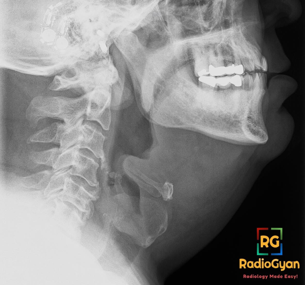
Why is it called so?
The oedematous epiglottis loses its thin, curved, leaf-like profile and projects into the airway with the broad rounded outline of a thumb. A double thumb sign has also been reported, in which the swollen aryepiglottic folds project as a second thumb behind the first [9].
Pathophysiology
Bacterial infection invades the loose submucosa of the supraglottis, a plane only loosely attached to the underlying cartilage, so oedema accumulates rapidly and expands the epiglottis and aryepiglottic folds in every direction at once. The swelling is therefore never confined to the epiglottis: the vallecula fills in, the aryepiglottic folds thicken, and the hypopharynx balloons above the narrowing. Obstruction can be sudden and complete, because the swollen supraglottis both narrows the airway and can prolapse into it.
Alternative names: Thumbprint sign
Other associated named signs: Vallecula sign, steeple sign of croup, ballooned hypopharynx, and retropharyngeal soft-tissue widening of retropharyngeal abscess
Measure the epiglottis, do not eyeball the thumb
The thumb shape is the memorable part of the sign and the least reliable part of the read. Measuring beats pattern matching, but the literature does not agree on one threshold: three case-control series give cutoffs of 5.02, 6.3 and 7 mm, sensitivity 75.8% to 100%.
| Parameter on the lateral view | Cutoff | Sensitivity | Specificity | Series |
|---|---|---|---|---|
| Epiglottis width at the base | 5.02 mm | 96.2% | 98.2% | 260 vs 1166 [1] |
| Epiglottis width | 6.3 mm | 75.8% | 97.8% | 91 vs 91 [3] |
| Epiglottis width | 7 mm | 100% | 100% | 30 vs 30 [4] |
| Epiglottis width at the tip | 4.84 mm | 91.2% | 97.3% | 260 vs 1166 [1] |
| Aryepiglottic fold width | 6.59 mm | 86.5% | 78.8% | 260 vs 1166 [1] |
| Aryepiglottic fold width | 4.5 mm | 83% | 100% | 30 vs 30 [4] |
Sensitivity is where the series diverge, and that matters because the qualitative read is reader-limited rather than image-limited. When a mixed group of emergency physicians, residents and medical students classified 26 epiglottitis and 26 control radiographs, accuracy was 80.5% before a five-minute tutorial on the vallecula sign and 98.8% afterwards, with sensitivity rising from 78.5% to 98.2% [2]. The films did not change; the readers did.
The film misses about a third of adult cases
In 91 patients with laryngoscopy-confirmed acute epiglottitis read against 91 controls, the false-negative rate for lateral neck radiography was 31.9%, and previous oral antibiotic use was an independent risk factor for a falsely negative film [3]. Interobserver agreement was excellent, so these were not reader disagreements; the finding was genuinely absent or too subtle. A partly treated patient with a convincing history and an unremarkable film is the trap.
Thumb sign vs steeple sign
Both signs appear in a patient with stridor and they sit on opposite sides of the glottis.
| Feature | Thumb sign | Steeple sign |
|---|---|---|
| Disease | Acute epiglottitis | Croup, laryngotracheobronchitis |
| Level | Supraglottic | Subglottic |
| Best view | Lateral soft-tissue neck | Frontal |
| What you see | Broad, rounded epiglottis | Tapered inverted V of the tracheal air column |
| Usual organism | Streptococci, formerly Hib | Parainfluenza virus |
| Objective measure | Epiglottis width, 5 to 7 mm [1,3,4] | Frontal and lateral narrowing ratios [8] |
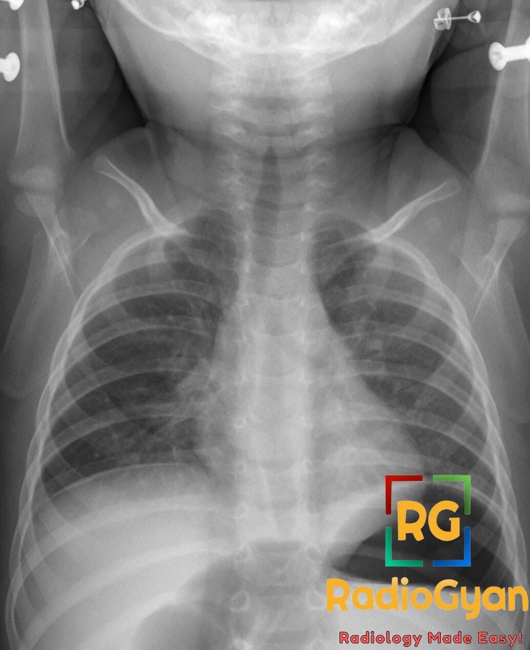
The same lesson repeats on the croup side. In 192 prospectively recruited patients, the measured narrowing ratios tracked the clinical course: a frontal ratio below 0.23 preceded a longer stay in about 71% of cases, while above 0.65 about 98% were discharged [8]. That study recorded whether the steeple sign was present but reports no performance figure for the sign itself.
Now an adult disease
Hib conjugate vaccination removed the childhood disease and left the adult one. Across 11 Sydney hospitals, adults over 20 made up 84% of correctly coded vaccine-era cases against 17% before vaccination, and Hib bacteraemia fell from 62% of cases to none [7]. An Icelandic series covering 1983 to 2005 found a mean age of 33.3 years, an annual incidence of 0.93 per 100,000, no Hib cultured after 1991, and streptococci as the leading organism [6].
What to do about the airway
The radiograph is a screening test. Direct inspection at laryngoscopy is the reference standard [5], and the film must not delay it.
- Do not send an unstable patient with stridor out of the resuscitation area for imaging: leaving a monitored setting carries its own risk [5].
- A normal-looking film does not exclude epiglottitis, and misses about a third of confirmed adult cases [3]. Measure the epiglottis before reporting it as normal, and state the measurement.
- Most adults never need an airway: across 56 studies and 10,630 patients the intervention rate was 15.6%, falling from 20% to 10% between 1980 and 2020. When it is needed it is high risk, with intubation in 10.2% and failed intubation in 4.2%, roughly one in 25 [10].
- Bedside ultrasound through the thyrohyoid membrane shows the swollen epiglottis as the alphabet P sign without moving the patient, though it is described in case reports, not validated [5].
Frequently asked questions
What is the thumb sign seen in?
The thumb sign is seen in acute epiglottitis. It is a swollen, rounded epiglottis on the lateral soft-tissue neck radiograph. In the post-vaccination era it is predominantly an adult finding, with streptococci rather than Haemophilus influenzae type b as the usual organism [6].
What is the difference between the steeple sign and the thumb sign?
The steeple sign is subglottic tracheal narrowing on the frontal view and indicates croup, which is viral. The thumb sign is supraglottic swelling of the epiglottis on the lateral view and indicates epiglottitis, which is bacterial. Different level, different view, different disease.
Does a normal lateral neck X-ray rule out epiglottitis?
No. In 91 laryngoscopy-confirmed adult cases the false-negative rate for lateral neck radiography was 31.9%, and prior oral antibiotic use made a negative film more likely [3]. If clinical suspicion is high, proceed to direct visualisation of the airway rather than trusting the radiograph.
How do you measure the epiglottis on a lateral neck radiograph?
Measure the width of the epiglottis on the lateral soft-tissue view. The normal mean is about 4 mm and the mean in supraglottitis about 16 mm [4]. Reported cutoffs are 5.02 mm at the base [1], 6.3 mm [3] and 7 mm [4]; specificity is high at all three, sensitivity is not.
References
- Kim KH, Kim YH, Lee JH, et al. Accuracy of objective parameters in acute epiglottitis diagnosis: a case-control study. Medicine (Baltimore). 2018;97(37):e12256. PMID: 30212960.
- Ducic Y, Hébert PC, MacLachlan L, et al. Description and evaluation of the vallecula sign: a new radiologic sign in the diagnosis of adult epiglottitis. Ann Emerg Med. 1997;30(1):1-6. PMID: 9209217.
- Lee SH, Yun SJ, Kim DH, et al. Do we need a change in ED diagnostic strategy for adult acute epiglottitis? Am J Emerg Med. 2017;35(10):1519-24. PMID: 28460811.
- Yong MG, Choo MJ, Yum CS, et al. Radiologic laryngeal parameters in acute supraglottitis in Korean adults. Yonsei Med J. 2001;42(4):367-70. PMID: 11519076.
- Hung TY, Li S, Chen PS, et al. Bedside ultrasonography as a safe and effective tool to diagnose acute epiglottitis. Am J Emerg Med. 2011;29(3):359.e1-3. PMID: 20674236.
- Briem B, Thorvardsson O, Petersen H. Acute epiglottitis in Iceland 1983-2005. Auris Nasus Larynx. 2009;36(1):46-52. PMID: 18502071.
- Wood N, Menzies R, McIntyre P. Epiglottitis in Sydney before and after the introduction of vaccination against Haemophilus influenzae type b disease. Intern Med J. 2005;35(9):530-5. PMID: 16105154.
- Yang WC, Hsu YL, Chen CY, et al. Initial radiographic tracheal ratio in predicting clinical outcomes in croup in children. Sci Rep. 2019;9(1):17893. PMID: 31784540.
- Lee J, Loh J, Lee TS. Double thumb sign in a case of epiglottitis. Radiol Case Rep. 2021;16(7):1810-4. PMID: 34025892.
- Booth AWG, Pungsornruk K, Llewellyn S, et al. Airway management of adult epiglottitis: a systematic review and meta-analysis. BJA Open. 2024;9:100250. PMID: 38230383.
Access all radiology signs posted so far: https://radiogyan.com/radiology-signs/

Improve Content And Help Your Colleagues!
Found an error in the post? Please let us know using our contact page and we will update it with due credits!
If you wish to contribute radiological images for this case (or any other article on the website), you can submit them here and you will be duly credited: Submit radiology case