


What causes Rigler’s sign in the bowel on abdominal radiography?
Let me know in the comments.
Click to reveal the answer
Answer:
Pneumoperitoneum – free gas in the peritoneal cavity. Gas inside the lumen outlines the mucosal surface of the bowel wall and free gas outside it outlines the serosal surface, so both sides of the same wall are silhouetted and the wall itself appears as a thin soft-tissue stripe. In more than 90% of patients this means a perforated hollow viscus, most often a perforated peptic ulcer, and it is a surgical emergency until proven otherwise.
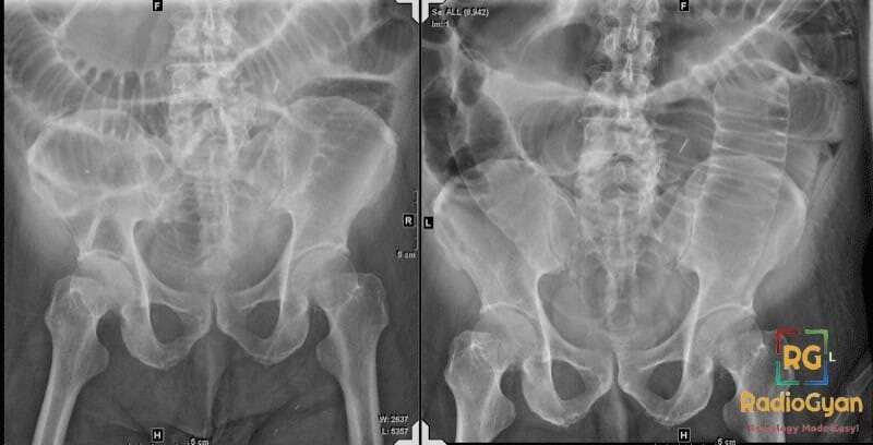
| At a glance | |
|---|---|
| What it is | Both the inner and outer surfaces of a bowel wall visible on an abdominal radiograph, so the wall is seen as a discrete stripe |
| What it means | Pneumoperitoneum, and in over 90% of patients a perforated hollow viscus |
| Also called | Double wall sign, bas-relief sign, double-lumen sign |
| Described | Leo George Rigler, Radiology 1941 – four supine cases |
| Gas needed | Around 1000 mL for confident recognition, versus about 1 mL under the diaphragm on a good erect chest film |
| Sensitivity | Present in about 32% of proven pneumoperitoneum on supine films |
| False positives | About 13% of films in the same series – apposed bowel loops (pseudo-Rigler) |
| Best next test | CT of the abdomen and pelvis, plus a surgical referral |
| Careful | Three other unrelated signs also carry Rigler’s name |
Why is it called so?
The sign is named after Leo George Rigler (1896–1979), the American radiologist who described it in 1941 in Spontaneous Pneumoperitoneum: A Roentgenologic Sign Found in the Supine Position (Radiology 1941;37:604–607). He reported four cases in which free intraperitoneal gas made the wall of a gas-containing viscus visible, and made two points that are still the most useful things anyone has said about the sign.
First, that it was seen only when large quantities of free gas were present, typically after colonic perforation. Second, that its value lay in the fact that it worked on a supine, bedside film – often the only projection obtainable in a patient too ill to sit up. Rigler was describing a rescue sign for a sick patient, not a sensitive test. Radiologists have tended to remember the first half of that message and forget the second.
Pathophysiology
A normal gas-filled loop of bowel is visible only from the inside. Intraluminal gas provides contrast against the mucosal surface, but the serosal surface is bounded by mesenteric fat, fluid and adjacent viscera of almost identical radiographic density, so the outer margin dissolves into the soft tissues. You see a lucent lumen with an indistinct edge, not a wall.
When a hollow viscus perforates, gas escapes into the peritoneal cavity and, on a supine patient, collects in the non-dependent anterior compartment and between loops. That gas now provides contrast against the serosal surface as well. The bowel wall, sandwiched between two lucencies, is rendered as a thin, sharply defined soft-tissue line – the double wall. The effect is essentially a silhouette-sign argument in reverse: the wall becomes visible not because it changed, but because what lies against it did.
That mechanism explains the sign’s main weakness. Gas must be present on both sides of the same segment of wall, and enough of it must be interposed to separate that loop from its neighbours. Small volumes of free gas rise anteriorly and lie in a sheet that never gets between loops, which is why a patient may have unequivocal free gas on CT and a completely unremarkable supine film.
Alternative names: Double wall sign, bas-relief sign, double-lumen sign
Other associated named signs: Football sign, falciform ligament sign, cupola sign, half-moon sign, continuous diaphragm sign (all additional signs of pneumoperitoneum).
How much free gas does Rigler’s sign actually need?
This is the single most useful thing to know about the sign, and it is usually left out. Confident recognition of Rigler’s sign needs on the order of a litre of free intraperitoneal gas. A well-exposed erect chest radiograph, by contrast, can show about 1 mL under the diaphragm. The two findings are separated by three orders of magnitude, so they are not interchangeable and should never be reported as though they were.
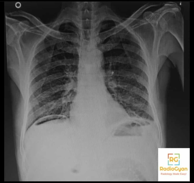
The practical consequences follow directly. A positive Rigler’s sign is informative twice over: it says there is free gas, and it says there is a lot of it, which in turn suggests a large or colonic perforation. A negative Rigler’s sign says almost nothing. Reporting “no Rigler’s sign” as reassurance is the commonest misuse of the finding.
How often is it actually seen? The numbers
Two studies do most of the work here. Levine and colleagues (AJR 1991) blinded readers to supine films from 44 patients with pneumoperitoneum mixed among 87 controls. Chiu and colleagues (American Journal of Emergency Medicine 2009) reviewed 527 radiographs from 250 patients with surgically proven perforation.
| Sign on the supine film | Frequency in proven pneumoperitoneum (Levine 1991) |
|---|---|
| Any supine sign of free gas | 59% (26 of 44) |
| Right upper quadrant gas sign | 41% (18 of 44) |
| Rigler’s sign | 32% (14 of 44) |
| Falciform ligament sign | 2% (1 of 44) |
| Football sign | 2% (1 of 44) |
| False-positive calls across the whole series | 13% (11 films) |
Read the last row again. In a blinded reading, radiologists called free air that was not there on 13% of films, and the errors clustered on the two commonest signs – right upper quadrant gas and Rigler’s sign. The sign is not only insensitive, it is also the one most likely to be called wrongly.
Chiu’s series answers the other half of the question: which film to ask for.
| Projection | Free gas detected in 250 proven perforations |
|---|---|
| Left lateral decubitus abdomen | 98.0% |
| Erect chest radiograph | 85.1% |
| Supine abdomen (KUB) | 80.4% |
| Supine chest radiograph | 78.7% |
The decisive variable is not the projection name but the horizontal beam. Free gas rises to the highest point in the peritoneal cavity; only a beam travelling parallel to the floor silhouettes it against a soft-tissue interface. A left lateral decubitus film, taken after the patient has lain on the left side for 10 to 20 minutes, collects gas over the right lobe of the liver where nothing else is lucent, and it is tolerated by patients who cannot stand. When Rigler’s sign is equivocal, this is the film to ask for – not a repeat supine.
A caveat on Chiu’s figures: 91% of those perforations were upper gastrointestinal, where free gas is common. The same numbers should not be assumed for small bowel or appendiceal perforation, where free gas is often absent altogether.
Pseudo-Rigler: the 1 mm rule that separates true from false
The classic false positive is the pseudo-Rigler sign: two gas-distended loops lying against each other. Their two apposed walls summate into a single stripe with gas on either side, which looks exactly like the real thing. Levine’s study produced a discriminator that is easy to apply at the workstation:
| Feature | True Rigler’s sign | Pseudo-Rigler / mimic |
|---|---|---|
| Apparent wall thickness | 1–8 mm (every true positive in the Levine series) | 1 mm or less (every false positive in that series) |
| What the stripe is | One bowel wall, thickened by oedema or inflammation | Two thin walls in contact, or a wall plus a fat plane |
| Extent | Traceable around the convex outer margin of a loop, often over several loops | Only where two loops happen to touch; disappears where they separate |
| Company it keeps | Other free-gas signs – right upper quadrant gas, football, falciform, triangle sign | Isolated finding on an otherwise unremarkable film |
| Response to a horizontal-beam film | Free gas confirmed on erect or left lateral decubitus | No free gas on the decubitus film |
Other things that reproduce the appearance: prominent perivascular or subserosal fat outlining a loop in a thin patient, pneumatosis intestinalis (where the gas is in the wall rather than outside it – look for a bubbly or curvilinear texture that follows the wall rather than a clean outer lucency), overlapping colonic haustra, and interposed colon between liver and diaphragm (the Chilaiditi position) mimicking free gas elsewhere on the film. When in doubt, the answer is another projection or a CT, not a harder stare.
The other supine free-gas signs worth checking
If you are looking at a supine film because the patient cannot sit up, Rigler’s sign is one of several patterns, and it is not the most frequent. Most free-gas signs on a supine film sit over the right upper abdomen, so that is where the search should start.
| Sign | What you see | Note |
|---|---|---|
| Right upper quadrant gas sign | Localised lucency over the liver, triangular or linear, running inferolateral to superomedial; if triangular, a concave superolateral border | The commonest supine sign (41%); the shape rules above separate true from false |
| Anterior superior oval sign | Oval lucency in the right upper abdomen from gas in the anterior subhepatic space | Commonest sign in the Chiu series – 44% on supine abdomen, 34% on supine chest |
| Rigler’s sign | Both sides of the bowel wall visible | Needs a large volume of gas |
| Triangle sign | Triangular lucency trapped between three loops, or two loops and the parietal peritoneum | Angular shape is the giveaway – bowel gas is never sharply triangular |
| Falciform ligament sign | Vertical soft-tissue line to the right of midline, from the xiphoid towards the umbilicus | Rare on supine films (about 2%), but highly specific |
| Football sign | The whole peritoneal cavity outlined as a large oval lucency | Classically in neonates; implies massive pneumoperitoneum |
| Inverted-V sign | Gas outlining the lateral umbilical folds and the inferior epigastric vessels in the pelvis | Look low on the film, below the usual search pattern |
| Continuous diaphragm sign | The whole diaphragm visible as one unbroken line across the midline | Gas between the diaphragm and the heart; also seen with pneumomediastinum |
| Doge’s cap sign | Triangular gas in the right upper quadrant in Morison’s pouch | Named for the hat of the Venetian Doge |
Does Rigler’s sign always mean laparotomy?
No, and this is the second trap. Free intraperitoneal gas indicates perforation in over 90% of patients, which leaves a real minority in whom it does not. Mularski and colleagues reviewed the literature on nonsurgical pneumoperitoneum and found 196 reported cases, of which 45 had a laparotomy that found no perforation. Most of those cases were procedural or iatrogenic.
| Route | Cause | How common |
|---|---|---|
| Abdominal | Retained air after laparotomy or laparoscopy | 25–60% of postoperative patients – by far the commonest cause |
| Abdominal | Peritoneal dialysis catheter insertion or exchange | 10–34% of patients |
| Abdominal | Gastrointestinal endoscopy, including colonoscopy and ERCP | 0.3–25%, varying by procedure |
| Abdominal | Pneumatosis intestinalis with rupture of a subserosal cyst | Uncommon; the bowel itself is intact |
| Thoracic | Mechanical ventilation and barotrauma | Gas tracks from the mediastinum through diaphragmatic defects |
| Thoracic | Cardiopulmonary resuscitation, pneumothorax, pneumomediastinum | Consider in any ventilated ICU patient |
| Gynaecological | Tubal insufflation, pelvic examination, vaginal douching, post-partum exercises, coitus | Gas enters through a patent genital tract |
| Idiopathic | No cause identified despite investigation | A diagnosis of exclusion |
The postoperative patient deserves its own rule of thumb, because that is where this comes up daily. After open colorectal resection, subdiaphragmatic gas had cleared in only 72% of patients by day 5, 80% by day 6 and 89% by day 7, and 5 of 75 patients still had visible gas beyond day 10. A small volume of free gas in the first postoperative week is expected. What matters is an increasing volume of gas, or free gas that appears alongside a clinical deterioration – in that series, the two patients with an anastomotic leak declared themselves as an increase in subdiaphragmatic gas, not as its mere presence.
Four unrelated signs called Rigler
Leo Rigler published over 200 papers and six textbooks, and his name attached to several findings that have nothing to do with each other. Two of them were described in the same year, which is where most of the confusion starts – published sources, including peer-reviewed ones, have treated “Rigler sign” and “Rigler triad” as synonyms. They are not.
| Eponym | Year | Finding | Means |
|---|---|---|---|
| Rigler’s sign (double wall) | 1941 | Both sides of the bowel wall visible on an abdominal radiograph | Pneumoperitoneum, usually hollow viscus perforation |
| Rigler’s triad | 1941 | Pneumobilia + small bowel obstruction + an ectopic gallstone | Gallstone ileus. Pathognomonic but insensitive: the full triad is seen on plain films in only about 15% of cases, and in roughly 14–53% across series, rising to about 78% on CT |
| Rigler notch sign | 1955 | A notched or umbilicated indentation on the border of a pulmonary nodule, thought to mark an entering vessel | Suggests malignancy in a solitary pulmonary nodule, but also occurs in granulomatous disease, so its discriminating value is limited |
| Hoffman-Rigler sign | 1965 | Posterior border of the left ventricle projecting more than 18 mm behind the inferior vena cava, measured 2 cm above their crossing, on a lateral chest radiograph | Left ventricular enlargement – but measured against echocardiography in a modern population it was only 55% sensitive and 59% specific, worse than the cardiothoracic ratio |
Two further extensions of the gallstone-ileus triad are occasionally quoted: Rigler’s tetrad adds a change in position of a previously identified gallstone, and Rigler’s pentad adds a dual air-fluid level in the right upper quadrant – one in the duodenum, one in the gallbladder.
What to do next: from the radiograph to CT
Rigler’s sign is a trigger, not a destination. The current standard of care is that suspected perforation is a CT question.
- Guideline position. The 2020 World Society of Emergency Surgery guideline on perforated peptic ulcer makes a strong recommendation for CT in a patient with an acute abdomen from suspected perforation, and recommends chest or abdominal radiographs as the initial assessment only when CT is not promptly available.
- ACR Appropriateness Criteria. For acute non-localised abdominal pain, CT of the abdomen and pelvis with intravenous contrast is rated usually appropriate (9 of 9), while abdominal radiography rates only may be appropriate (4 of 9), and is usually not appropriate in the neutropenic patient.
- A normal CT does not exclude perforation. Up to 12% of proven perforations have a normal CT. Where suspicion remains, water-soluble contrast given orally or via a nasogastric tube improves sensitivity. A negative radiograph is far weaker still.
- Ultrasound has a real role at the bedside. The enhanced peritoneal stripe sign – a thickened, brightly echogenic peritoneal line with reverberation and no bowel movement deep to it – was positive in all 21 patients with pneumoperitoneum in a prospective series of 600 patients with acute abdominal pain (100% sensitivity, 99% specificity, 100% negative predictive value in that cohort). Worth knowing when the patient cannot be moved.
Where CT adds what the radiograph cannot: the site of perforation
Knowing where the bowel has perforated changes the operation, and CT can often say. Two things to hold in the right order: the direct signs beat the gas-distribution signs.
| Finding on CT | Points to |
|---|---|
| Focal bowel wall thickening | The strongest single predictor of gastroduodenal perforation – accuracy 96%, better than any gas-distribution sign |
| Focal wall defect; concentrated extraluminal bubbles hugging the wall | Direct signs; localise the perforation better than free-gas distribution |
| Periportal, perihepatic, perigastric gas | Gastroduodenal perforation |
| Falciform ligament sign on CT | Gastroduodenal perforation, but chiefly a marker of a large volume of gas |
| Mesenteric gas | Small bowel or distal colonic perforation |
| Pelvic gas | Distal colonic perforation |
| Periappendiceal gas | Perforated appendicitis – a strong predictor |
The nuance is worth carrying: in the series that tested all three of the classic upper-abdominal gas-distribution signs, all three were present together in only 21% of gastroduodenal perforations, and every direct sign outperformed them. Treat the distribution of gas as a supporting clue and go looking for the wall defect.
How to report it
- Say pneumoperitoneum, not “Rigler’s sign”. The eponym is a description of pixels; the referrer needs the diagnosis.
- Say roughly how much. Visible Rigler’s sign implies a large volume, which is itself clinically meaningful – volume of pneumoperitoneum was one of the factors predicting failure of non-operative management of perforated peptic ulcer.
- Say what you cannot exclude. If the film is negative, state explicitly that a normal radiograph does not exclude perforation and recommend CT.
- Check the surgical history before calling a postoperative patient perforated. Free gas within a week of laparotomy is usually retained air.
- Communicate it directly. Unexpected pneumoperitoneum is a critical result.
Frequently asked questions
What does Rigler’s sign mean on an abdominal X-ray?
It means pneumoperitoneum, free gas inside the peritoneal cavity. Gas inside the bowel outlines the mucosal surface of the wall and free gas outside the bowel outlines the serosal surface, so both sides of the same wall become visible as a thin soft-tissue stripe. In more than 90 percent of patients free intraperitoneal gas means a perforated hollow viscus, so the sign is an emergency finding until proven otherwise.
How much free air is needed to see Rigler’s sign?
A lot. Rigler himself stressed in 1941 that the sign appeared only with large quantities of free gas, typically after colonic perforation, and modern sources put the volume needed for confident recognition at roughly 1000 mL. By comparison, as little as 1 mL of free gas can be seen under the diaphragm on a properly exposed erect chest radiograph. Rigler’s sign is therefore a late, insensitive sign, not a screening test.
How sensitive is Rigler’s sign for pneumoperitoneum?
Low. In a blinded study of supine films from 44 patients with pneumoperitoneum, Rigler’s sign was present in only 14 cases, 32 percent, and any supine sign of free air was present in 59 percent. Right upper quadrant gas was commoner, at 41 percent. Absence of Rigler’s sign says nothing useful about whether the patient has perforated.
What is the pseudo-Rigler sign and how do I avoid it?
A pseudo-Rigler sign occurs when two gas-filled bowel loops lie against each other, so their two apposed walls form a single stripe with gas on both sides and mimic free air. Wall thickness is the practical discriminator. In the Levine study every true positive Rigler’s sign had an apparent wall 1 to 8 mm thick, while every false positive had a wall of 1 mm or less. In that same series, 13 percent of all films were called falsely positive, so the error is common.
Which radiographic projection is best for detecting free air?
The left lateral decubitus abdominal radiograph. In 250 patients with surgically proven perforation the positive rate for free gas was 98 percent on left lateral decubitus, 85 percent on erect chest radiograph, 80 percent on supine abdominal film and 79 percent on supine chest film. A horizontal beam is what makes the difference, because free gas has to rise to a non-dependent surface to be silhouetted.
Does Rigler’s sign always mean the patient needs surgery?
No. Free gas indicates perforation in over 90 percent of patients, but the remainder have nonsurgical pneumoperitoneum. The commonest cause is simply retained postoperative air, present in 25 to 60 percent of patients after laparotomy; others include peritoneal dialysis catheter placement, endoscopy, mechanical ventilation, cardiopulmonary resuscitation and gynaecological procedures. In one review of 196 published cases of nonsurgical pneumoperitoneum, 45 patients underwent a laparotomy that found no perforation.
How long does free air last after abdominal surgery?
Longer than most people expect. After open colorectal resection, subdiaphragmatic gas had disappeared in 72 percent of patients by the fifth postoperative day, 80 percent by the sixth and 89 percent by the seventh, and five of 75 patients still had visible gas beyond day 10. A small volume of free gas in the first postoperative week is therefore expected; what matters is an increase in volume or a change in the clinical picture.
What should I do when I see Rigler’s sign?
Treat it as a positive result, not as the end of the work-up. Contact the surgical team and get a CT of the abdomen and pelvis, which is both far more sensitive for free gas and able to show the site of perforation. The 2020 WSES guideline on perforated peptic ulcer makes a strong recommendation for CT in suspected perforation, with chest and abdominal radiographs reserved for when CT is not promptly available.
Can a normal CT exclude perforation?
Not completely. The WSES guideline notes that up to 12 percent of patients with a proven perforation have a normal CT. Where clinical suspicion stays high and no free gas is seen, adding water-soluble oral contrast or contrast via a nasogastric tube improves sensitivity. A negative radiograph is far weaker still and never excludes perforation.
Can free gas be diagnosed on ultrasound?
Yes. The enhanced peritoneal stripe sign, a thickened, brightly echogenic peritoneal line with reverberation artefact and no bowel movement behind it, was prospectively assessed in 600 patients with acute abdominal pain: all 21 patients with pneumoperitoneum were detected, giving 100 percent sensitivity, 99 percent specificity and a 100 percent negative predictive value in that series. This is useful at the bedside in a patient too unstable to move.
How many different signs are named after Rigler?
Four. Rigler’s sign is the double-wall sign of pneumoperitoneum, described in 1941. Rigler’s triad, also 1941, is pneumobilia plus small bowel obstruction plus an ectopic gallstone in gallstone ileus. The Rigler notch sign, from 1955, is a notched or umbilicated border on a pulmonary nodule suggesting malignancy. The Hoffman-Rigler sign indicates left ventricular enlargement on a lateral chest radiograph. They are unrelated and are frequently confused in print.
Does the distribution of free gas on CT show where the perforation is?
It helps, but it is a secondary clue. Periportal, perihepatic and perigastric gas favour gastroduodenal perforation; mesenteric gas favours small bowel or distal colon; pelvic gas favours distal colon; periappendiceal gas strongly favours perforated appendicitis. Direct signs remain better: focal bowel wall thickening was the single most accurate predictor of gastroduodenal perforation at 96 percent accuracy, ahead of any specific air distribution.
Related radiology signs
- Pseudo-Rigler’s sign – the apposed-loops mimic
- Cupola sign – free gas under the central tendon on a supine film
- Football sign – massive pneumoperitoneum in the neonate
- Falciform ligament sign – gas outlining the ligament
- Half-moon sign – subphrenic free gas
- String-of-pearls sign – small bowel obstruction on the erect film
- Peritoneal anatomy – where free gas actually goes
References
- Rigler LG. Spontaneous pneumoperitoneum: a roentgenologic sign found in the supine position. Radiology. 1941;37:604-607.
- Lewicki AM. The Rigler sign and Leo G. Rigler. Radiology. 2004;233(1):7-12. PMID: 15333763
- Levine MS, Scheiner JD, Rubesin SE, Laufer I, Herlinger H. Diagnosis of pneumoperitoneum on supine abdominal radiographs. AJR Am J Roentgenol. 1991;156(4):731-735. PMID: 2003436
- Chiu YH, Chen JD, Tiu CM, et al. Reappraisal of radiographic signs of pneumoperitoneum at emergency department. Am J Emerg Med. 2009;27(3):320-327. PMID: 19328377
- Mularski RA, Sippel JM, Osborne ML. Pneumoperitoneum: a review of nonsurgical causes. Crit Care Med. 2000;28(7):2638-2644. PMID: 10921609
- Karaman A, Demirbilek S, Akin M, Gürünlüoğlu K, Irşi C. Does pneumoperitoneum always require laparotomy? Report of six cases and review of the literature. Pediatr Surg Int. 2005;21(10):819-824. PMID: 16096797
- Tang CL, Yeong KY, Nyam DC, et al. Postoperative intra-abdominal free gas after open colorectal resection. Dis Colon Rectum. 2000;43(8):1116-1120. PMID: 10950010
- Tarasconi A, Coccolini F, Biffl WL, et al. Perforated and bleeding peptic ulcer: WSES guidelines. World J Emerg Surg. 2020;15:3. PMID: 31921329
- Scheirey CD, Fowler KJ, Therrien JA, et al. ACR Appropriateness Criteria: Acute Nonlocalized Abdominal Pain. J Am Coll Radiol. 2018;15(11S):S217-S231. PMID: 30392591
- Asrani A. Sonographic diagnosis of pneumoperitoneum using the “enhancement of the peritoneal stripe sign.” A prospective study. Emerg Radiol. 2007;14(1):29-39. PMID: 17347768
- Muradali D, Wilson S, Burns PN, Shapiro H, Hope-Simpson D. A specific sign of pneumoperitoneum on sonography: enhancement of the peritoneal stripe. AJR Am J Roentgenol. 1999;173(5):1257-1262. PMID: 10541100
- Celik H, Kamar MA, Altay C, Basara Akin I, Secil M. Accuracy of specific free air distributions in predicting the localization of gastrointestinal perforations. Emerg Radiol. 2022;29(1):99-105. PMID: 34633581
- Toprak H, Yilmaz TF, Yurtsever I, et al. Multidetector CT findings in gastrointestinal tract perforation that can help prediction of perforation site accurately. Clin Radiol. 2019;74(9):736.e1-736.e7. PMID: 31303326
- Choi AL, Jang KM, Kim MJ, et al. What determines the periportal free air, and ligamentum teres and falciform ligament signs on CT: can these specific air distributions be valuable predictors of gastroduodenal perforation? Eur J Radiol. 2011;77(2):319-324. PMID: 19716250
- Chang L, Chang M, Chang HM, Chang AI, Chang F. Clinical and radiological diagnosis of gallstone ileus: a mini review. Emerg Radiol. 2018;25(2):189-196. PMID: 29147883
- Rigler LG, Heitzman ER. Planigraphy in the differential diagnosis of the pulmonary nodule, with particular reference to the notch sign of malignancy. Radiology. 1955;65(5):692-702. PMID: 13273641
- Spaziano M, Marquis-Gravel G, Ramsay I, Romanelli G, Marchand É, Tournoux F. Left ventricular dilatation assessed on the lateral chest radiograph: the classic Hoffman and Rigler sign falls short in a modern-day population. Can J Cardiol. 2016;32(3):378-383. PMID: 26481080
Access all radiology signs posted so far: https://radiogyan.com/radiology-signs/

Improve Content And Help Your Colleagues!
Found an error in the post? Please let us know using our contact page and we will update it with due credits!
If you wish to contribute radiological images for this case (or any other article on the website), you can submit them here and you will be duly credited: Submit radiology case