


What causes the jackstone appearance of a calculus on radiography and CT?
Let me know in the comments.
Click to reveal the answer
Answer:
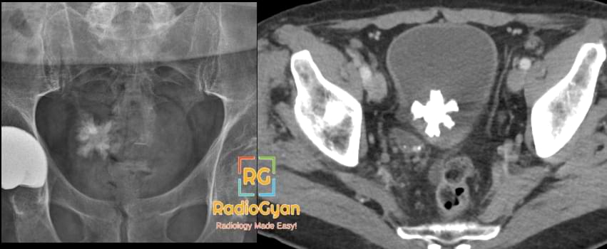
A jackstone calculus is a solitary, spiculated, star-shaped bladder stone with a dense central core and blunt radiating spicules, named for its resemblance to the metal jack of the children’s game. It is usually composed of calcium oxalate dihydrate, whose bipyramidal crystal habit produces the sharp projections; described specimens also show concentric calcium oxalate monohydrate laminations around a protein-rich radiolucent nidus. On plain radiography it is a dense spiculated opacity in the pelvis, and CT characterises the morphology and composition definitively. Formation requires urinary stasis, so the great majority arise in a bladder with outlet obstruction from benign prostatic hyperplasia, urethral stricture or a neurogenic bladder, and the remainder around a foreign body or in chronic infection. The loose crystalline architecture generally fragments readily at cystolitholapaxy or lithotripsy, but the underlying obstruction must be treated or the stone recurs. Rarely a jackstone forms within a calyceal diverticulum or renal cyst.
Why is it called so?
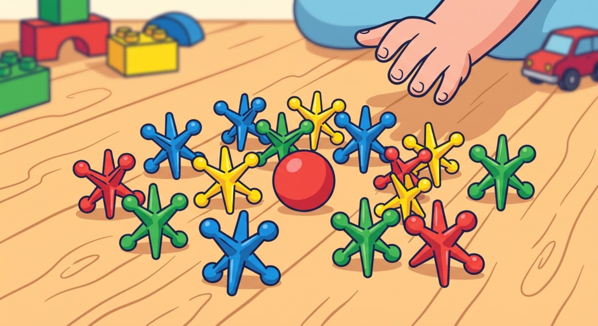
The central body with its blunt radiating projections looks like the six-pointed metal jack used in the game of jacks.
Pathophysiology
Jackstone calculi form in stagnant, supersaturated bladder urine as calcium oxalate dihydrate crystals precipitate around a nidus. The spiky rather than smooth shape is thought to arise because the growing arms repeatedly contact and abrade against the bladder wall: this friction strips freshly deposited material from the tips while leaving the shafts undisturbed, so growth continues preferentially at the exposed ends rather than uniformly over the surface. Morphometric and compositional studies have found that the arm tips contain a protein-rich, radiolucent core, believed to bind urinary proteins preferentially and drive faster linear extension of the arms relative to the body of the stone. Urinary stasis from bladder outlet obstruction supplies the prolonged residence time and supersaturation needed for this slow, tip-driven growth, which is why the morphology is characteristic of bladder stones rather than of rapidly transiting upper tract stones.
Alternative names: Jack stone, jackstone bladder calculus
Other associated named signs: Staghorn calculus, milk of calcium in a calyceal diverticulum, and the laminated bladder stone
References
Access all radiology signs posted so far: https://radiogyan.com/radiology-signs/

Improve Content And Help Your Colleagues!
Found an error in the post? Please let us know using our contact page and we will update it with due credits!
If you wish to contribute radiological images for this case (or any other article on the website), you can submit them here and you will be duly credited: Submit radiology case