


What causes the vacuum joint sign in a synovial joint on radiography?
Let me know in the comments.
Click to reveal the answer
Answer:
The vacuum joint sign is gas, mostly nitrogen, appearing within a joint space when traction, degeneration or instability lowers the intra-articular pressure and dissolved gas comes out of solution. It is seen most often in the large joints, the hip, shoulder and knee, on radiographs and CT, and is also called the pneumoarthrogram sign when the gas outlines the articular surfaces like a contrast arthrogram. Associations include degenerative joint disease, ligamentous laxity, femoroacetabular impingement and trauma, and it appears transiently during traction or hip reduction manoeuvres. In a child or an adult with a painful hip the sign has traditionally been read as reassuring, on the argument that a significant effusion would displace the gas, but it is not reliable as a negative predictor and does not exclude a joint effusion or septic arthritis when the clinical picture suggests them. The spinal equivalent within an intervertebral disc is the vacuum disc sign, and it should not be confused with the vacuum cleft sign, which is intravertebral gas within an ischaemic collapsed vertebral body.
Why is it called so?
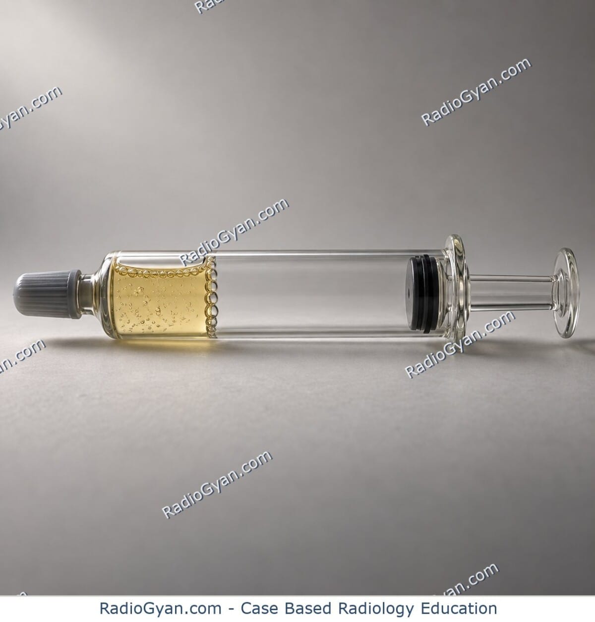
The lucency looks like an empty space in the joint, and is attributed to the negative pressure, or vacuum, created when the joint surfaces are pulled apart; when the gas outlines the cartilage surfaces the result mimics an air arthrogram, hence pneumoarthrogram.
Pathophysiology
Traction, degenerative loss of cartilage or disc hydration, or instability separates the joint surfaces and increases the volume of a closed space, so local pressure falls. Gas dissolved in the synovial fluid and adjacent tissues, predominantly nitrogen because it is the least soluble, then comes out of solution and collects in the clefts as a radiolucent line or collection. The gas is reabsorbed when the pressure normalises, which is why the finding is transient and position dependent. In the hip a lever mechanism during physiological leg positioning can generate the same effect, and in the spine annular tears and disc degeneration create the cleft in which the gas accumulates. A joint filled with fluid cannot generate the low pressure as readily and tends to obscure the gas, which is the basis, though an unreliable one, for reading the sign as evidence against an effusion.
Alternative names: Vacuum phenomenon; pneumoarthrogram sign; pneumoarthrosis; vacuum arthrogram
Other associated named signs: Vacuum disc sign in the intervertebral disc and the vacuum cleft sign of ischaemic vertebral collapse, which is a different entity
References
Access all radiology signs posted so far: https://radiogyan.com/radiology-signs/

Improve Content And Help Your Colleagues!
Found an error in the post? Please let us know using our contact page and we will update it with due credits!
If you wish to contribute radiological images for this case (or any other article on the website), you can submit them here and you will be duly credited: Submit radiology case