


What causes the small bowel feces sign in the small bowel on CT imaging?
Let me know in the comments.
Click to reveal the answer
The small bowel feces sign is caused by stasis. When small bowel contents sit still for long enough, water is resorbed across the bowel wall and bacterial fermentation adds gas, so the residue becomes a mottled, particulate, gas-speckled column that looks exactly like colonic stool – except it is sitting in the small bowel, where stool does not belong. The commonest reason for that stasis is a subacute or low-grade small bowel obstruction, most often adhesive, and the feces column usually tapers down to the transition zone, which is why the sign is so useful for finding the level of the block.
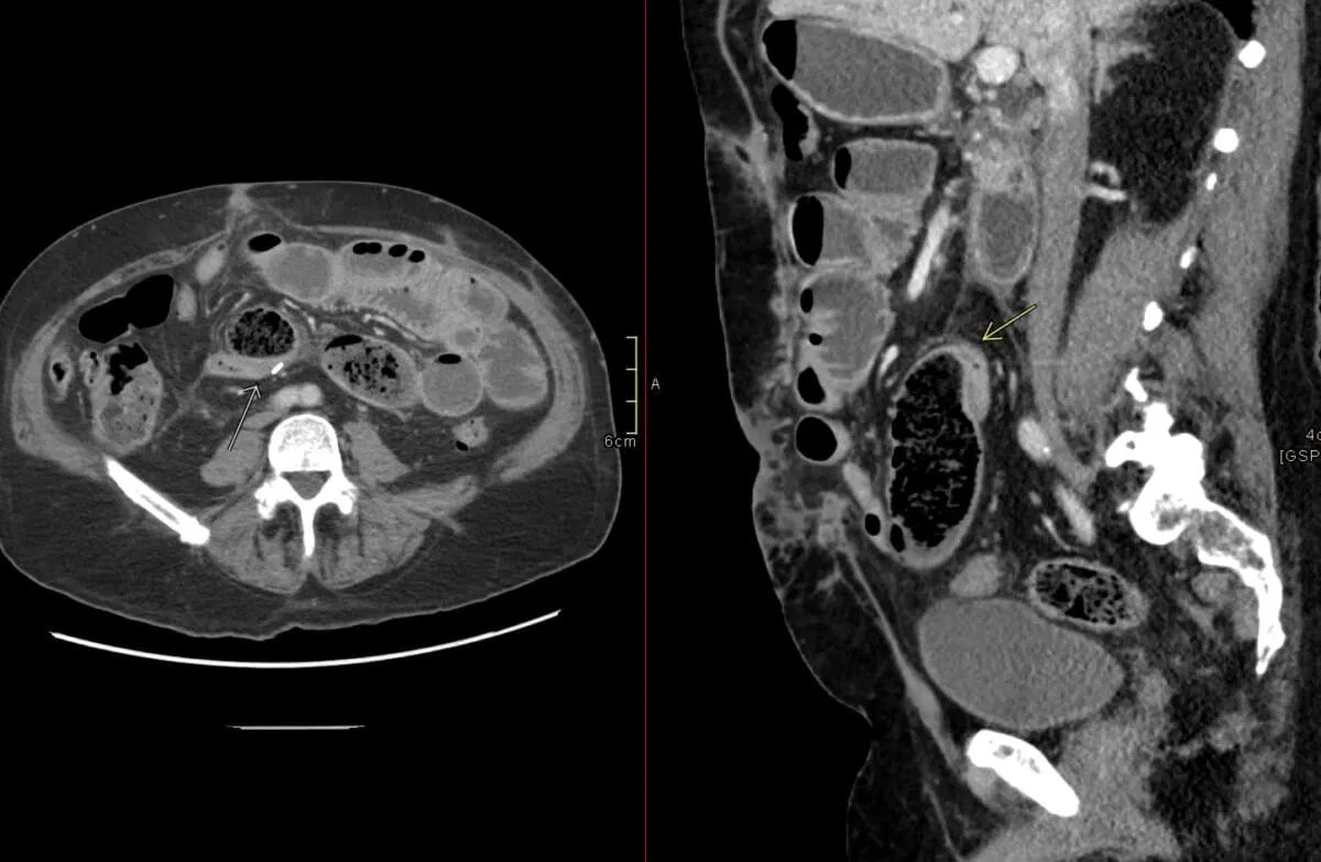
But the sign is not the diagnosis. In the largest unselected series, two-thirds of patients with a small bowel feces sign had no obstruction at all. What separates the two groups is not the feces – it is the calibre of the bowel and whether the distal loops are collapsed.
| At a glance | |
|---|---|
| What it is | Particulate, faeces-like material mottled with gas bubbles inside a dilated small bowel loop on CT |
| What it means | Prolonged intraluminal stasis with water resorption – usually subacute or low-grade small bowel obstruction |
| Also called | Feces sign, small bowel faeces sign, fecalization (faecalisation) of small bowel, feces-like content |
| Where it is seen | Almost always the ileum, immediately proximal to the transition zone; described length 2–25 cm |
| Commonest cause | Adhesive small bowel obstruction; then hernia, Crohn disease stricture, tumour, bezoar |
| Specificity for obstruction | Poor on its own. Only specific when combined with moderate-to-severe dilatation and distal collapse |
| Main value | It points to the transition zone, so it tells you where to look, not whether there is a block |
| Main mimic | Small bowel bezoar or impacted vegetal foreign body – a discrete encapsulated mass, not a tapering column |
| First described | Mayo-Smith and colleagues, Clinical Radiology, 1995 |
Why is it called so?
The term “small bowel feces sign” is used because the appearance of the intraluminal contents – heterogeneous, particulate material mixed with gas bubbles – mimics the typical appearance of feces seen in the colon, despite its location in the small intestine. The designation highlights the radiological similarity to colonic stool, even though the material is actually partially digested food residue, desquamated epithelial cells and mucus rather than true faeces. Mayo-Smith and colleagues described it in 22 patients in 1995, and found it in none of 120 control CT examinations – which is where its reputation for being “always abnormal” comes from.
Pathophysiology
Three things have to happen together for small bowel content to look like stool.
- Transit has to slow down. A mechanical block, a stricture, or severe dysmotility keeps chyme in one segment instead of moving it on.
- Water has to be resorbed. The small bowel mucosa keeps absorbing fluid from the stagnant column, so the residue thickens and becomes particulate.
- Gas has to be produced. Stasis allows bacterial overgrowth and fermentation, which is what produces the scattered bubbles that give the mottled texture.
That sequence takes time, which is the single most useful thing to remember about the sign: it belongs to subacute, incomplete or low-grade obstruction rather than to acute, high-grade strangulating obstruction. A loop that twisted an hour ago has not had time to fecalize. This is also why the sign has a reputation as a marker of “non-severity” – and why that reputation is only partly deserved, as the evidence below shows.
The one situation where the sign appears faster than the time course predicts is mesenteric ischaemia, where sudden loss of motility and mural perfusion can produce fecalization early. Cases have been reported in occlusive mesenteric ischaemia, and a Mayo Clinic series found a statistically significant association between the feces sign and surgically confirmed small bowel ischaemia.
How to recognise it on CT

Four features together make the call:
- Particulate, heterogeneous material – not simple fluid, not simple gas, but a mottled mixture of the two, low in attenuation.
- Inside a small bowel loop – confirm on coronal reformats that you are looking at small bowel and not a redundant sigmoid or a caecum sitting in an unusual position.
- The loop is dilated. This is the step that is most often skipped. A small bowel calibre under 2.5 cm with particulate content is not an obstruction.
- The column tapers to a point. Follow it distally; where the fecalized content stops is almost always the transition zone.
The caveat that matters most: the sign alone is not obstruction
A single-centre review of 1,642 consecutive emergency department abdominal CT studies found the small bowel feces sign in 100 of them (6 per cent). Only 32 of those 100 patients had a documented small bowel obstruction. The other 68 had something else entirely – enteritis, dysmotility, chronic diarrhoeal states, or no gastrointestinal diagnosis at all. What separated the two groups was not the feces.
| CT feature accompanying the feces sign | Patients with obstruction (n = 32) | Patients without obstruction (n = 68) |
|---|---|---|
| Normal or only mildly dilated small bowel | 19% | 100% |
| Moderate or severe small bowel dilatation | 81% | 0% |
| Collapsed distal small bowel | 84% | 0% |
| All three together (feces sign, dilatation, distal collapse) | 72% | 0% |
Read that table the other way round and it becomes a reporting rule: if the bowel carrying the fecalized content is not dilated, the patient almost certainly does not have an obstruction. The sign is a prompt to measure the calibre and check the distal loops, not a substitute for doing so.
How often does it actually appear?
Reported frequency varies enormously, and almost all of the variation comes from how the population was selected and how strictly the sign was defined.
| Study | Population | Frequency of the feces sign |
|---|---|---|
| Mayo-Smith 1995 | 120 control CT examinations | 0% |
| Catalano 1997 | 94 small bowel obstructions | 7.4% |
| Lazarus 2004 | 34 consecutive small bowel obstructions | 55.9% |
| Jacobs 2007 | 1,642 unselected emergency abdominal CTs | 6% overall (only a third of these had obstruction) |
| Khaled 2018 | 237 CTs in adhesive small bowel obstruction | 37.1% |
| Guiraud 2026 | 263 patients with adhesive small bowel obstruction | 47.5% |
The early Italian series reporting 7 per cent used a strict definition in ileal loops only; the modern series using thin-section multidetector CT and a broader definition land somewhere between a third and a half of obstructed patients. In practice, expect to see it in roughly one in three patients with adhesive obstruction.
Frequency also tracks the grade of the block. In the Lazarus series the sign was present in 1 of 6 mild obstructions (17 per cent), 8 of 11 moderate obstructions (73 per cent) and 10 of 17 high-grade obstructions (59 per cent), and the fecalized segment was longer in moderate and high-grade disease.
Its real strength: finding the transition zone
This is the one thing every study agrees on. In the Lazarus series, the particulate material could be traced to the point of transition in every single patient in whom the sign was present, and it was most conspicuous at the transition zone itself. In the 237-CT Paris series, the sign sat at the transition zone in 93 per cent of positive cases.
That series also drew a distinction that is worth adopting in reports, because the two patterns carry opposite implications:
| Pattern | What it looks like | Frequency | Association (univariate) |
|---|---|---|---|
| Transition zone feces sign | Fecalized column immediately proximal to a single transition zone | 93% of positive cases | Successful non-operative treatment, odds ratio 3.37; lower odds of ischaemia, odds ratio 0.33 |
| Trapped feces sign | Fecalized content lying between two transition zones – a closed loop | 16% of positive cases | When combined with a transition zone feces sign, ischaemia with odds ratio 24.16 |
Fecalized content trapped between two transition points is a closed-loop obstruction until proven otherwise, and it should change the urgency of your report even though the individual odds ratio comes from a small number of patients. Separately, in closed-loop obstruction managed conservatively, a distance of more than 1 cm between the two transition zones and a low-grade block were the independent predictors of conservative treatment succeeding.
Does the feces sign predict who avoids surgery?
This is where the literature is genuinely contradictory, and where most online summaries oversimplify. The original French work called it “the first criterion of non-severity” in adhesive obstruction, and that framing has stuck. The data are messier.
| Study | Patients | What the analysis showed |
|---|---|---|
| Zielinski 2010 (Mayo Clinic) | 100 | Absence of the sign was an independent predictor of needing an operation. Vomiting, no feces sign, free intraperitoneal fluid and mesenteric oedema together gave 96% sensitivity and 90% positive predictive value for requiring exploration |
| Khaled 2018 | 237 CTs | Significant on univariate analysis, but on multivariate analysis the feces sign did not independently predict either non-operative success or ischaemia |
| Kim 2021 | 189 | Lack of the sign independently predicted failure of conservative treatment, odds ratio 5.23, alongside mesenteric haziness and mesenteric fluid |
| Yamamoto 2021 | 92 | Positive patients resumed diet and were discharged sooner; independent predictor of diet resumption (odds ratio 1.685) and discharge (odds ratio 1.861) |
| van Veen 2023 | 360 | Lack of the sign associated with operative management, adjusted odds ratio 2.25 |
| Morelli 2024 | 168 laparoscopic operations | In patients who did reach theatre, the presence of the sign independently predicted conversion to open surgery, odds ratio 1.965 |
Two conclusions survive all of this. First, the absence of the feces sign is a more consistent warning than its presence is a reassurance – when a patient has a clear obstruction with no fecalization at all, the block is more likely to be recent, high-grade and surgical. Second, when the feces sign is entered into a multivariate model alongside mesenteric fluid, mesenteric oedema and mural enhancement, it usually stops being an independent predictor. It is a supporting finding, not a decision rule. Treat it as one piece of a CT gestalt.
The feces sign and ischaemia
It would be convenient if fecalization simply excluded ischaemia. It does not.
- A Mayo Clinic study of 61 CT examinations in patients who went to surgery within seven days found a significant association between the feces sign and surgically confirmed small bowel ischaemia. The same study is a caution about CT in general: prospective sensitivity for ischaemia was only 14.8 per cent, and the most specific sign was decreased segmental mural enhancement.
- Fecalization has been reported appearing early in occlusive mesenteric ischaemia, where the proposed mechanism is abrupt loss of motility rather than slow mechanical stasis.
- The trapped-feces pattern between two transition zones carried a strongly raised odds of ischaemia.
So the ischaemia checklist runs regardless of whether the feces sign is there: reduced or absent mural enhancement, increased unenhanced mural attenuation, mesenteric oedema and fluid, free intraperitoneal fluid, pneumatosis intestinalis and portal venous gas.
Causes of the small bowel feces sign
| Category | Causes | Notes |
|---|---|---|
| Mechanical obstruction (most cases) | Adhesions; external and internal hernia; Crohn disease stricture; primary or metastatic small bowel tumour; gallstone ileus; intussusception; radiation enteritis | Adhesions accounted for 20 of 34 obstructions in the Lazarus series, hernia 4, Crohn disease 4, tumour 3 |
| Intraluminal obstructing mass | Phytobezoar, trichobezoar, impacted vegetal foreign body, worm bolus | The mass itself mimics the sign – see the next section |
| No obstruction (a third of positive cases) | Infectious or inflammatory enteritis, chronic dysmotility, scleroderma, coeliac disease, cystic fibrosis, opioid-related slow transit, paralytic ileus | Bowel calibre is normal or only mildly dilated; distal loops are not collapsed |
| Vascular | Occlusive mesenteric ischaemia | Uncommon, but appears earlier than the usual time course predicts |
Mimics: bezoar and impacted foreign body
This is the differential that changes management, because a bezoar is not a consequence of the obstruction – it is the obstruction, and it usually needs to be removed.
| Feature | Small bowel feces sign | Small bowel bezoar or foreign body |
|---|---|---|
| Shape | Amorphous column, ill-defined margins, tapers along the loop | Discrete, well-defined ovoid mass |
| Capsule | None | Encapsulating wall, often visible |
| Floating fat-density debris | Absent | Present – described as characteristic of bezoar |
| Similar lesion in the stomach | No | Frequently, and finding one should prompt a second look |
| Relationship to transition point | Lies proximal to it and tapers into it | Sits at the transition point and is the obstructing lesion |
| Attenuation (paediatric data) | Mean 8.2 HU | Mean 12.6 HU; a cut-off above 9 HU combined with a clinical severity score above 3 gave a positive predictive value of 80% and a negative predictive value of 84% for phytobezoar |
A 2026 case report of an impacted cork foreign body makes the practical point well: a mottled, gas-containing intraluminal mass at the transition zone should not automatically be labelled the feces sign. If it is discrete and there is identical material in the stomach, think ingested material.
What to do when you see it
Reporting checklist
- Measure the bowel. Under 2.5 cm with no distal collapse: describe the fecalization, do not diagnose obstruction.
- Check the distal small bowel and colon. Collapse distally is what converts the finding into an obstruction.
- Follow the column distally and name the transition zone. This is the single most clinically useful sentence you can write about the sign.
- Count the transition zones. Fecalized content between two of them equals closed loop; say so explicitly and raise the urgency.
- Exclude a bezoar. Discrete mass, capsule, gastric counterpart.
- Run the ischaemia checklist anyway. Mural enhancement, mesenteric oedema and fluid, free fluid, pneumatosis, portal venous gas.
- Do not use the sign as a reason to reassure. Say what it shows and where; leave the operative decision to the combined picture.
How the surgical team will use it
The World Society of Emergency Surgery Bologna guidelines for adhesive small bowel obstruction recommend non-operative management when there is no strangulation, peritonitis or ischaemia, with a water-soluble contrast study used both to predict resolution and to help achieve it. Two more recent data points sharpen that:
- A prospective multicentre cohort of 982 patients with adhesive obstruction found non-operative management succeeded in 57 per cent, with a mean length of stay of 5.3 days. When non-operative management was tried and failed, the mean stay rose to 12.9 days. Full guideline compliance was achieved in only 17 per cent of even the successful group.
- A 360-patient cohort found the median time to symptom resolution in the non-operative group was about two days, and that the odds of needing bowel resection rose roughly 20 per cent for each additional day surgery was delayed – concluding that the conservative trial should not exceed three days.
In other words: the feces sign may nudge the team towards an initial non-operative trial, but it does not extend the clock.
Alternative names: Feces sign, small bowel faeces sign, small bowel faecal sign, fecalization (faecalisation) of small bowel, feces-like content of the small bowel.
Other associated named signs: The beak sign (tapered transition point), the fat notch sign (extraluminal indentation by an adhesive band), the whirl sign (twisted mesentery) and the recently described flower bouquet sign (radially arranged closed-loop segments with converging mesenteric vessels) are the other CT signs of adhesive small bowel obstruction. The main radiological differential is a small bowel bezoar, which mimics the feces sign on CT but represents a discrete obstructing intraluminal mass rather than stasis-related particulate content.
Frequently asked questions
What does the small bowel feces sign mean on CT?
It means intraluminal contents have been stagnant long enough for water to be resorbed and gas to accumulate, producing a mottled, stool-like column inside the small bowel. Most often the cause is a subacute or low-grade small bowel obstruction, and the column typically tapers down to the transition zone. It is a marker of slow transit, not a diagnosis in itself.
Does fecalization of the small bowel always mean obstruction?
No. In a review of 1,642 unselected emergency abdominal CT studies, the sign was present in 6 per cent, and only about a third of those patients had an obstruction. The discriminating features were bowel calibre and distal collapse: every patient without obstruction had normal or only mildly dilated small bowel, whereas 81 per cent of those with obstruction had moderate or severe dilatation.
Where is the small bowel feces sign usually seen?
Almost always in the ileum, immediately proximal to the transition zone. Reported lengths range from 2 to 25 cm, and the segment is longer in moderate and high-grade obstruction. In one series the fecalized material could be traced to the transition point in every positive case, which is the sign’s main practical value.
Does the feces sign mean the patient can avoid surgery?
Not reliably. Several cohorts found that the absence of the sign predicts needing an operation, and one found patients with the sign resumed diet and were discharged sooner. But the largest blinded study found that on multivariate analysis the sign did not independently predict either non-operative success or ischaemia, and one surgical series found the sign predicted conversion from laparoscopic to open surgery. Use it as a supporting finding alongside mesenteric fluid, mesenteric oedema and mural enhancement, not as a decision rule.
Can the small bowel feces sign occur with bowel ischaemia?
Yes. A Mayo Clinic study found a significant association between the feces sign and surgically confirmed small bowel ischaemia, and fecalization has been reported appearing early in occlusive mesenteric ischaemia. Fecalized content trapped between two transition zones, indicating a closed loop, carries a particularly high odds of ischaemia. The standard ischaemia checklist should be applied whether or not the sign is present.
How do you tell a small bowel bezoar from the feces sign?
A bezoar is a discrete, well-defined ovoid mass with an encapsulating wall, often shows floating fat-density debris, frequently has an identical lesion in the stomach, and sits at the transition point as the obstructing lesion. The feces sign is an amorphous, ill-defined column that lies proximal to the transition point and tapers into it. In children, a debris attenuation above 9 HU combined with a high clinical severity score favours phytobezoar.
Why is there faeces in the small bowel at all?
Strictly, there is not. The material is partially digested food residue, desquamated epithelial cells and mucus that has been concentrated by water resorption and speckled with gas from bacterial fermentation. It only resembles stool; the name describes the appearance, not the content.
Related radiology signs
- Stack of coins appearance – regular thickened valvulae conniventes in small bowel wall oedema or haemorrhage
- Misty mesentery – increased mesenteric fat attenuation, one of the findings to assess alongside an obstruction
- Thumbprinting – the large bowel counterpart of mural oedema on plain radiography
- Apple-core sign – the annular malignant lesion, a cause of obstruction further downstream
References
- Mayo-Smith WW, Wittenberg J, Bennett GL, Gervais DA, Gazelle GS, Mueller PR. The CT small bowel faeces sign: description and clinical significance. Clinical Radiology. 1995;50(11):765-767. PMID: 7489626
- Catalano O. The faeces sign. A CT finding in small-bowel obstruction. Radiologe. 1997;37(5):417-419. PMID: 9312785
- Fuchsjäger MH. The small-bowel feces sign. Radiology. 2002;225(2):378-379. PMID: 12409569
- Lazarus DE, Slywotsky C, Bennett GL, Megibow AJ, Macari M. Frequency and relevance of the “small-bowel feces” sign on CT in patients with small-bowel obstruction. American Journal of Roentgenology. 2004;183(5):1361-1366. PMID: 15505304
- Delabrousse E, Baulard R, Sarliève P, Michalakis D, Rodière E, Kastler B. Value of the small bowel feces sign at CT in adhesive small bowel obstruction. Journal de Radiologie. 2005;86(4):393-398. PMID: 15959431
- Sheedy SP, Earnest F 4th, Fletcher JG, Fidler JL, Hoskin TL. CT of small-bowel ischemia associated with obstruction in emergency department patients: diagnostic performance evaluation. Radiology. 2006;241(3):729-736. PMID: 17114622
- Jacobs SL, Rozenblit A, Ricci Z, et al. Small bowel faeces sign in patients without small bowel obstruction. Clinical Radiology. 2007;62(4):353-357. PMID: 17331829
- Delabrousse E, Lubrano J, Sailley N, Aubry S, Mantion GA, Kastler BA. Small-bowel bezoar versus small-bowel feces: CT evaluation. American Journal of Roentgenology. 2008;191(5):1465-1468. PMID: 18941086
- Zielinski MD, Eiken PW, Bannon MP, et al. Small bowel obstruction – who needs an operation? A multivariate prediction model. World Journal of Surgery. 2010;34(5):910-919. PMID: 20217412
- Abu-Hmeidan JH, Bismar HA, Hamid AM. Small bowel feces sign in association with occlusive mesenteric ischemia. Acta Radiologica Short Reports. 2014;3(7):2047981614540142. PMID: 25298875
- Berl S, Dawkins A, DiSantis D. The small bowel feces sign. Abdominal Radiology. 2016;41(4):794-795. PMID: 27112778
- Khaled W, Millet I, Corno L, et al. Clinical relevance of the feces sign in small-bowel obstruction due to adhesions depends on its location. American Journal of Roentgenology. 2018;210(1):78-84. PMID: 29045179
- Ten Broek RPG, Krielen P, Di Saverio S, et al. Bologna guidelines for diagnosis and management of adhesive small bowel obstruction (ASBO): 2017 update of the evidence-based guidelines from the World Society of Emergency Surgery ASBO working group. World Journal of Emergency Surgery. 2018;13:24. PMID: 29946347
- Chang KJ, Marin D, Kim DH, et al; Expert Panel on Gastrointestinal Imaging. ACR Appropriateness Criteria® suspected small-bowel obstruction. Journal of the American College of Radiology. 2020;17(5S):S305-S314. PMID: 32370974
- Kim J, Lee Y, Yoon JH, et al. Non-strangulated adhesive small bowel obstruction: CT findings predicting outcome of conservative treatment. European Radiology. 2021;31(3):1597-1607. PMID: 33128599
- Yamamoto Y, Miyagawa Y, Kitazawa M, et al. Association of feces sign with prognosis of non-emergency adhesive small bowel obstruction. Asian Journal of Surgery. 2021;44(1):292-297. PMID: 32732062
- Kim HR, Lee Y, Kim J, et al. Closed loop obstruction of small bowel: CT signs predicting successful non-surgical treatment. European Journal of Radiology. 2023;161:110716. PMID: 36758277
- van Veen T, Ramanathan P, Ramsey L, Dort J, Tabello D. Predictive factors for operative intervention and ideal length of non-operative trial in adhesive small bowel obstruction. Surgical Endoscopy. 2023;37(11):8628-8635. PMID: 37495847
- Wang N, Wu X, Lin X, Zhang S, Shen W. Computed tomography with clinical scoring to differentiate phytobezoar from feces in childhood small bowel obstruction. Turkish Journal of Pediatrics. 2023;65(6):1002-1011. PMID: 38204315
- Morelli M, Strambi S, Cremonini C, et al. Adhesive small bowel obstruction: predictive factors of laparoscopic failure. Updates in Surgery. 2024;76(2):705-712. PMID: 38151681
- Kaplan LJ, Martinez-Casas I, Mohseni S, et al; SnapSBO Collaborators. Small bowel obstruction outcomes according to compliance with the World Society of Emergency Surgery Bologna guidelines. British Journal of Surgery. 2025;112(4):znaf080. PMID: 40246692
- Guiraud A, Vadot V, Fournel I, Loffroy R, Gozalichvili D, Ortega-Deballon P. Value of the feces sign in predicting successful conservative management of adhesive small bowel obstruction: a retrospective cohort study. Current Problems in Surgery. 2026;78:102022. PMID: 42000347
- Sakhy Y, Azhari I, Raoui FZ, et al. Small bowel obstruction caused by impacted cork foreign body mimicking the small bowel feces sign: an educational imaging case. Radiology Case Reports. 2026;21(9):3708-3710. PMID: 42293497
- Hokama A, Morioka H, Matayoshi T. The flower bouquet sign and the fat notch sign in adhesive small bowel obstruction. Revista Española de Enfermedades Digestivas. 2026. PMID: 41879012
Access all radiology signs posted so far: https://radiogyan.com/radiology-signs/

Improve Content And Help Your Colleagues!
Found an error in the post? Please let us know using our contact page and we will update it with due credits!
If you wish to contribute radiological images for this case (or any other article on the website), you can submit them here and you will be duly credited: Submit radiology case