


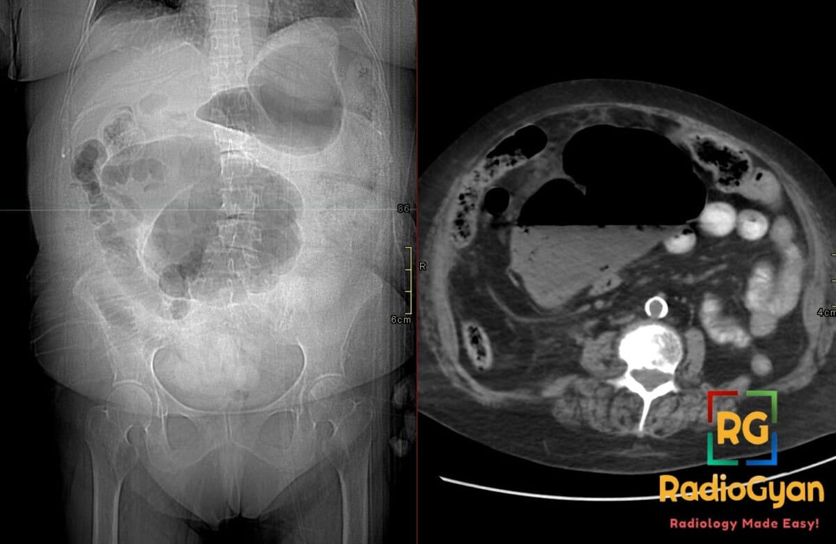
What causes the kidney bean sign in the colon on abdominal radiographs?
Let me know in the comments.
Click to reveal the answer
Answer:
The kidney bean sign is a radiologic sign observed on abdominal radiographs that indicates cecal volvulus, a form of bowel obstruction where the cecum twists around its mesenteric attachment. In cecal volvulus, the haustra of the dilated cecum create a characteristic kidney bean-shaped appearance, typically displaced toward the mid-abdomen or left upper quadrant. This sign indicates torsion of the cecum and represents a potentially life-threatening bowel obstruction that may require emergent medical intervention to prevent complications such as ischemia or perforation.
Why is it called so?
The sign is named for its visual resemblance to a kidney bean on radiographic imaging. The haustra (the natural segmentations of the colon) combined with the twisted, dilated configuration of the cecum create a shape reminiscent of a kidney bean rather than the typical appearance of normal bowel loops.
Pathophysiology
The kidney bean sign develops when the cecum rotates around its mesenteric axis, causing the organ to twist upon itself. This torsion displaces the cecum from its normal right lower quadrant position, most often toward the mid-abdomen or left upper quadrant. The volvulus causes luminal obstruction and subsequent gas accumulation and dilation of the cecum proximal to the twist. The haustra become prominent due to this distension, and when the dilated, twisted cecum is viewed on abdominal radiographs, the combination of the haustral pattern and the organ’s bean-like contour creates the characteristic radiographic appearance.
Alternative names:
None. This sign is specifically distinct from the coffee bean sign, which occurs in sigmoid volvulus — some sources use the two terms loosely interchangeably for sigmoid volvulus, but in the context of cecal volvulus the “kidney bean sign” refers specifically to the displaced, haustrated cecum.
Other associated named signs:
Coffee bean sign (the analogous appearance in sigmoid volvulus); Whirl sign (or whirlpool sign) – seen when bowel rotates around its mesentery, producing a whirling pattern of mesenteric vessels on CT imaging; Bird’s beak sign – the focal tapering at the point of torsion on contrast enema studies.
References
Access all radiology signs posted so far: https://radiogyan.com/radiology-signs/

Improve Content And Help Your Colleagues!
Found an error in the post? Please let us know using our contact page and we will update it with due credits!
If you wish to contribute radiological images for this case (or any other article on the website), you can submit them here and you will be duly credited: Submit radiology case