


What causes Steeple sign in airway on frontal neck radiograph?
Let me know in the comments.
Click to reveal the answer
Answer:
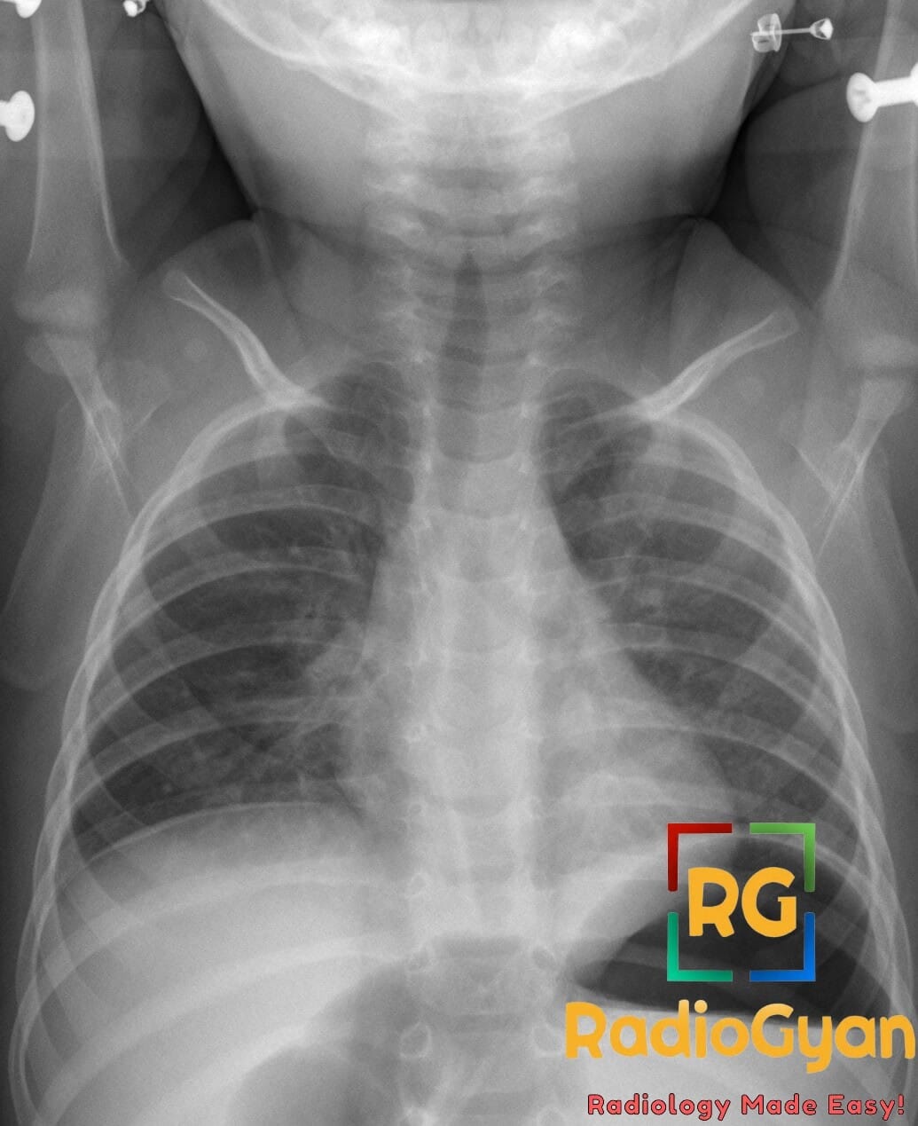
Steeple sign: Tapered narrowing of the subglottic trachea due to mucosal edema and inflammation in croup (acute viral laryngotracheobronchitis), most commonly caused by parainfluenza virus.
Why is it called so?
The Steeple sign is named for the tapered, cone-shaped narrowing of the subglottic trachea that resembles the pointed silhouette of a church steeple on frontal radiograph.
Pathophysiology
Viral infection causes submucosal edema and inflammatory thickening in the subglottic region, which is prone to swelling due to its loosely supported structure; this results in symmetrical conical narrowing just below the vocal cords, replacing the normal squared-shoulder appearance.
Alternative names:
Wine bottle sign, inverted V sign, pencil-tip sign
Other associated named signs:
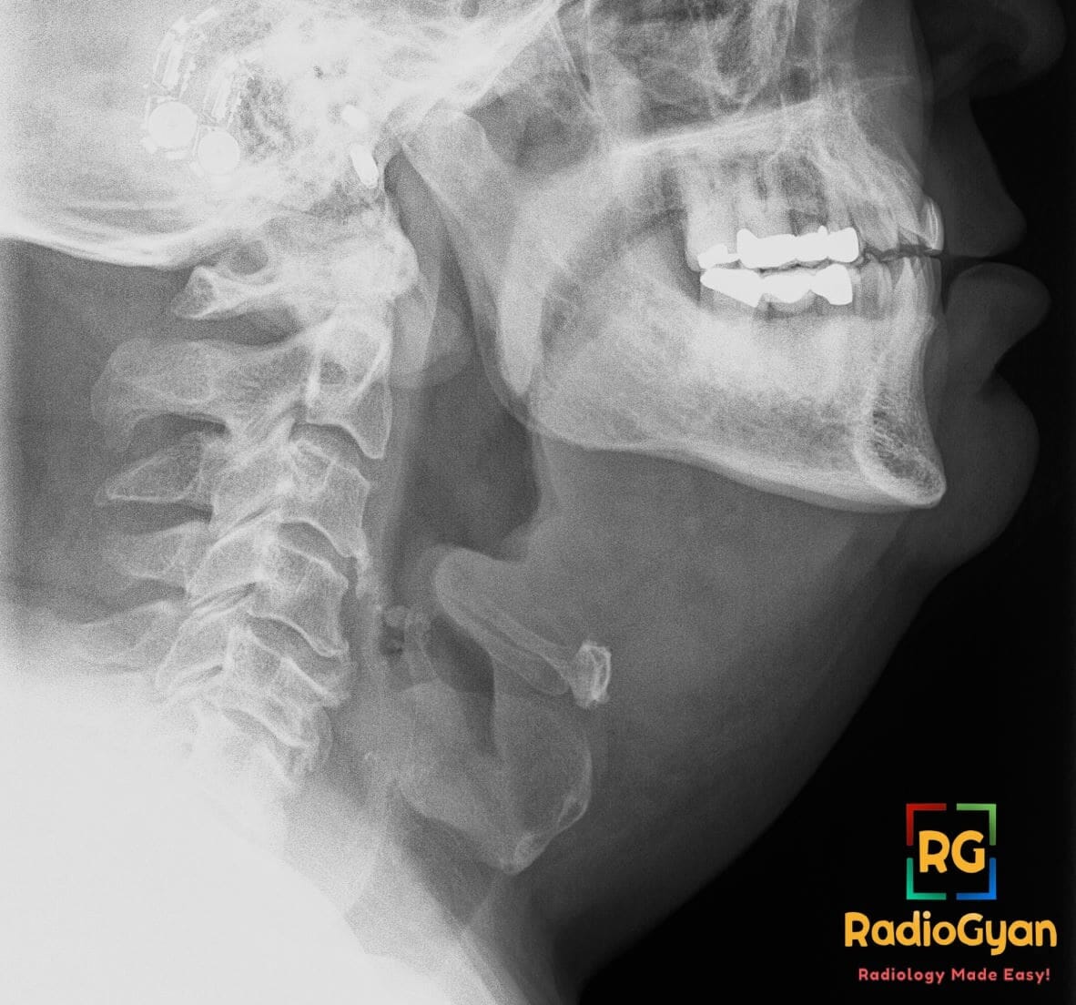
Thumb sign (thickened, rounded epiglottis on lateral neck radiograph, seen in acute epiglottitis – the key mimic to distinguish from croup)
The steeple sign is a symmetrical, tapered narrowing of the subglottic trachea seen on a frontal (anteroposterior) radiograph of the neck or upper chest. The normal subglottic airway has squared-off lateral margins, sometimes called the shoulders; when the submucosa swells, those shoulders are lost and the air column narrows into a cone that resembles the pointed silhouette of a church steeple. It is the classic radiographic appearance of croup (acute viral laryngotracheobronchitis), most often caused by parainfluenza virus. Two things about the sign matter more than the pattern itself: croup is a clinical diagnosis that does not require a radiograph, and the steeple sign is neither sensitive nor specific enough to confirm or exclude it.
What is the steeple sign?
On a well-positioned frontal radiograph the tracheal air column below the vocal cords normally has parallel walls with a slight lateral bulge on each side, produced by the distensible mucosa within the rigid cricoid ring. This gives the subglottis a squared-off, shouldered outline. In croup, submucosal oedema fills that space from both sides, obliterating the shoulders and leaving a narrow, smoothly tapering column of air that comes to a point. That inverted-V or cone shape, extending roughly 5 to 10 mm below the level of the cords, is the steeple sign.
- View: frontal (AP) soft-tissue radiograph of the neck, or the upper trachea on a frontal chest radiograph.
- Location: subglottic trachea, immediately below the vocal cords and within the cricoid ring, the narrowest and least distensible part of the paediatric airway.
- Morphology: symmetrical, smooth, tapered narrowing. Asymmetry, an irregular margin or an intraluminal membrane is a red flag for a different diagnosis.
- Classic association: croup, most commonly caused by parainfluenza virus types 1 and 3, typically in children aged 6 months to 3 years.
Why is it called the steeple sign?
The narrowed air column tapers upward to a point in the same way a church spire tapers above its tower, and the two normal subglottic shoulders that disappear correspond to the wider base beneath the spire. Several other names describe the identical shape from different angles, and all of them are used interchangeably in reports and textbooks.
| Name | What the name describes |
|---|---|
| Steeple sign (church steeple sign) | The tapering point of the subglottic air column, like a spire. |
| Wine bottle sign | The narrow neck of a wine bottle rising from a wider body. |
| Inverted V sign | The converging lateral walls of the narrowed air column. |
| Pencil-tip sign (pencil-point sign) | The sharpened, tapered tip of the tracheal air column. |
| Hourglass sign | Occasionally used when the narrowing is bounded above and below by wider airway. |
Pathophysiology: why the subglottis is the bottleneck
Croup begins as an ordinary upper respiratory infection that spreads down to the larynx and trachea. The virus provokes mucosal and submucosal inflammation with oedema, cellular infiltration and increased secretions. The subglottis suffers most because it is the only part of the airway completely encircled by a rigid, complete cartilage ring, the cricoid. Swelling there cannot expand outward, so it expands inward into the lumen.
The consequences are disproportionate in small children. Resistance to laminar airflow rises with the fourth power of the reduction in radius, so a millimetre of circumferential oedema in an infant subglottis, which is only about 5 to 7 mm across, can cut the cross-sectional area dramatically and multiply the work of breathing. That is the physiological reason a young child develops stridor from an infection that produces nothing more than hoarseness in an adult, and the anatomical reason the radiographic narrowing appears where it does.
How to look for the steeple sign on the radiograph
- Use the frontal view for the subglottis, the lateral for everything else. The tapered subglottic narrowing is a frontal finding. The lateral soft-tissue neck view is what excludes the dangerous mimics: a swollen epiglottis, thickened aryepiglottic folds, widened prevertebral soft tissues or a radiopaque foreign body.
- Insist on inspiration and extension. Expiration, neck flexion and phonation all narrow and buckle the normal subglottic and cervical trachea and can manufacture a false steeple appearance. Assess the airway on a well-inspired film with the neck extended before calling it abnormal.
- Check for symmetry and a smooth margin. Croup narrows the subglottis smoothly and symmetrically. An eccentric, irregular or shaggy tracheal wall, or a linear intraluminal membrane, points to bacterial tracheitis rather than croup.
- Follow the whole air column. Trace the airway from the nasopharynx to the carina on both views. Ballooning of the hypopharynx, an abrupt cut-off, unilateral hyperinflation or air trapping suggest an aspirated foreign body.
- Do not put an unstable child in the radiology department. A child with severe distress, drooling or a tripod posture needs airway assessment by an experienced team, not a trip to the X-ray table. In a distressed child, a single lateral view may be all that is appropriate.
How reliable is the steeple sign?
This is the part most often missed when the sign is learned as a spotter. The steeple sign is a useful illustration of croup pathology, but it performs poorly as a diagnostic test, and imaging is not part of the routine diagnostic pathway.
| Question | Practical answer |
|---|---|
| Does a normal radiograph exclude croup? | No. A substantial proportion of children with unequivocal clinical croup have a normal-looking subglottis, so the absence of a steeple sign does not change the diagnosis. |
| Does a steeple sign confirm croup? | No. Subglottic narrowing also occurs in bacterial tracheitis, epiglottitis, angioedema, thermal or caustic airway injury, subglottic haemangioma and subglottic stenosis, and it can be simulated by expiration or phonation in a normal child. |
| Is a radiograph needed to diagnose croup? | No. Barking cough, hoarseness and inspiratory stridor in a child of the right age is a clinical diagnosis. Current evidence reviews state that radiography and laboratory testing are typically unnecessary and should be reserved for cases where the diagnosis is unclear. |
| How accurate are airway radiographs generally? | Variable by disease. In a paediatric series correlating airway radiographs with microlaryngoscopy and bronchoscopy, sensitivity exceeded 86% for exudative tracheitis, airway foreign body and innominate artery compression, but fell to 62% for tracheomalacia and 5% for laryngomalacia. |
| Is there a downside to imaging? | Yes. It costs time, delivers radiation, may remove an unstable child from a monitored area, and in hospitalised croup the use of a radiograph has been associated with a greater need for significant subsequent intervention, reflecting a sicker or more diagnostically uncertain group. |
The practical position: treat the steeple sign as supportive when it appears on a film obtained for another reason, and never let its absence override a convincing clinical picture. When you do report it, say what it is, and say what has been excluded on the lateral view.
Croup: the clinical picture behind the sign
| Feature | Typical findings |
|---|---|
| Age | 6 months to 3 years, with the peak in the second year of life. It affects roughly 3% of children per year. |
| Organism | Parainfluenza virus (types 1 and 3) accounts for the majority. Influenza A, RSV, human metapneumovirus, adenovirus, rhinovirus and SARS-CoV-2 also cause it. |
| Seasonality | Peak incidence in autumn, classically October and November in the northern hemisphere. |
| Onset | One to two days of coryza, then abrupt onset of the classic triad, characteristically at night. |
| Cardinal features | Barking (seal-like) cough, hoarse voice or cry, inspiratory stridor, variable respiratory distress. Low-grade fever is common but not required. |
| Course | Usually self-limiting, with symptoms resolving within about 48 hours. Severe upper airway obstruction and respiratory failure are rare. |
| Red flags against croup | High fever with toxicity, drooling, tripod posture, absent cough, rapid deterioration, or age outside the usual range. |
Westley croup score
Severity is graded clinically, not radiologically. The Westley score, derived from the 1978 racemic epinephrine trial, remains the standard research instrument and is widely used at the bedside. It scores five clinical items, with a maximum of 17.
| Item | Scoring |
|---|---|
| Level of consciousness | Normal, including asleep = 0; disoriented = 5 |
| Cyanosis | None = 0; with agitation = 4; at rest = 5 |
| Stridor | None = 0; with agitation = 1; at rest = 2 |
| Air entry | Normal = 0; decreased = 1; markedly decreased = 2 |
| Retractions | None = 0; mild = 1; moderate = 2; severe = 3 |
| Total score | Severity | Typical implication |
|---|---|---|
| 0 to 2 | Mild | Barking cough, no stridor at rest. Single dose of corticosteroid, discharge with advice. |
| 3 to 5 | Moderate | Stridor at rest with mild retractions. Corticosteroid, observe. |
| 6 to 11 | Severe | Stridor at rest with marked retractions and agitation. Corticosteroid plus nebulised epinephrine, observe for several hours. |
| 12 to 17 | Impending respiratory failure | Lethargy, decreased air entry, cyanosis. Urgent airway team involvement and critical care. |
Differential diagnosis of the narrowed paediatric airway
A tapered subglottis is a pattern, not a diagnosis. The task in a child with stridor is to separate self-limiting croup from the conditions that threaten the airway.
| Condition | Clinical clue | Imaging discriminator |
|---|---|---|
| Croup (viral laryngotracheobronchitis) | Barking cough, hoarseness, night onset, well between spasms | Symmetrical smooth subglottic tapering (steeple sign); normal epiglottis and prevertebral soft tissues |
| Acute epiglottitis | Toxic child, high fever, drooling, muffled voice, no barking cough, tripod posture | Thumb sign: enlarged rounded epiglottis with thickened aryepiglottic folds on the lateral view |
| Bacterial tracheitis | Croup-like prodrome then high fever, toxicity and no response to epinephrine | Ragged, irregular tracheal wall with intraluminal membranes; subglottic narrowing may coexist |
| Retropharyngeal abscess | Fever, neck stiffness, pain on swallowing, older toddler or young child | Widened prevertebral soft tissues on a true lateral in extension; contrast CT for a drainable collection |
| Inhaled foreign body | Abrupt onset, choking episode, often no preceding coryza | Radiopaque object, focal air trapping, unilateral hyperinflation on expiratory or decubitus views |
| Angioedema or anaphylaxis | Very rapid onset, urticaria, lip or tongue swelling, known trigger | Diffuse supraglottic and subglottic soft-tissue swelling; imaging rarely needed or appropriate |
| Subglottic haemangioma | Recurrent or biphasic stridor under 6 months, sometimes cutaneous haemangiomas | Asymmetric, eccentric subglottic soft-tissue mass; confirmed on MRI and endoscopy |
| Subglottic stenosis | Recurrent croup, previous intubation or prematurity | Fixed narrowing unchanged between studies; diagnosis at bronchoscopy |
When imaging is actually indicated
- The diagnosis is unclear. Atypical age, absent barking cough, high fever with toxicity, or features that do not fit viral croup.
- A foreign body is suspected. A choking episode or abrupt onset without a viral prodrome.
- The child is not responding as expected. Persisting or worsening stridor after corticosteroid and nebulised epinephrine raises bacterial tracheitis, abscess or a fixed structural lesion.
- Croup is recurrent. Repeated episodes warrant evaluation for an underlying anatomic or medical cause rather than a repeat film during each attack.
Management, in one place
Radiologists are asked about this constantly, so it is worth knowing what happens after the film. Current evidence reviews and the Cochrane review of glucocorticoids for croup support the following.
- Corticosteroids for every severity. A single dose of dexamethasone, 0.6 mg/kg to a usual maximum of 12 mg, given orally, intramuscularly or intravenously, is first-line for croup of any severity. It reduces symptom scores, return visits and length of stay. Prednisolone is an alternative, and nebulised budesonide is an option when the oral route is refused.
- Nebulised epinephrine for moderate to severe croup. Added to the corticosteroid when there is stridor at rest, it produces rapid but temporary improvement, so the child needs observation for several hours afterwards for rebound symptoms.
- Supportive care. Keep the child calm and with the caregiver, since agitation worsens obstruction. Humidified air, long the traditional remedy, has not been shown to help.
- Escalation. Persistent severe obstruction or impending failure requires an experienced airway team; intubation is uncommon and usually needs a tube half a size to a full size smaller than predicted for age.
Recurrent croup and when to look further
Repeated croup-like episodes shift the question from “which virus” to “which airway”. A systematic review and meta-analysis of bronchoscopy in recurrent croup found that the commonest findings were subglottic stenosis, reflux-related changes and broncho- or tracheomalacia, and that clinically significant findings were present in 8.7% of patients overall. The risk of a significant finding was higher with a history of intubation (OR 5.17), inpatient consultation (OR 4.01), age under 3 years (OR 3.22), age under 1 year (OR 2.86) and prematurity. Those are the children in whom a fixed narrowing, rather than a viral one, should be actively sought.
The steeple sign in adults
Croup is overwhelmingly a disease of small children, but adult cases are reported, including influenza A-associated and SARS-CoV-2-associated laryngotracheitis, and they can produce the same tapered subglottic narrowing. The clinical stakes differ: because the adult subglottis starts wider, an adult who does develop stridor has usually lost a great deal of lumen, and the differential expands to include angioedema, inhalation injury, post-intubation stenosis, granulomatosis with polyangiitis and subglottic malignancy. A steeple sign in an adult is a prompt to look for those, not a shortcut to a diagnosis of croup.
Key points and pitfalls
- Croup is a clinical diagnosis. Radiography is not required and should be reserved for diagnostic uncertainty, suspected foreign body or failure to respond.
- A normal subglottis does not exclude croup, and a steeple sign does not confirm it.
- Expiration, flexion and phonation mimic the sign. Judge the airway only on an inspiratory film with the neck extended.
- Always read the lateral view. The epiglottis, aryepiglottic folds and prevertebral soft tissues are where the dangerous mimics declare themselves.
- An irregular tracheal wall is not croup. Ragged margins or intraluminal membranes with a toxic child mean bacterial tracheitis until proven otherwise.
- Never delay airway management for a film. The unstable child belongs with the airway team, not in the imaging queue.
Frequently asked questions
What is the steeple sign in radiology?
The steeple sign is a symmetrical, tapered narrowing of the subglottic trachea on a frontal radiograph of the neck or upper chest. Submucosal oedema within the rigid cricoid ring obliterates the normal squared subglottic shoulders, leaving a cone-shaped air column that resembles a church spire. It is the classic radiographic appearance of croup.
What causes the steeple sign?
Croup, or acute viral laryngotracheobronchitis, is the classic cause, most often due to parainfluenza virus types 1 and 3. Influenza A, RSV, human metapneumovirus, adenovirus and SARS-CoV-2 can also cause it. Other causes of subglottic narrowing include bacterial tracheitis, epiglottitis, angioedema, thermal or caustic airway injury, subglottic haemangioma and subglottic stenosis.
Is a chest X-ray needed to diagnose croup?
No. Croup is a clinical diagnosis based on a barking cough, hoarseness and inspiratory stridor in a child aged roughly 6 months to 3 years. Current evidence reviews state that radiography and laboratory testing are typically unnecessary and should be reserved for cases where the diagnosis is unclear, a foreign body is suspected, or the child fails to respond to treatment.
How sensitive and specific is the steeple sign for croup?
It is neither sensitive nor specific. Many children with unequivocal clinical croup have a normal-looking subglottis, so a normal radiograph does not exclude the diagnosis. Conversely, a steeple appearance can be produced by expiration, neck flexion or phonation in a normal child, and by bacterial tracheitis, epiglottitis, angioedema and fixed subglottic narrowing. The sign supports the diagnosis but cannot make or refute it.
What is the difference between the steeple sign and the thumb sign?
The steeple sign is subglottic narrowing on the frontal view and indicates croup, an infraglottic illness. The thumb sign is a swollen, rounded epiglottis on the lateral view and indicates acute epiglottitis, a supraglottic emergency. Croup gives a barking cough with a well-looking child between spasms; epiglottitis gives a toxic, drooling child with a muffled voice and no barking cough.
Which view shows the steeple sign?
The frontal, or anteroposterior, soft-tissue radiograph of the neck, or the upper trachea on a frontal chest radiograph. The film must be taken in inspiration with the neck extended, because expiration, flexion and phonation narrow the normal subglottic and cervical trachea and can simulate the sign. The lateral view is added to exclude epiglottitis, retropharyngeal abscess and a radiopaque foreign body.
How is croup treated?
A single dose of dexamethasone, 0.6 mg/kg up to a usual maximum of 12 mg, given orally, intramuscularly or intravenously, is first-line for croup of any severity. Nebulised epinephrine is added for moderate to severe croup with stridor at rest, and the child is then observed for several hours because the benefit is temporary. Humidified air has not been shown to help.
What should be done for recurrent croup?
Recurrent episodes should prompt evaluation for an underlying anatomic abnormality or medical condition rather than repeated radiographs. In a meta-analysis of bronchoscopy for recurrent croup, the commonest findings were subglottic stenosis, reflux changes and tracheobronchomalacia, with clinically significant findings in 8.7% of patients. Risk was higher with previous intubation, age under 3 years and prematurity.
References
- Cooke A, Conway S, Griffin L. Croup: Rapid Evidence Review. Am Fam Physician. 2026;113(3):254-258. PMID: 41839076.
- Aregbesola A, Tam CM, Kothari A, Le ML, Ragheb M, Klassen TP. Glucocorticoids for croup in children. Cochrane Database Syst Rev. 2023;1(1):CD001955. PMID: 36626194.
- Darras KE, Roston AT, Yewchuk LK. Imaging Acute Airway Obstruction in Infants and Children. RadioGraphics. 2015;35(7):2064-2079. PMID: 26495798.
- Smith DK, McDermott AJ, Sullivan JF. Croup: Diagnosis and Management. Am Fam Physician. 2018;97(9):575-580. PMID: 29763253.
- Walner DL, Ouanounou S, Donnelly LF, Cotton RT. Utility of radiographs in the evaluation of pediatric upper airway obstruction. Ann Otol Rhinol Laryngol. 1999;108(4):378-383. PMID: 10214786.
- Westley CR, Cotton EK, Brooks JG. Nebulized racemic epinephrine by IPPB for the treatment of croup: a double-blind study. Am J Dis Child. 1978;132(5):484-487. PMID: 347921.
- Hiebert JC, Zhao YD, Willis EB. Bronchoscopy findings in recurrent croup: A systematic review and meta-analysis. Int J Pediatr Otorhinolaryngol. 2016;90:86-90. PMID: 27729160.
- Huang CT. Steeple sign: not specific for croup. J Emerg Med. 2012;43(5):e333-e334. PMID: 21742458.
- Asmundsson AS, Arms J, Kaila R, et al. Hospital Course of Croup After Emergency Department Management. Hosp Pediatr. 2019;9(5):326-332. PMID: 30988017.
- Kuo CY, Parikh SR. Bacterial tracheitis. Pediatr Rev. 2014;35(11):497-499. PMID: 25361911.
- Salour M. The steeple sign. Radiology. 2000;216(2):428-429. PMID: 10924564.
- Kashiura M, Amagasa S, Moriya T. The Steeple Sign of Croup in an Adult. Intern Med. 2022;61(18):2825. PMID: 35185054.
Access all radiology signs posted so far: https://radiogyan.com/radiology-signs/

Improve Content And Help Your Colleagues!
Found an error in the post? Please let us know using our contact page and we will update it with due credits!
If you wish to contribute radiological images for this case (or any other article on the website), you can submit them here and you will be duly credited: Submit radiology case