


What causes the Split Pleura Sign in the pleural space on contrast-enhanced CT?
Let me know in the comments.
Click to reveal the answer
Answer:
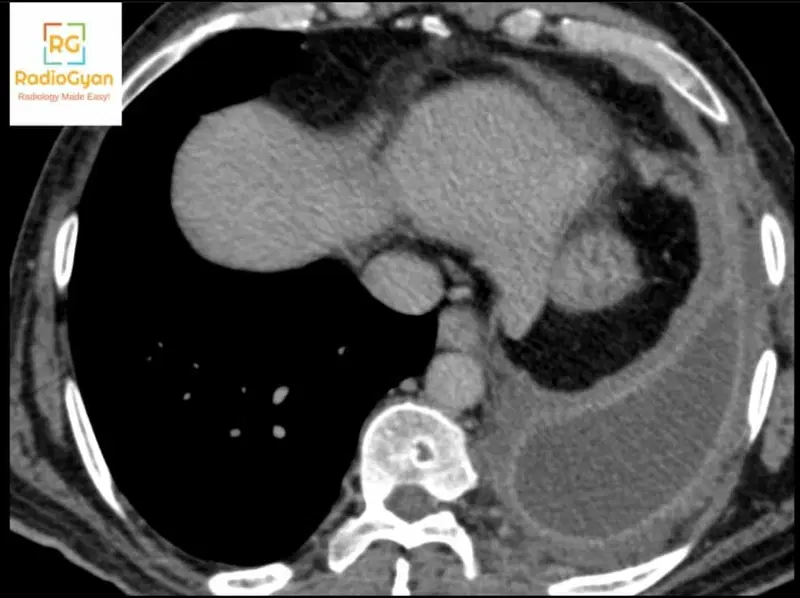
The Split Pleura Sign represents separation and thickening of both the visceral pleura and parietal pleura layers by an accumulation of pleural fluid, most commonly indicating pleural empyema. This sign develops when infected pleural fluid accumulates between the two pleural layers, causing them to become individually visible, thickened, and enhancing on contrast-enhanced CT, and separated on imaging.
The sign has high diagnostic utility for distinguishing complicated parapneumonic effusion and empyema from simple parapneumonic effusion, with one study reporting a sensitivity of 80.6% and specificity of 74.5%. When only one pleural layer demonstrates thickening without separation of both layers, this is termed the hemi-split pleura sign. The Split Pleura Sign is a reliable CT finding for differentiating empyema from noninfectious pleural effusion, though it is not entirely specific and can occasionally be seen with other exudative or hemorrhagic effusions.
Why is it called so?
The sign is named for its appearance of the pleural membrane appearing as two distinct split layers rather than a single continuous structure. The descriptive term reflects the visual separation of the visceral pleura (innermost layer) from the parietal pleura (outer layer) by the intervening pleural effusion, creating a characteristic split appearance on cross-sectional imaging. The sign was first described by Waite et al. in a 1990 Radiology paper on the CT appearances of parietal pleural changes in empyema.
Pathophysiology
In pleural empyema, bacterial or fungal infection triggers an inflammatory response within the pleural space. This inflammatory process causes both the visceral pleura and parietal pleura layers to become edematous and hyperenhanced on contrast-enhanced CT. As purulent fluid accumulates and exerts pressure between these two thickened pleural layers, they are mechanically separated and pushed apart. The combination of pleural thickening with fluid separation makes both layers individually conspicuous on imaging, whereas in simple pleural effusion without infection, pleural thickening is typically absent or minimal.
Alternative names:
There are no significantly different alternative names for this sign, though the incomplete form is designated as the hemi-split pleura sign when only single pleural layer thickening occurs.
Other associated named signs:
Claw sign, which helps distinguish empyema (forming an acute angle with the chest wall) from a peripheral lung/pulmonary abscess (forming a round shape) — the Split Pleura Sign remains the primary radiological indicator for distinguishing complicated parapneumonic effusion from uncomplicated effusion.
References
- Waite RJ et al. Parietal pleural changes in empyema: appearances at CT. Radiology. 1990.
- Tsujimoto N et al. A Simple Method for Differentiating Complicated Parapneumonic Effusion/Empyema from Parapneumonic Effusion Using the Split Pleura Sign and the Amount of Pleural Effusion on Thoracic CT. PLoS One. 2015.
Access all radiology signs posted so far: https://radiogyan.com/radiology-signs/

Improve Content And Help Your Colleagues!
Found an error in the post? Please let us know using our contact page and we will update it with due credits!
If you wish to contribute radiological images for this case (or any other article on the website), you can submit them here and you will be duly credited: Submit radiology case