


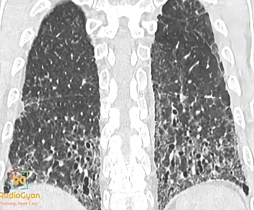
What causes honeycombing on HRCT?
Let me know in the comments.
Click to reveal the answer
Answer:
Honeycombing is defined by the Fleischner Society as clustered cystic airspaces of comparable diameter, typically 3-10 mm but occasionally up to 25 mm, with well-defined thick walls that are shared between adjacent cysts. The cysts are characteristically subpleural and basal, and stack in several layers, a feature that distinguishes true honeycombing from a single subpleural row of paraseptal emphysema or from traction bronchiectasis seen in cross-section. It is the imaging hallmark of the usual interstitial pneumonia (UIP) pattern, and in the appropriate clinical setting its presence together with basal subpleural reticulation and traction bronchiectasis permits a confident diagnosis of idiopathic pulmonary fibrosis without surgical lung biopsy. Honeycombing also occurs in fibrotic hypersensitivity pneumonitis, connective tissue disease-related fibrosis, asbestosis and end-stage sarcoidosis, so the distribution and accompanying findings, not the cysts alone, determine the diagnosis.
Why is it called so?
The clustered, thick-walled, similarly sized cysts sharing their walls resemble the packed hexagonal cells of a honeycomb.
Pathophysiology
Honeycombing represents end-stage pulmonary fibrosis with complete destruction of normal acinar architecture. Dense collagen deposition and fibroblastic remodelling obliterate the alveoli and distort the distal airways, which dilate and become lined by bronchiolar epithelium. The resulting enlarged airspaces are separated by thick fibrous walls and are stacked in layers in the subpleural, basal lung where the fibrotic injury is greatest, producing the characteristic clustered cystic appearance.
Alternative names: Honeycomb lung
Other associated named signs: Traction bronchiectasis, reticular pattern, subpleural reticulation, and the straight-edge sign and exuberant honeycombing sign used to separate fibrotic hypersensitivity pneumonitis from IPF
References
Access all radiology signs posted so far: https://radiogyan.com/radiology-signs/

Improve Content And Help Your Colleagues!
Found an error in the post? Please let us know using our contact page and we will update it with due credits!
If you wish to contribute radiological images for this case (or any other article on the website), you can submit them here and you will be duly credited: Submit radiology case