


What causes the Stripe sign in the brain on MRI?
Let me know in the comments.
Click to reveal the answer
Answer:
The Stripe sign is caused by a specific pattern of demyelination characterized by sparing of the perivascular white matter amidst widespread demyelination of the surrounding periventricular white matter and centrum semiovale. It is most commonly associated with metachromatic leukodystrophy (MLD), an autosomal recessive lysosomal storage disorder due to deficiency of the arylsulfatase A enzyme. The enzyme deficiency leads to accumulation of sulfatides, essential components of myelin, resulting in progressive demyelination affecting the central and peripheral nervous systems. Clinically, MLD presents with progressive neurological deterioration including peripheral neuropathy, cognitive decline, speech and coordination impairment, and ultimately severe disability and death in early childhood in the common late infantile form. This pattern is not specific to MLD alone and can also be seen in other dysmyelinating disorders such as Krabbe disease and Pelizaeus-Merzbacher disease.
Why is it called so?
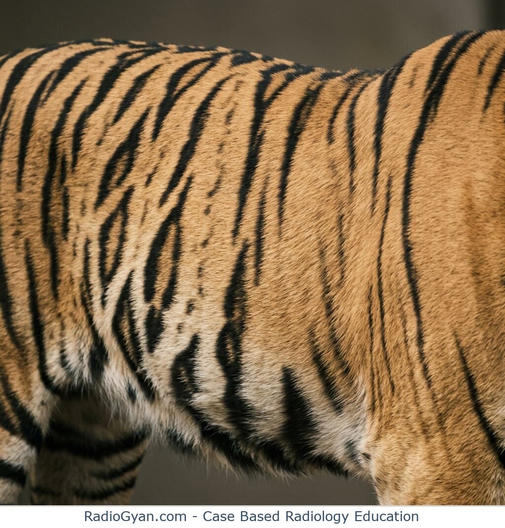
The name “Stripe sign” refers to the appearance on T2-weighted MRI images where dark linear hypointense stripes radiate from the periventricular ventricular margins through the hyperintense demyelinated white matter. These stripes correspond to spared perivascular white matter, creating a striped or “tigroid” pattern resembling the skin pattern of a leopard, also known as the “leopard skin sign.” This brain white-matter usage is a distinct entity from other unrelated “stripe sign” terms used elsewhere in radiology (e.g., orbital fat stripe, pleural stripe, psoas stripe signs).
Pathophysiology
Demyelination in MLD predominantly affects the periventricular white matter but sparing of the perivascular white matter occurs because these perivascular regions resist the sulfatide accumulation injurious process. The differential involvement leads to interspersed normal myelin around blood vessels appearing as dark stripes on T2-weighted MRI against the background of bright demyelinated white matter, producing the characteristic striped pattern. This reflects the patchy but structured nature of demyelination and myelin loss secondary to sulfatide accumulation and oligodendrocyte dysfunction caused by arylsulfatase deficiency.
Alternative names:
Tigroid pattern/sign (radiating stripes, typically on axial images); leopard skin sign (dot-like variant, typically on sagittal images)
Other associated named signs:
None specific, though the same tigroid/stripe pattern of dysmyelination is also described in Krabbe disease and Pelizaeus-Merzbacher disease.
References
Access all radiology signs posted so far: https://radiogyan.com/radiology-signs/

Improve Content And Help Your Colleagues!
Found an error in the post? Please let us know using our contact page and we will update it with due credits!
If you wish to contribute radiological images for this case (or any other article on the website), you can submit them here and you will be duly credited: Submit radiology case