


What causes the snowstorm appearance on ultrasound?
Let me know in the comments.
Click to reveal the answer
- Hydatidiform mole (uterus)
- Breast: free silicone from implant rupture
- Thyroid: diffuse sclerosing variant of PTC
- Crystal arthropathy (gout and CPPD)
- Subcutaneous emphysema and soft-tissue gas
The snowstorm appearance is a sonographic pattern of innumerable tiny, densely packed bright echoes on a darker background, shimmering as the probe is moved, like snow falling in a storm. It is a pattern, not a diagnosis. What it means depends on the organ you are scanning.
On pelvic ultrasound, which is where most people meet the term, a snowstorm in the uterine cavity means a complete hydatidiform mole. In the breast it means free silicone from an extracapsular implant rupture. In the thyroid it points to the diffuse sclerosing variant of papillary carcinoma. In a joint it means crystal aggregates, and in soft tissue it usually means gas.
Snowstorm appearance at a glance
| At a glance | |
|---|---|
| What it is | Countless small hyperechoic foci filling a region on ultrasound, with a shimmering or moving quality |
| Physical basis | Innumerable sub-resolution acoustic interfaces acting as point scatterers |
| Commonest use | Complete hydatidiform mole on pelvic ultrasound |
| Second commonest | Free silicone from extracapsular breast implant rupture |
| Also called | Snowstorm sign, blizzard appearance, echogenic noise (breast); cluster of grapes appearance (uterus) |
| Specificity | High within a given organ. Silicone snowstorm in a node is 100 percent specific for silicone lymphadenopathy |
| Sensitivity | Low to moderate. Pre-evacuation ultrasound identifies only 44 percent of hydatidiform moles overall |
| Biggest pitfall | Expecting the classic blizzard in the first trimester, when a complete mole usually looks like a complex echogenic mass with small cysts |
| Not a real use | Chest radiographs. Innumerable micronodules are a miliary pattern, not a snowstorm |
Where the snowstorm appearance is seen
| Where | Cause | What you actually see | Confirm with |
|---|---|---|---|
| Uterus (pelvic US) | Complete hydatidiform mole | Echogenic intrauterine mass studded with innumerable small anechoic cysts, no fetus, often bilateral theca lutein cysts | Serum hCG, evacuation with histopathology |
| Breast (US) | Free silicone, extracapsular implant rupture or direct injection | Intensely echogenic field with dirty posterior shadowing that obscures deeper tissue | MRI without contrast, or targeted US of the implant |
| Axillary node (US) | Silicone lymphadenopathy | Echogenic snowstorm replacing the node hilum in a patient with implants | Correlate with implant integrity, avoid calling metastasis |
| Thyroid (US) | Diffuse sclerosing variant of papillary thyroid carcinoma | Diffuse microcalcifications scattered through a heterogeneous lobe or gland, usually with abnormal nodes | FNA, plus node mapping of both compartments |
| Joint or bursa (US) | Crystal aggregates in gout or CPPD | Mobile hyperechoic specks swirling within an effusion under probe pressure | Look for double contour sign, tophus, chondrocalcinosis; joint aspiration |
| Soft tissue (US) | Subcutaneous gas | Bright echoes with ring-down and comet-tail artifact, dirty shadowing, complete loss of deeper structures | Radiograph or CT; ultrasound cannot see past the gas |
Why is it called so?
In every one of these settings the ultrasound beam meets innumerable tiny, densely packed reflectors: hydropic chorionic villi, silicone droplets, psammomatous microcalcifications, crystal aggregates or gas microbubbles. Each one scatters sound back to the transducer. Because the individual scatterers are at or below the resolution of the beam, they are not resolved as discrete structures but blur into a field of countless small bright echoes, which shift and shimmer as the probe moves. That is the snowstorm.
Hydatidiform mole (uterus)
This is the classic obstetric usage and the reason most searches for this sign exist.
A complete hydatidiform mole fills the uterine cavity with a heterogeneous, echogenic mass containing innumerable small anechoic cystic spaces, which are grossly hydropic chorionic villi. There is no fetus and no normal amniotic sac. The uterus is often large for dates, and about a quarter to a half of patients have bilateral theca lutein cysts, multiseptate ovarian cysts driven by very high hCG.
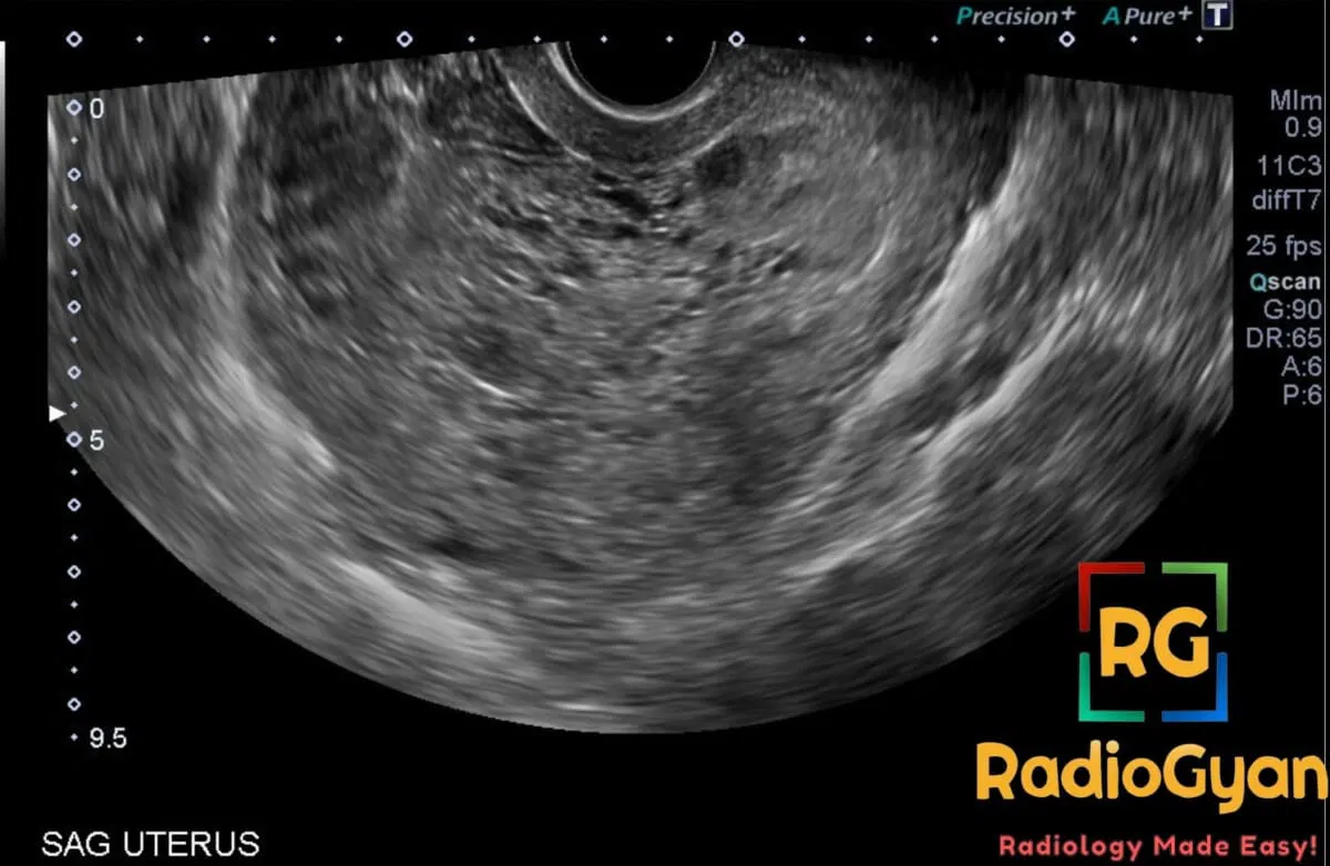
Complete versus partial mole on ultrasound
| Feature | Complete mole | Partial mole |
|---|---|---|
| Karyotype | Diploid, entirely paternal (46,XX in about 90 percent) | Triploid (69,XXX or 69,XXY) |
| Fetus | Absent | Present, growth restricted, often with anomalies |
| Placenta | Cavity filled by cystic vesicular tissue | Enlarged placenta with focal cystic spaces |
| Snowstorm | Typical in the second trimester | Rarely convincing |
| Serum hCG | Very high, often above 100,000 mIU/mL | Normal or only modestly raised |
| Theca lutein cysts | Common | Uncommon |
| Ultrasound detection | 79 percent in the largest series | 29 percent in the same series |
| Risk of post-molar GTN | Roughly 15 to 20 percent | Roughly 1 to 5 percent |
| p57 immunostain | Negative in villous cytotrophoblast and stroma | Positive |
How reliable is the sign? Not as reliable as its fame suggests
This is the single most useful thing to know about the snowstorm appearance, and it is the part most references leave out.
| Evidence | What it showed |
|---|---|
| Fowler 2006, 1053 consecutive cases referred to a regional trophoblastic disease centre | Pre-evacuation ultrasound suggested a mole in 44 percent overall: 79 percent of complete moles, 29 percent of partial moles. Sensitivity 44 percent, specificity 74 percent, PPV 88 percent, NPV 23 percent. Detection was 35 to 40 percent before 14 weeks and around 60 percent after. |
| Kirk 2007, 90 women scanned in an early pregnancy unit | Overall sensitivity 44 percent with a PPV of 48 percent. Complete moles: sensitivity 95 percent but PPV only 40 percent. Partial moles: sensitivity 20 percent, PPV 22 percent. |
| Benson 2000, 24 first-trimester complete moles, mean 8.7 weeks | Prospectively called a complete mole in 71 percent, and in 82 percent on blinded review. The typical first-trimester appearance was a complex echogenic intrauterine mass with many small cystic spaces, not a florid blizzard. |
Three practical conclusions follow. First, a normal-looking scan does not exclude a mole, and most missed moles look like a missed or incomplete miscarriage, which is exactly why histopathology of evacuated products of conception is mandatory. Second, the classic snowstorm is a late sign; at 8 to 9 weeks the villi are only 1 to 2 mm and the cavity contains a complex echogenic mass rather than a blizzard, so do not talk yourself out of the diagnosis because it is not florid. Third, a suspicious scan is far more useful for a complete than a partial mole, because ultrasound sees almost all complete moles but only one partial mole in five.
What else to look for on the same scan
- Theca lutein cysts — bilateral, multiseptate, often large ovarian cysts. Their presence supports the diagnosis and flags a higher risk of post-molar neoplasia.
- Myometrial invasion — molar tissue extending into the myometrium, with disorganised, low-resistance vascularity on colour Doppler, suggests an invasive mole rather than a simple complete mole.
- Uterine size for dates — large-for-dates is classic but far from universal now that moles present earlier.
- Clinical context — hyperemesis, early pre-eclampsia before 20 weeks, and clinical or biochemical hyperthyroidism are all driven by extreme hCG levels and should prompt a second look at the cavity.
Mimics of the uterine snowstorm
| Mimic | Overlap | Discriminator |
|---|---|---|
| Hydropic (non-molar) miscarriage | Villous hydrops without trophoblastic proliferation; accounted for 18 percent of referrals in the Fowler series | Lower hCG, no theca lutein cysts; separated only on histology with p57 |
| Retained products of conception | Echogenic endometrial material, often vascular | Preceding delivery or miscarriage, hCG falling not rising |
| Degenerating fibroid | Heterogeneous myometrial mass with cystic change | Arises from the myometrium, not the cavity; hCG normal |
| Incomplete or missed miscarriage | Heterogeneous cavity contents | Most commonly what a missed mole looks like, which is why histology is compulsory |
| Endometrial cystic change or hyperplasia | Small cysts within the endometrium | Non-pregnant patient, hCG negative |
What happens next
Suspected molar pregnancy goes to uterine evacuation with histopathological examination, and p57 immunostaining separates complete from partial mole. Serial hCG follow-up afterwards is what actually detects post-molar gestational trophoblastic neoplasia; the duration of monitoring varies with the histological type and the rate of hCG regression. Under the FIGO framework, low-risk gestational trophoblastic neoplasia (Stages I to III with a score under 7) is treated with single-agent chemotherapy with survival approaching 100 percent, while high-risk disease (score of 7 or more, or Stage IV) needs multiagent chemotherapy, with survival around 90 percent.
Breast: free silicone from implant rupture
Very common breast ultrasound usage, and the one most likely to appear in routine practice.
In the breast, a snowstorm appearance means free silicone: an intensely echogenic, noisy field with marked posterior attenuation and dirty shadowing that obscures everything deeper. It indicates that silicone has escaped outside the fibrous capsule, that is an extracapsular rupture, or that liquid silicone was injected directly into the breast, a practice still encountered in patients treated decades ago or abroad. The same pattern in a lymph node means silicone has migrated there.
Snowstorm versus stepladder versus linguine
| Sign | Modality | What it means | Appearance |
|---|---|---|---|
| Snowstorm (echogenic noise) | US | Extracapsular rupture or free injected silicone | Echogenic blizzard with dirty shadowing in the parenchyma or a node |
| Stepladder sign | US | Intracapsular rupture | Multiple parallel echogenic lines floating within the implant lumen |
| Linguine sign | MRI | Intracapsular rupture | Collapsed implant shell as wavy low-signal lines within the silicone |
| Keyhole / noose / teardrop sign | MRI | Early or incomplete intracapsular rupture | Silicone tracking into a fold of the shell |
| Water-droplet sign | MRI | Normal variant or early rupture | Small fluid foci within silicone, interpret with care |
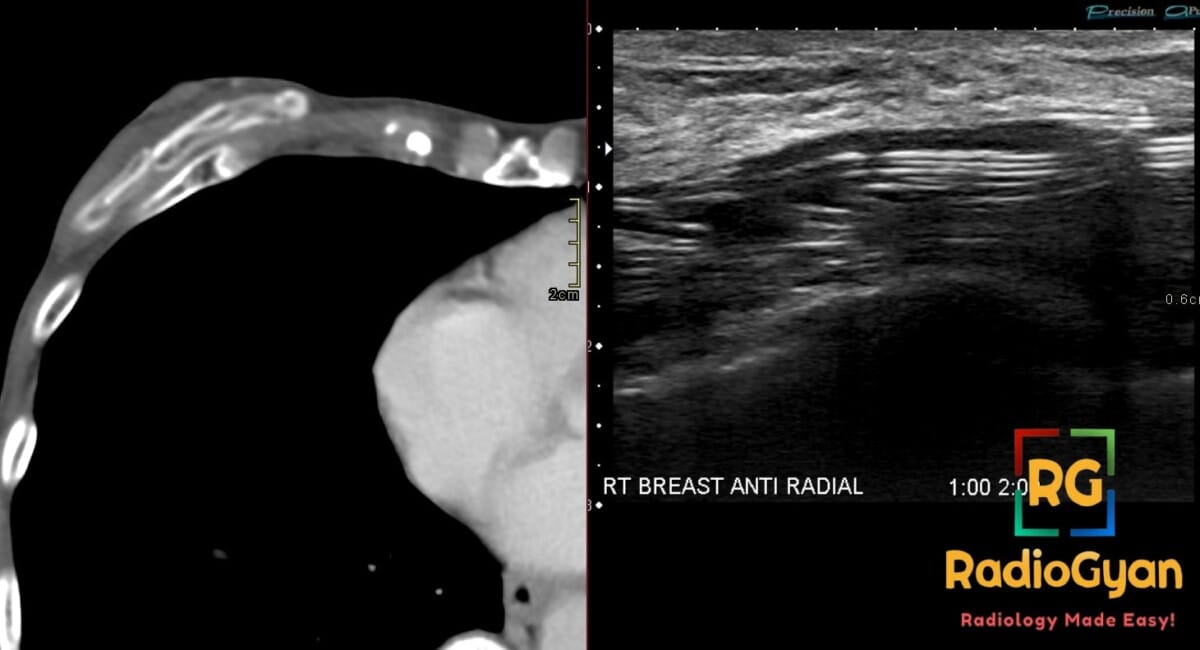
Silicone lymphadenopathy
An echogenic snowstorm node in the axilla of a woman with implants is silicone until proven otherwise, and it should not be reported as suspicious for metastasis. In a series of 41 biopsied intramammary and axillary nodes, the snowstorm sign was present in 7 of 8 pathologically proven silicone-containing nodes and in none of the 29 reactive or 4 malignant nodes: sensitivity 87.5 percent, specificity 100 percent. The MRI silicone signal in the same nodes was only 20 percent sensitive, so ultrasound outperforms MRI for this specific question.
When to image implants
The FDA recommends an initial ultrasound or MRI 5 to 6 years after silicone implant surgery, then every 2 to 3 years. The 2023 ACR Appropriateness Criteria update on breast implant evaluation reflects this, and the practically important change is the acceptance of ultrasound, not MRI alone, as a screening examination for silent rupture. Asymptomatic saline implants need no imaging, and saline rupture is usually clinically obvious. In a patient with unexplained axillary adenopathy and current or prior silicone implants, ultrasound with or without mammography is usually appropriate.
Thyroid: diffuse sclerosing variant of papillary carcinoma
In the thyroid, a snowstorm means innumerable microcalcifications scattered diffusely through a lobe or the whole gland rather than confined to a discrete nodule. The classic cause is the diffuse sclerosing variant of papillary thyroid carcinoma (DSVPTC), an aggressive variant that typically affects younger patients, is frequently associated with Hashimoto thyroiditis, and presents with nodal disease.
| Sonographic feature | Frequency | Source |
|---|---|---|
| Snowstorm pattern | Present in 6 of 10 cases (60 percent); the remaining 40 percent showed only a hypoechoic solid nodule | Li 2024 |
| Vague, infiltrative borders | 100 percent of DSVPTC versus 18.5 percent of conventional PTC | Li 2024 |
| Abundant microcalcifications | 66.7 percent versus 10.9 percent of conventional PTC | Li 2024 |
| Heterogeneous gland background | 80 percent | Li 2024 |
| Suspicious cervical nodes on US | 80 percent; every case had histologically proven nodal metastasis | Li 2024 |
| Lymph node metastasis overall | 95.9 percent of patients | Wang 2022 |
| Coexisting Hashimoto thyroiditis | 69.1 percent | Wang 2022 |
| Poor internal vascularity | 71.6 percent (Doppler grade 0 to 1) | Wang 2022 |
| US sensitivity for contralateral nodes | 50 percent central, 66.7 percent lateral | Wang 2022 |
Two reporting points matter here. First, ACR TI-RADS scores discrete nodules; a gland studded with diffuse microcalcifications and no dominant mass gives the system nothing to score, so a purely nodule-based read can under-call DSVPTC. Describe the diffuse pattern explicitly and recommend fine-needle aspiration regardless of whether a scoreable nodule exists. Second, scan both sides of the neck properly: ultrasound misses roughly a third to a half of contralateral nodal disease, so a negative contralateral compartment on ultrasound does not clear it.
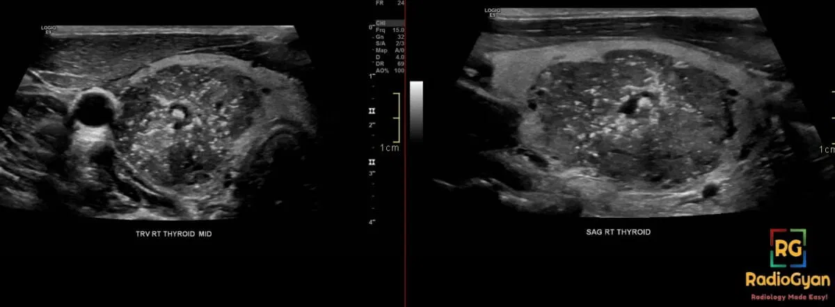
Similar diffuse microcalcification can occasionally be seen in extensively metastatic or chronically inflamed glands, so correlate with nodes, thyroid function and antibody status before committing.
Crystal arthropathy (gout and CPPD)
Within a joint or bursa, mobile hyperechoic specks suspended in an effusion, swirling when the probe presses or the joint moves, are informally called a snowstorm. They represent aggregated monosodium urate or calcium pyrophosphate crystals acting as point scatterers.
Worth knowing: snowstorm is not a defined term in the ultrasound literature on gout. The OMERACT international consensus defines four elementary lesions, and none of them is called a snowstorm:
| OMERACT lesion | Definition |
|---|---|
| Double contour sign | A hyperechoic band over the surface of the hyaline cartilage, independent of the angle of insonation |
| Tophus | A circumscribed, heterogeneous, hyperechoic mass, sometimes with an anechoic rim |
| Aggregates | Heterogeneous hyperechoic foci that retain their brightness at low gain or when the angle changes — this is the lesion a snowstorm actually describes |
| Bone erosion | An intra-articular discontinuity of the bone surface seen in two perpendicular planes |
So describe what you see as hyperechoic aggregates within the effusion and look deliberately for the double contour sign, tophi and erosions, which carry the diagnostic weight. In CPPD, look additionally for chondrocalcinosis: hyperechoic deposits within the substance of the cartilage rather than on its surface. Aspiration remains the reference standard.
Subcutaneous emphysema and soft-tissue gas
Gas in the soft tissues produces countless bright echoes with ring-down and comet-tail artifact and dirty shadowing. The key practical point is that this is an acoustic barrier, not a finding to characterise: gas reflects almost all of the incident beam, so everything deep to it is invisible. Subcutaneous emphysema will defeat an attempted ultrasound-guided vascular access, nerve block or FAST scan, and in that situation the answer is a radiograph or CT, not more gain.
Ultrasound is nonetheless extremely sensitive to small volumes of soft-tissue gas, which can be useful when the question is necrotising soft-tissue infection or surgical emphysema tracking from an airway injury. Report it, then image the source another way.
What about a snowstorm on a chest radiograph?
“Snowstorm” is sometimes used colloquially for the innumerable micronodules of a miliary distribution on chest imaging. This is not a formal radiological sign and it should not appear in a report. The established term is miliary pattern, defined by innumerable 1 to 3 mm nodules in a random distribution, and the differential is worth stating explicitly: miliary tuberculosis, miliary metastases from thyroid, renal cell or breast primaries, fungal infection, sarcoidosis and hypersensitivity pneumonitis. CT, not the radiograph, decides the distribution.
Pitfalls and reporting
| Pitfall | Why it happens | What to do |
|---|---|---|
| Calling a mole only when the blizzard is florid | The classic appearance is second-trimester. First-trimester complete moles look like a complex echogenic mass with small cysts | Suspect and refer for evacuation with histology even when the appearance is subtle |
| Excluding a mole because the scan looks like a miscarriage | Most missed moles are reported as missed or incomplete miscarriage | Histopathology of all products of conception, every time |
| Trusting the sign for a partial mole | Ultrasound detects only about a fifth to a third of partial moles | Do not reassure on the basis of a normal-looking scan |
| Calling a snowstorm axillary node metastatic | In an implant patient it is almost always silicone | Check implant integrity; the sign is 100 percent specific for silicone in this setting |
| Using TI-RADS alone in the thyroid | TI-RADS scores discrete nodules and has nothing to score in diffuse disease | Describe the diffuse microcalcification pattern and recommend FNA on its own merits |
| Pushing through subcutaneous gas | Gas is an absolute acoustic barrier | Stop and get a radiograph or CT |
| Writing snowstorm for a miliary chest pattern | Not an accepted term, and it loses the differential | Write miliary pattern |
Reporting checklist
- Name the location and the presumed scatterer, not just the pattern: “echogenic material with innumerable small cystic spaces filling the endometrial cavity” beats “snowstorm appearance”.
- State the most likely cause and the confidence, and say plainly that ultrasound cannot exclude the alternative.
- Record the supporting findings you looked for: theca lutein cysts and myometrial invasion in the uterus; implant shell integrity and nodes in the breast; nodal compartments in the thyroid; double contour and erosions in a joint.
- Give the next step: serum hCG and evacuation with histopathology; MRI or targeted implant US; FNA; aspiration; radiograph or CT.
- If deeper structures are obscured by silicone or gas, say so explicitly so the limitation is on record.
Related radiology signs
- Stepladder sign — intracapsular implant rupture, the counterpart of the breast snowstorm.
- Double contour sign and tophus — the OMERACT-defined lesions that carry the diagnostic weight in gout.
- Cluster of grapes appearance — the macroscopic counterpart of the uterine snowstorm.
- Browse the full radiology signs collection.
Frequently asked questions
What does a snowstorm appearance on ultrasound mean?
It means the beam is meeting innumerable tiny, densely packed reflectors, so the image fills with countless small bright echoes on a darker background. It is a pattern, not a diagnosis, and what it means depends entirely on where you see it: in the uterus it suggests a complete hydatidiform mole, in the breast it means free silicone from an extracapsular implant rupture, in the thyroid it suggests the diffuse sclerosing variant of papillary carcinoma, in a joint it suggests crystal aggregates, and in soft tissue it usually means gas.
Is the snowstorm appearance diagnostic of a molar pregnancy?
No. Routine pre-evacuation ultrasound identifies fewer than half of all hydatidiform moles. In the largest series, 1053 cases from a regional trophoblastic disease centre, ultrasound suggested a mole in 44 percent overall: 79 percent of complete moles but only 29 percent of partial moles. Most of the rest looked like a missed or incomplete miscarriage. Histopathology of the evacuated products of conception, not ultrasound, remains the diagnostic standard.
Why do first-trimester molar pregnancies not look like a snowstorm?
The classic vesicular snowstorm is a second-trimester appearance, produced by villi that have had time to become grossly hydropic. At 8 to 9 weeks the villi are only 1 to 2 mm, so a complete mole usually presents as a complex echogenic intrauterine mass containing many small cystic spaces rather than a true blizzard of echoes. Detection improves after 14 weeks, from roughly 35 to 40 percent before that point to around 60 percent after.
What is the difference between the snowstorm sign and the stepladder sign in breast implants?
They indicate opposite types of rupture. The snowstorm, or echogenic noise, is free silicone outside the fibrous capsule, meaning extracapsular rupture, and looks like an intensely echogenic field with dirty posterior shadowing. The stepladder sign is multiple parallel echogenic lines floating inside the implant lumen, which is the collapsed shell of an intracapsular rupture with the silicone still contained. The linguine sign is the MRI equivalent of the stepladder.
How often should silicone breast implants be imaged for rupture?
The US Food and Drug Administration recommends an initial ultrasound or MRI 5 to 6 years after silicone implant placement and then every 2 to 3 years, and the 2023 ACR Appropriateness Criteria update reflects this. The addition of ultrasound as an acceptable screening test, rather than MRI alone, is the most practical change for radiology departments. Asymptomatic saline implants need no imaging.
Does a snowstorm axillary lymph node mean cancer?
No, it usually means silicone. In a series of 41 biopsied nodes in women with silicone implants, the snowstorm sign was present in 7 of 8 pathologically proven silicone-containing nodes and in none of the reactive or malignant nodes, giving a sensitivity of 87.5 percent and a specificity of 100 percent. The MRI silicone signal was far less sensitive at 20 percent. So an echogenic snowstorm node in an implant patient argues for silicone lymphadenopathy, not metastasis.
Is a snowstorm appearance on a chest radiograph the same thing?
It is a loose, informal usage for innumerable micronodules, and the accepted term there is miliary pattern. Keep it out of your report. Write miliary pattern and give the differential, which includes miliary tuberculosis, miliary metastases from thyroid, renal or breast primaries, and hypersensitivity pneumonitis.
What should I do next when I see a snowstorm pattern in the uterus?
Correlate with serum hCG, look for theca lutein cysts and for myometrial invasion or abnormal vascularity, and refer for uterine evacuation with histopathology, including p57 immunostaining to separate complete from partial mole. Serial hCG follow-up afterwards is what detects post-molar gestational trophoblastic neoplasia, and its duration depends on the histological type and the rate of hCG regression.
References
- Fowler DJ, Lindsay I, Seckl MJ, Sebire NJ. Routine pre-evacuation ultrasound diagnosis of hydatidiform mole: experience of more than 1000 cases from a regional referral center. Ultrasound Obstet Gynecol. 2006;27(1):56-60. PMID: 16273594.
- Kirk E, Papageorghiou AT, Condous G, Bottomley C, Bourne T. The accuracy of first trimester ultrasound in the diagnosis of hydatidiform mole. Ultrasound Obstet Gynecol. 2007;29(1):70-75. PMID: 17201012.
- Benson CB, Genest DR, Bernstein MR, Soto-Wright V, Goldstein DP, Berkowitz RS. Sonographic appearance of first trimester complete hydatidiform moles. Ultrasound Obstet Gynecol. 2000;16(2):188-191. PMID: 11117091.
- Shaaban AM, Rezvani M, Haroun RR, et al. Gestational Trophoblastic Disease: Clinical and Imaging Features. RadioGraphics. 2017;37(2):681-700. PMID: 28287945.
- Ngan HYS, Seckl MJ, Berkowitz RS, et al. Diagnosis and management of gestational trophoblastic disease: 2021 update. Int J Gynaecol Obstet. 2021;155(Suppl 1):86-93. PMID: 34669197.
- Harris KM, Ganott MA, Shestak KC, Losken HW, Tobon H. Silicone implant rupture: detection with US. Radiology. 1993;187(3):761-768. PMID: 8497626.
- Seiler SJ, Sharma PB, Hayes JC, et al. Multimodality Imaging-based Evaluation of Single-Lumen Silicone Breast Implants for Rupture. RadioGraphics. 2017;37(2):366-382. PMID: 28186859.
- Expert Panel on Breast Imaging; Chetlen A, Niell BL, et al. ACR Appropriateness Criteria Breast Implant Evaluation: 2023 Update. J Am Coll Radiol. 2023;20(11S):S329-S350. PMID: 38040459.
- Le-Petross HT, Scoggins ME, Clemens MW. Assessment, Complications, and Surveillance of Breast Implants: Making Sense of 2022 FDA Breast Implant Guidance. J Breast Imaging. 2023;5(3):360-372. PMID: 38416893.
- Klang E, Yosepovich A, Krosser A, et al. Detection of Pathologically Proven Silicone Lymphadenopathy: Ultrasonography Versus Magnetic Resonance Imaging. J Ultrasound Med. 2018;37(4):969-975. PMID: 28960388.
- Li W, Wang Y, Gao L, et al. Sonographic characteristics of diffuse sclerosing variant of papillary thyroid carcinoma with histopathological correlation: a preliminary study. Orphanet J Rare Dis. 2024;19(1):136. PMID: 38532506.
- Wang Q, Chang Q, Zhang R, et al. Diffuse sclerosing variant of papillary thyroid carcinoma: ultrasonographic and clinicopathological features in children/adolescents and adults. Clin Radiol. 2022;77(5):e356-e362. PMID: 35197192.
- Lei R, Yang H. Thyroid papillary carcinoma with the ‘snowstorm appearance’: a clinicopathological analysis of three cases. J Int Med Res. 2022;50(7):3000605221099465. PMID: 35866423.
- Tessler FN, Middleton WD, Grant EG, et al. ACR Thyroid Imaging, Reporting and Data System (TI-RADS): White Paper of the ACR TI-RADS Committee. J Am Coll Radiol. 2017;14(5):587-595. PMID: 28372962.
- Gutierrez M, Schmidt WA, Thiele RG, et al. International Consensus for ultrasound lesions in gout: results of Delphi process and web-reliability exercise. Rheumatology (Oxford). 2015;54(10):1797-1805. PMID: 25972391.
- Verniquet A, Kakel R. Subcutaneous emphysema: ultrasound barrier. Can J Anaesth. 2011;58(3):336-337. PMID: 21132473.
Access all radiology signs posted so far: https://radiogyan.com/radiology-signs/

Improve Content And Help Your Colleagues!
Found an error in the post? Please let us know using our contact page and we will update it with due credits!
If you wish to contribute radiological images for this case (or any other article on the website), you can submit them here and you will be duly credited: Submit radiology case