


What causes the Terry-Thomas sign on wrist radiographs
Let me know in the comments.
Click to reveal the answer
The Terry-Thomas sign is a widened gap between the scaphoid and lunate on a posteroanterior (PA) wrist radiograph, caused by rupture of the scapholunate interosseous ligament. No single measurement proves it: normal wrists reach 5.0 mm, so the finding is only meaningful against the opposite wrist.
Why is it called so?
It is named after the British comedian Terry-Thomas, who had a conspicuous gap between his two upper front teeth. The widened scapholunate interval resembles that dental diastema. The eponym enters the literature in a 1977 note by the orthopaedic surgeon Victor H. Frankel [1].
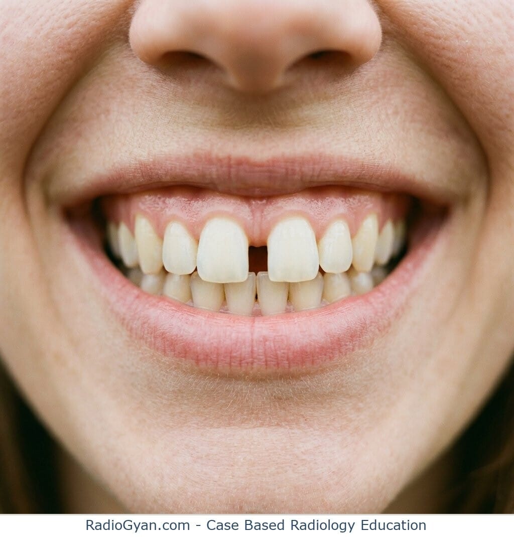
Pathophysiology
The scapholunate interosseous ligament ties the proximal scaphoid to the lunate and resists the scaphoid’s tendency to flex while the lunate extends. When it tears, usually after a fall onto the outstretched hand, the scaphoid rotates into flexion and the lunate into extension. The two bones separate, which widens the interval on the PA view, and the lunate tilts dorsally, which is the dorsal intercalated segment instability (DISI) pattern seen on the lateral view.
Alternative names: David Letterman sign; Lauren Hutton sign (informal later celebrity comparisons for the same widened gap, neither indexed in the medical literature).
Other associated named signs: Scaphoid cortical ring sign (the foreshortened, flexed scaphoid seen end-on), and in the wider spectrum of carpal instability the piece-of-pie sign and crowded carpal sign of lunate and perilunate dislocation.
How wide is too wide? The scapholunate gap on X-ray
There is no threshold that separates a torn ligament from an intact one. The widely quoted “under 2 mm is normal” comes from mid-joint measurement on MRI and on cadaver specimens, not from clinical radiographs [2]. On actual PA films of normal wrists the gap is substantially wider.
| Series | What was measured | Result |
|---|---|---|
| Schimmerl-Metz, MRI, 40 normal wrists [2] | Mid-joint interval | 1.45 mm coronal, 1.00 mm axial; stated as under 2 mm |
| Cautilli, PA radiographs, 100 normal wrists [3] | Scapholunate distance | Mean 3.7 mm, range 2.5 to 5.0 mm |
| Megerle, 849 wrists against arthroscopy [4] | Best discriminating cut-off | 2.5 mm |
| Picha, 124 patients [5] | Gap of 5 mm or more | 81% of symptomatic and 52% of asymptomatic wrists |
Read those last two rows together. The cut-off that best predicts an arthroscopic tear is 2.5 mm [4], yet half of asymptomatic wrists exceed twice that [5]. Picha also found the abnormality was bilateral in 80% of patients, while only 11% had a clinical instability pattern [5]. A wide scapholunate interval is often simply that person’s carpal morphology.
This is why the contralateral PA view matters more than any absolute number. Measure both wrists at the mid-joint, in the same position, and report the side-to-side difference.
The scapholunate angle and the cortical ring sign
The two supporting signs carry the same problem: the published thresholds overlap heavily with normal wrists.
| Sign | Normal | Threshold in use | The catch |
|---|---|---|---|
| Scapholunate gap, PA view | Up to 5.0 mm [3] | 2.5 mm optimal against arthroscopy [4] | 52% of asymptomatic wrists exceed 5 mm [5] |
| Scapholunate angle, lateral view | 36 to 66 degrees, mean 50.8 [6] | 60 degrees [5], or 62.5 degrees as the optimum [4] | 70% of asymptomatic wrists exceed 60 degrees [5] |
| Scaphoid cortical ring sign | Present in 25% of normal wrists [7] | Pathological if present at 13 degrees or more of ulnar deviation [7] | Depends on wrist positioning, not on the ligament |
Two practical consequences. First, grade the lateral view by the angle but do not call DISI on a number alone; Megerle’s 62.5 degree cut-off reached a specificity of 0.93 but the normal range runs to 66 degrees [4][6]. Second, a cortical ring sign on a radially deviated film means little, because Pirela-Cruz found it in a quarter of normal wrists and showed it appears and disappears with wrist position [7].
Does a normal wrist X-ray rule out a scapholunate tear?
No, and the stress view does not rescue it. Validated against arthroscopy in 88 patients, the clenched-fist view had a sensitivity of only 40% (specificity 82%), and 42% of patients with a negative clenched-fist view had a Geissler grade 3 or higher tear [8]. A normal radiograph, static or stressed, cannot exclude the injury.
If clinical suspicion persists, the imaging decision is about field strength and contrast, not about more plain films.
| Technique | Sensitivity | Specificity |
|---|---|---|
| 1.5 T MRI | 45.7% | 80.5% |
| 3.0 T MRI | 75.7% | 97.1% |
| MR arthrography | 82.1% | 92.8% |
Pooled from 24 studies and 1,902 examinations against surgical reference [9]. A negative 1.5 T study is close to uninformative; 3 T or MR arthrography is the study worth requesting.
What untreated scapholunate dissociation becomes
Scapholunate advanced collapse (SLAC) is the degenerative endpoint. Reviewing 4,000 wrist films, Watson and Ballet found 210 cases of degenerative arthritis once other arthritides were excluded, and the scaphoid-lunate-radius pattern accounted for 57% of them, making it the commonest arthritic pattern of the wrist [10]. Scaphotrapeziotrapezoid disease accounted for 27% and a combination for 15% [10].
How to report it
- Give the scapholunate distance in millimetres, measured at the mid-joint on the PA view, rather than the word “widened”.
- Measure the contralateral wrist in the same projection and report the side-to-side difference. This is the most useful number on the study.
- Assess the lateral view for a scapholunate angle above 60 degrees and for dorsal lunate tilt.
- State explicitly that a normal radiograph does not exclude the injury, and recommend 3 T MRI or MR arthrography if suspicion persists.
- In chronic presentations, look for the scaphoid-lunate-radius degenerative pattern of SLAC wrist.
What is the Terry-Thomas sign?
A widened gap between the scaphoid and lunate on a posteroanterior wrist radiograph, indicating rupture of the scapholunate interosseous ligament and scapholunate dissociation. It is named for a British comedian’s gap-toothed grin and is usually the result of a fall onto the outstretched hand.
How many millimetres is a widened scapholunate gap?
There is no reliable cut-off. The value that best discriminates an arthroscopic tear is 2.5 mm [4], but normal wrists measure a mean of 3.7 mm with a range up to 5.0 mm on PA films [3], and 52% of asymptomatic wrists exceed 5 mm [5]. Compare with the other wrist instead of applying a threshold.
Why is it called the Terry-Thomas sign, and what is the David Letterman sign?
Both names refer to the same finding and to the same resemblance, a conspicuous gap between the upper front teeth. Terry-Thomas was a British comedian and the eponym appears in a 1977 note by Frankel [1]. David Letterman sign and Lauren Hutton sign are informal later substitutions of a more familiar gap-toothed celebrity; neither appears in the indexed medical literature.
Can the Terry-Thomas sign be a normal finding?
Yes. A gap of 5 mm or more was present in 52% of asymptomatic wrists in one series of 124 patients, the finding was bilateral in 80%, and only 11% had a clinical instability pattern [5]. A wide scapholunate interval on its own, without trauma, tenderness or a side-to-side difference, is frequently normal carpal morphology.
References
- Frankel VH. The Terry-Thomas sign. Clin Orthop Relat Res. 1977;(129):321-2.
- Schimmerl-Metz SM, Metz VM, Totterman SM, Mann FA, Gilula LA. Radiologic measurement of the scapholunate joint: implications of biologic variation in scapholunate joint morphology. J Hand Surg Am. 1999;24(6):1237-44.
- Cautilli GP, Wehbe MA. Scapho-lunate distance and cortical ring sign. J Hand Surg Am. 1991;16(3):501-3.
- Megerle K, Pohlmann S, Kloeters O, Germann G, Sauerbier M. The significance of conventional radiographic parameters in the diagnosis of scapholunate ligament lesions. Eur Radiol. 2011;21(1):176-81.
- Picha BM, Konstantakos EK, Gordon DA. Incidence of bilateral scapholunate dissociation in symptomatic and asymptomatic wrists. J Hand Surg Am. 2012;37(6):1130-5.
- Larsen CF, Mathiesen FK, Lindequist S. Measurements of carpal bone angles on lateral wrist radiographs. J Hand Surg Am. 1991;16(5):888-93.
- Pirela-Cruz MA, Hilton ME, Faillace J. Frequency and characteristics of the scaphoid cortical ring sign. Surg Radiol Anat. 2003;25(5-6):451-4.
- Oh C, Fort MW, Kakar S. Validation of the clenched fist view in detecting scapholunate ligamentous injury. Hand (N Y). 2025;20(4):607-11.
- Hafezi-Nejad N, Carrino JA, Eng J, Blackmore C, Shores J, Lifchez SD, et al. Scapholunate interosseous ligament tears: diagnostic performance of 1.5 T, 3 T MRI, and MR arthrography: a systematic review and meta-analysis. Acad Radiol. 2016;23(9):1091-103.
- Watson HK, Ballet FL. The SLAC wrist: scapholunate advanced collapse pattern of degenerative arthritis. J Hand Surg Am. 1984;9(3):358-65.
Access all radiology signs posted so far: https://radiogyan.com/radiology-signs/

Improve Content And Help Your Colleagues!
Found an error in the post? Please let us know using our contact page and we will update it with due credits!
If you wish to contribute radiological images for this case (or any other article on the website), you can submit them here and you will be duly credited: Submit radiology case