


A brain vascular territory is the volume of brain parenchyma supplied by a single artery. Assigning an infarct to a territory is the first thing a radiologist does with a positive diffusion-weighted study, because the pattern, not the size, points to the mechanism: one territory means an occluded artery, several territories mean a proximal or cardiac embolic source, and a lesion that ignores territorial boundaries altogether usually is not arterial ischaemia at all.
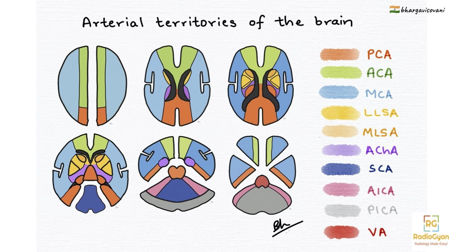
The map below covers the three cerebral arteries, the deep perforators that supply the basal ganglia, internal capsule and thalamus, the three cerebellar arteries and the brainstem, followed by the watershed zones between them and the patterns that mimic a territory.
| At a glance | |
|---|---|
| Anterior circulation | Internal carotid artery to ACA and MCA plus their perforators |
| Posterior circulation | Vertebral arteries to basilar artery to PCA, plus PICA, AICA and SCA |
| Cortical territories | ACA medial, MCA lateral, PCA posterior and inferomedial |
| Deep territories | Lenticulostriate, recurrent artery of Heubner, anterior choroidal, thalamoperforators |
| Watershed zones | Cortical ACA-MCA and MCA-PCA borders; internal borderzone in corona radiata |
| Commonest infarct | MCA territory, roughly two-thirds of all supratentorial infarcts |
| Key variant | Fetal PCA, in which the PCA is fed by the internal carotid rather than the basilar |
| Why it matters | Territorial pattern determines mechanism, workup and thrombectomy decision |
Cerebral artery territories: ACA, MCA and PCA
The three cerebral arteries divide the hemisphere into a medial strip, a large lateral convexity and a posterior-inferomedial block. Territorial boundaries are individual rather than fixed. The anatomical work behind the standard maps is Tatu and colleagues, who sectioned brains and correlated the supply territories slice by slice; their two Neurology papers remain the reference for hemispheric and for brainstem-cerebellar territories, and the 2023 Digital 3D Brain MRI Arterial Territories Atlas has since translated that work into a template that can be overlaid on a patient study.
| Artery | Segments | Territory supplied | Classic clinical deficit |
|---|---|---|---|
| Anterior cerebral artery (ACA) | A1 (precommunicating), A2-A5 (postcommunicating, pericallosal and callosomarginal) | Medial frontal and medial parietal cortex, paracentral lobule, anterior four-fifths of the corpus callosum, medial orbitofrontal cortex | Contralateral leg-predominant weakness, abulia, transcortical motor aphasia, urinary incontinence |
| Middle cerebral artery (MCA) | M1 (horizontal), M2 (insular), M3 (opercular), M4 (cortical) | Lateral frontal, parietal and superior temporal cortex, insula, most of the centrum semiovale | Contralateral face and arm-predominant weakness, hemianopia, gaze deviation to the side of the lesion, aphasia (dominant) or neglect (non-dominant) |
| Posterior cerebral artery (PCA) | P1 (precommunicating), P2 (ambient), P3 (quadrigeminal), P4 (calcarine) | Occipital lobe and calcarine cortex, inferomedial temporal lobe, splenium, midbrain and thalamus via perforators | Contralateral homonymous hemianopia with macular sparing, alexia without agraphia (dominant), memory impairment with hippocampal involvement |
Two practical points. First, the MCA territory is the one most often infarcted, and the insular ribbon and lentiform nucleus are where the earliest CT changes appear, before any of the convexity looks abnormal; see the insular ribbon sign and the dense MCA sign. Second, the PCA is posterior circulation by origin only in most people: in a substantial minority of hemispheres the P1 segment is hypoplastic or absent and the PCA is supplied by the internal carotid through the posterior communicating artery. In this fetal-type configuration an occipital infarct has a carotid source, and the carotid, not the vertebrobasilar system, is what needs imaging.
Deep perforating artery territories
The perforators are small, end-arterial and unforgiving. They produce small infarcts with deficits far out of proportion to lesion volume, and each one has a signature location.
| Perforating artery | Parent vessel | Territory supplied | Infarct signature |
|---|---|---|---|
| Lateral lenticulostriate arteries | M1 segment of MCA | Putamen, globus pallidus, body of caudate, superior part of the internal capsule, adjacent corona radiata | Pure motor hemiparesis; a striatocapsular infarct implies M1 occlusion, not small-vessel disease |
| Medial lenticulostriate arteries | A1 segment of ACA | Anteroinferior internal capsule, part of the globus pallidus | Small deep infarcts, often clinically silent |
| Recurrent artery of Heubner | A2 segment of ACA (near the anterior communicating artery) | Head of the caudate, anterior limb of the internal capsule, anterior putamen | Caudate infarct with abulia, dysarthria and faciobrachial weakness |
| Anterior choroidal artery (AChA) | Supraclinoid internal carotid artery | Posterior limb of the internal capsule, optic tract, medial temporal lobe including uncus and hippocampus, choroid plexus, part of the globus pallidus | Classic triad of contralateral hemiparesis, hemisensory loss and homonymous hemianopia from one small lesion |
| Tuberothalamic (polar) artery | Posterior communicating artery | Anterior thalamus | Absent in about a third of people, in which case the paramedian artery takes over its territory |
| Paramedian thalamoperforators | P1 segment of PCA | Medial thalamus and rostral midbrain | Impaired consciousness, vertical gaze palsy, memory loss |
| Artery of Percheron | Single dominant perforator arising from one P1 segment | Both paramedian thalami, with or without the rostral midbrain | Symmetrical bilateral thalamic infarct that looks like a metabolic or venous process but is a single arterial territory |
| Thalamogeniculate arteries | P2 segment of PCA | Inferolateral thalamus including the ventral posterior nucleus | Pure sensory stroke, later Dejerine-Roussy thalamic pain syndrome |
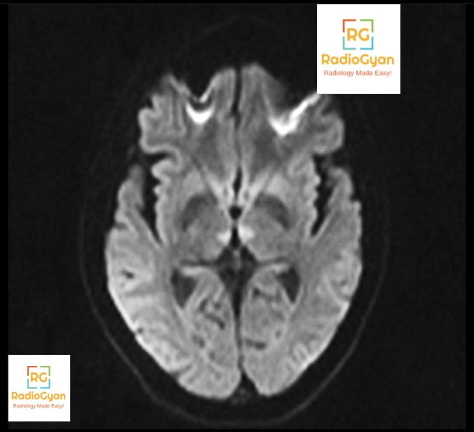
The thalamus is worth learning as four territories rather than one, because the clinical syndrome follows the territory closely, a point set out in the Stroke review by Schmahmann. The artery of Percheron is the variant that catches people out: a bilateral symmetrical thalamic lesion is far more often read as deep venous thrombosis, Wernicke encephalopathy or a toxic-metabolic insult than as one occluded perforator. Check the internal cerebral veins and the straight sinus before you commit.
Brainstem arterial territories
The brainstem follows one rule at every level. Paramedian perforators, arising directly off the basilar or vertebral artery, supply the medial structures; short and long circumferential branches supply the lateral structures. A medial infarct therefore takes out the corticospinal tract, medial lemniscus and the exiting motor cranial nerve, while a lateral infarct takes out the spinothalamic tract, sympathetic fibres, the vestibular nuclei and the sensory cranial nerves.
| Level | Medial (paramedian) supply | Lateral (circumferential) supply | Named syndrome |
|---|---|---|---|
| Medulla | Anterior spinal artery and vertebral perforators | Vertebral artery, with a variable contribution from PICA | Medial medullary (Dejerine) syndrome; lateral medullary (Wallenberg) syndrome |
| Pons | Basilar paramedian perforators | Short and long circumferential branches of the basilar artery, AICA | Medial pontine (Foville, Millard-Gubler) syndromes; lateral pontine syndrome |
| Midbrain | Basilar and P1 perforators, including the artery of Percheron | Collicular and posterior choroidal branches, SCA | Weber, Claude and Benedikt syndromes |
One correction worth making explicitly, because the older tables get it wrong: lateral medullary (Wallenberg) syndrome is most often caused by occlusion of the intracranial vertebral artery, not of PICA itself. PICA gets the eponym; the vertebral artery usually gets the clot.
Cerebellar vascular territories: PICA, AICA and SCA
Three paired arteries divide the cerebellum into horizontal slabs rather than the wedges the textbook diagrams suggest. The boundaries are among the most variable in the brain, and PICA and AICA are reciprocal: where one is dominant the other is small.
| Artery | Origin | Cerebellar territory | Additional structures | Typical presentation |
|---|---|---|---|---|
| Posterior inferior cerebellar artery (PICA) | Intracranial vertebral artery | Inferior surface of the cerebellar hemisphere, inferior vermis, cerebellar tonsil | Lateral medulla via its medial branch | Vertigo, ipsilateral ataxia, and with a large infarct, fourth ventricular compression and hydrocephalus |
| Anterior inferior cerebellar artery (AICA) | Lower basilar artery | Flocculus, middle cerebellar peduncle, anterolateral part of the inferior cerebellar surface | Lateral pons; the labyrinthine artery arises from AICA in most people | Acute vertigo with sudden ipsilateral hearing loss, the combination that separates AICA from PICA |
| Superior cerebellar artery (SCA) | Distal basilar artery, just below the PCA origin | Superior surface of the cerebellar hemisphere, superior vermis, dentate nucleus | Superior cerebellar peduncle, lateral upper pons | Ipsilateral limb ataxia and dysarthria; often the cleanest wedge on CT |
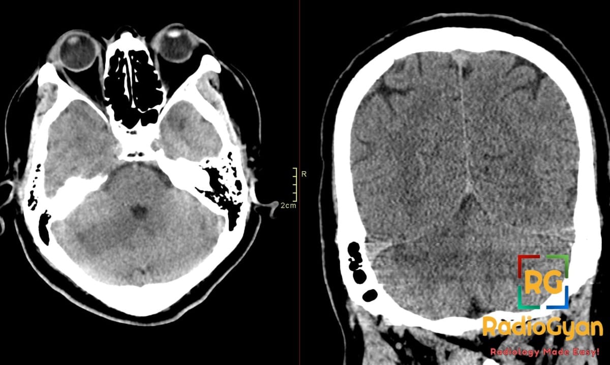
In the New England Medical Center Posterior Circulation Registry the majority of cerebellar infarcts fell in the PICA and SCA territories, with isolated AICA infarction distinctly uncommon. Two practical habits follow. Any cerebellar infarct that crosses the horizontal boundary between the SCA and PICA territories should raise the question of a proximal vertebrobasilar occlusion rather than a distal branch event, and a large cerebellar infarct needs a stated comment on fourth ventricular effacement and tonsillar herniation, since posterior fossa swelling, not the infarct itself, is what kills these patients.
Watershed (borderzone) territories
Borderzones are the strips of brain sitting at the far end of two arterial trees, perfused last and at the lowest pressure. They come in two forms with different mechanisms, set out in the RadioGraphics review by Mangla and colleagues.
| Borderzone | Location | Imaging appearance | Usual mechanism |
|---|---|---|---|
| Anterior cortical (ACA-MCA) | Parasagittal frontal cortex, superior frontal sulcus region | Wedge of cortical and subcortical infarction pointing towards the ventricle | Haemodynamic failure with severe carotid stenosis or occlusion; also microembolic |
| Posterior cortical (MCA-PCA) | Parieto-occipital junction | Wedge or band of infarction at the posterior convexity | Global hypotension, cardiac arrest, carotid disease |
| Internal (subcortical) | Corona radiata and centrum semiovale, between deep and superficial MCA perforators | Chain of small infarcts in a line parallel to the lateral ventricle, the rosary bead or string of pearls pattern | Strongly associated with haemodynamic compromise from proximal large-artery stenosis |
| Cerebellar borderzone | Between SCA, AICA and PICA territories | Small scattered infarcts near the territory boundaries | Hypoperfusion or embolic showers in vertebrobasilar disease |
The reason this classification earns its place in a report is that it changes the workup. A borderzone pattern, particularly the internal rosary-bead pattern, is a prompt to interrogate the cervical and intracranial vessels for a flow-limiting stenosis and to review blood pressure management, whereas a single cortical territorial infarct sends the team looking for an embolic source.
Territorial versus non-territorial patterns
Before naming a territory, decide whether the lesion respects one at all. This single question separates arterial ischaemia from the processes that imitate it.
| Pattern | What it looks like | What it means |
|---|---|---|
| Single territory, wedge-shaped, cortex plus subcortical white matter | Sharp boundary along a known arterial territory, grey and white matter involved together | Occlusion of that artery, embolic or thrombotic |
| Multiple territories, both hemispheres or both circulations | Scattered lesions of differing ages in ACA, MCA and PCA territories | Cardioembolic source, aortic arch atheroma, vasculitis or a hypercoagulable state |
| Borderzone chain or wedge | Rosary-bead lesions in the corona radiata, or parasagittal and parieto-occipital wedges | Haemodynamic failure from proximal stenosis or a systemic hypotensive event |
| Deep, small, single perforator lesion under 15 mm | Lacune in the basal ganglia, internal capsule, thalamus or pons | Small-vessel disease, unless the lesion is striatocapsular and large, which implies M1 occlusion |
| Crosses arterial boundaries, involves subcortical white matter with a cortical spared rim | Lesion centred on white matter, often haemorrhagic, not conforming to any artery | Venous infarction; look for cortical vein or dural sinus thrombosis |
| Symmetrical, bilateral, non-arterial distribution | Basal ganglia, thalami, cortical ribbon or watershed grey matter symmetrically involved | Hypoxic-ischaemic injury, toxic or metabolic insult, or the artery of Percheron variant |
| Posterior parieto-occipital, mainly vasogenic oedema, largely reversible | Symmetrical subcortical FLAIR hyperintensity without restricted diffusion | Posterior reversible encephalopathy syndrome, not PCA infarction |
The symmetrical bilateral pattern is the one to slow down on. Global hypoxic-ischaemic injury, deep venous thrombosis and an artery of Percheron infarct can look alike on a single DWI sequence and are separated by the sequences around it: check ADC, check susceptibility imaging for venous thrombus and check the deep venous system. See our case of hypoxic-ischaemic encephalopathy for the global-hypoxia pattern.
How to assign a vascular territory on CT or MRI
- Confirm the lesion is ischaemic: restricted diffusion with a low ADC, or established hypodensity with loss of grey-white differentiation on CT.
- Ask whether cortex is involved. Cortex plus underlying white matter indicates a cortical branch territory; pure deep involvement indicates a perforator.
- Match the cortical lesion to a territory: medial surface for ACA, lateral convexity and insula for MCA, occipital and inferomedial temporal for PCA.
- Check the deep structures separately. Caudate head points to Heubner, putamen and superior internal capsule to the lenticulostriates, posterior limb with the optic tract to the anterior choroidal artery.
- In the posterior fossa, decide the horizontal slab first: superior surface for SCA, inferior surface for PICA, anterolateral with the middle cerebellar peduncle for AICA.
- Count the territories. More than one territory, or bilateral involvement, moves the search proximally to the heart, arch or a vasculopathy.
- Look at the borderzones before finishing. A missed internal watershed chain changes the diagnosis from embolic stroke to haemodynamic failure.
- Correlate with the angiographic study. A territory without a matching occlusion should prompt a second look at variants, particularly a fetal PCA or a dominant vertebral artery.
The MCA territory and ASPECTS
Because the MCA territory carries most acute stroke work, it has its own scoring system. The Alberta Stroke Program Early CT Score divides the MCA territory into 10 regions on two axial CT levels: caudate, lentiform nucleus, internal capsule, insular ribbon and the M1 to M3 cortical regions at ganglionic level, plus M4 to M6 at the supraganglionic level. One point is subtracted for each region showing early ischaemic change, so a normal scan scores 10.
ASPECTS is a territory map used as a triage tool, and the thresholds have moved. Large-core thrombectomy trials published in 2023 showed benefit in patients who would previously have been excluded on a low score, and the 2026 American Heart Association and American Stroke Association acute ischaemic stroke guideline, which replaces the 2018 guideline and its 2019 update, broadened endovascular thrombectomy eligibility, expanded tenecteplase use within 4.5 hours and streamlined the imaging pathway. Score the regions and report them; do not use the number alone to declare a patient ineligible. Our ASPECTS calculator scores the 10 regions for you.
Quiz case
Look at the non-contrast CT below. Is there an acute infarct, and which vascular territory is involved? Answer in the comments.
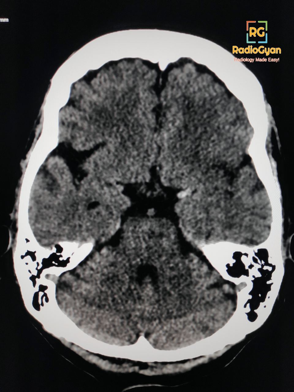
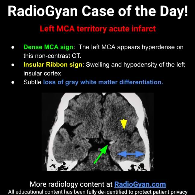
Read more: dense MCA sign.
Frequently asked questions
References
- Tatu L, Moulin T, Bogousslavsky J, Duvernoy H. Arterial territories of the human brain: cerebral hemispheres. Neurology. 1998;50(6):1699-1708. PMID: 9633714.
- Tatu L, Moulin T, Bogousslavsky J, Duvernoy H. Arterial territories of human brain: brainstem and cerebellum. Neurology. 1996;47(5):1125-1135. PMID: 8909417.
- Liu CF, Hsu J, Xu X, et al. Digital 3D brain MRI arterial territories atlas. Sci Data. 2023;10(1):74. PMID: 36739282.
- Mangla R, Kolar B, Almast J, Ekholm SE. Border zone infarcts: pathophysiologic and imaging characteristics. RadioGraphics. 2011;31(5):1201-1214. PMID: 21918038.
- Schmahmann JD. Vascular syndromes of the thalamus. Stroke. 2003;34(9):2264-2278. PMID: 12933968.
- Chaves CJ, Caplan LR, Chung CS, et al. Cerebellar infarcts in the New England Medical Center Posterior Circulation Stroke Registry. Neurology. 1994;44(8):1385-1390. PMID: 8058134.
- Jantre M, Howlett DC. Imaging the artery of Percheron: a pictorial review of associated pathology with important mimics of bithalamic disease. Neuroradiology. 2025;67(4):785-798. PMID: 40063261.
- Barber PA, Demchuk AM, Zhang J, Buchan AM. Validity and reliability of a quantitative computed tomography score in predicting outcome of hyperacute stroke before thrombolytic therapy (ASPECTS). Lancet. 2000;355(9216):1670-1674. PMID: 10905241.
- Prabhakaran S, Gonzalez NR, Zachrison KS, et al. 2026 guideline for the early management of patients with acute ischemic stroke: a guideline from the American Heart Association/American Stroke Association. Stroke. 2026;57(8):e316-e436. PMID: 41582814.
Related reading on RadioGyan: insular ribbon sign, dense MCA sign, persistent trigeminal artery and radiological anatomy.
Illustration by #TeamGyan member Dr. Bhargavi Sovani. Reviewed, expanded and updated 2026.
Check out more radiological illustrations:
- Cirrhosis and Portal Hypertension: Radiology and Imaging
- Rickets Radiology: X-Ray Findings, Cupping, Fraying, Splaying
- Carpal Boss
- Dorsal Triquetral Avulsion Fracture
- Torus Fracture (Buckle Fracture): X-ray Findings, Diagnosis and Treatment

Improve Content And Help Your Colleagues!
Found an error in the post? Please let us know using our contact page and we will update it with due credits!
If you wish to contribute radiological images for this case (or any other article on the website), you can submit them here and you will be duly credited: Submit radiology case