
Quiz
What is the most common mechanism of this fracture ?
- Blunt trauma to the wrist
- Fall from height
- Fall on the outstretched hand
- Penetrating trauma to the wrist
Pathophysiology
- Triquetrum is the second most commonly fractured carpal bone followed by scaphoid.
- Dorsal cortical fracture being the most common type of triquetrum fracture.
- Fall on the outstretched hand with wrist in extension is the most common cause of fracture
- Various mechanism involved in causing the fracture include impaction by ulnar styloid or hamate, avulsion by the dorsal radiotriquetral or dorsal scaphotriquetral ligaments.
Key Imaging Features
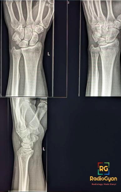
- Radiography– Dorsal cortical fractures are best evaluated on the 45-degree pronated oblique and lateral views- avulsed bony fragment seen posterior to the triquetral bone
- CT- To rule out occult triquetral fractures with high clinical suspicion.
- MRI – In suspected carpal instability to look for extrinsic carpal ligament injury and bone marrow edema in occult fracture.
Imaging Recommendation:
Radiograph with AP and lateral view or CT \ MRI for suspected occult fracture
Top Differential Diagnosis:
- Os triangulare- accessory ossicle between ulnar styloid, lunate and triquetrum
- Pisiform fracture- pisiform fractures are uncommon and 30 degrees supination view is required to visualise a pisotriquetral joint.
Clinical Features:
- Symptoms- ulnar-sided wrist pain worsening on wrist flexion and extension, dorsal wrist swelling and tenderness over the dorsal aspect of the triquetrum
Classification System:
Classification system developed by Garcia-Elias to differentiate the patterns of dorsal cortical fractures
Type 1 -nondisplaced- most common type
Type 2 -partially displaced at the proximal end
Type 3 – partially displaced at the distal end
Type 4 -completely displaced fragment
Type 5 -multiple completely displaced fragments
Type 6- fracture line through the coronal plane in which the dorsal side is displaced
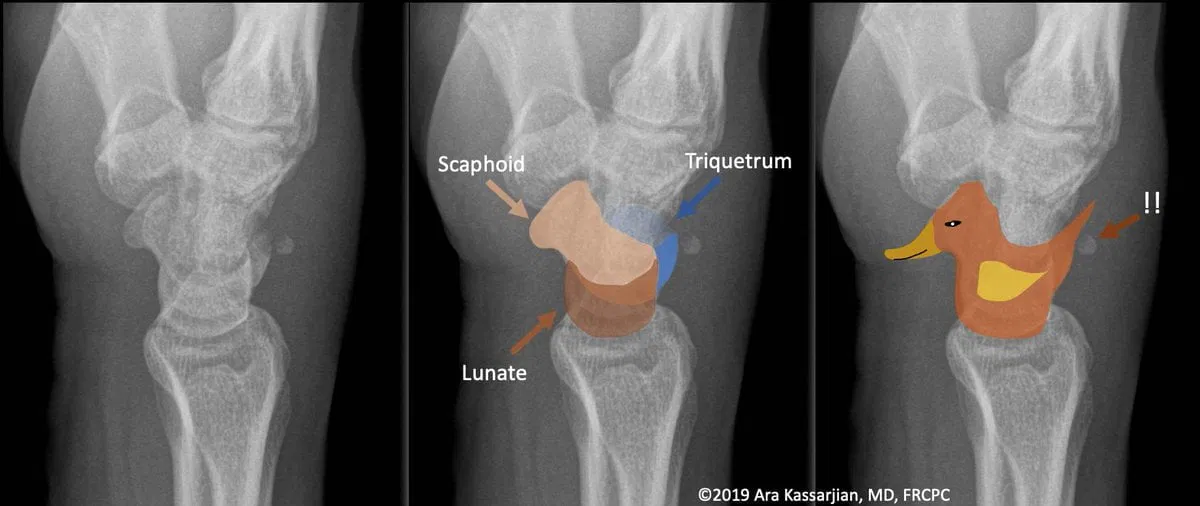
Etymology and synonyms :
The pooping duck sign derives its name from the resemblance of the combined outline of the scaphoid, lunate and the dorsal portion of the triquetrum to a duck and the avulsed triquetral fracture fragment to a poop from the duck.
Treatment:
- Non-surgical management – immobilisation for 4-6 weeks
- Surgical management- presence of significant displacement of the fracture fragments or fracture-dislocation concerning for instability
References:
Single best review article:
Case co-authored by TeamGyan Member Dr.Mansi

Improve Content And Help Your Colleagues!
Found an error in the post? Please let us know using our contact page and we will update it with due credits!
If you wish to contribute radiological images for this case (or any other article on the website), you can submit them here and you will be duly credited: Submit radiology case