


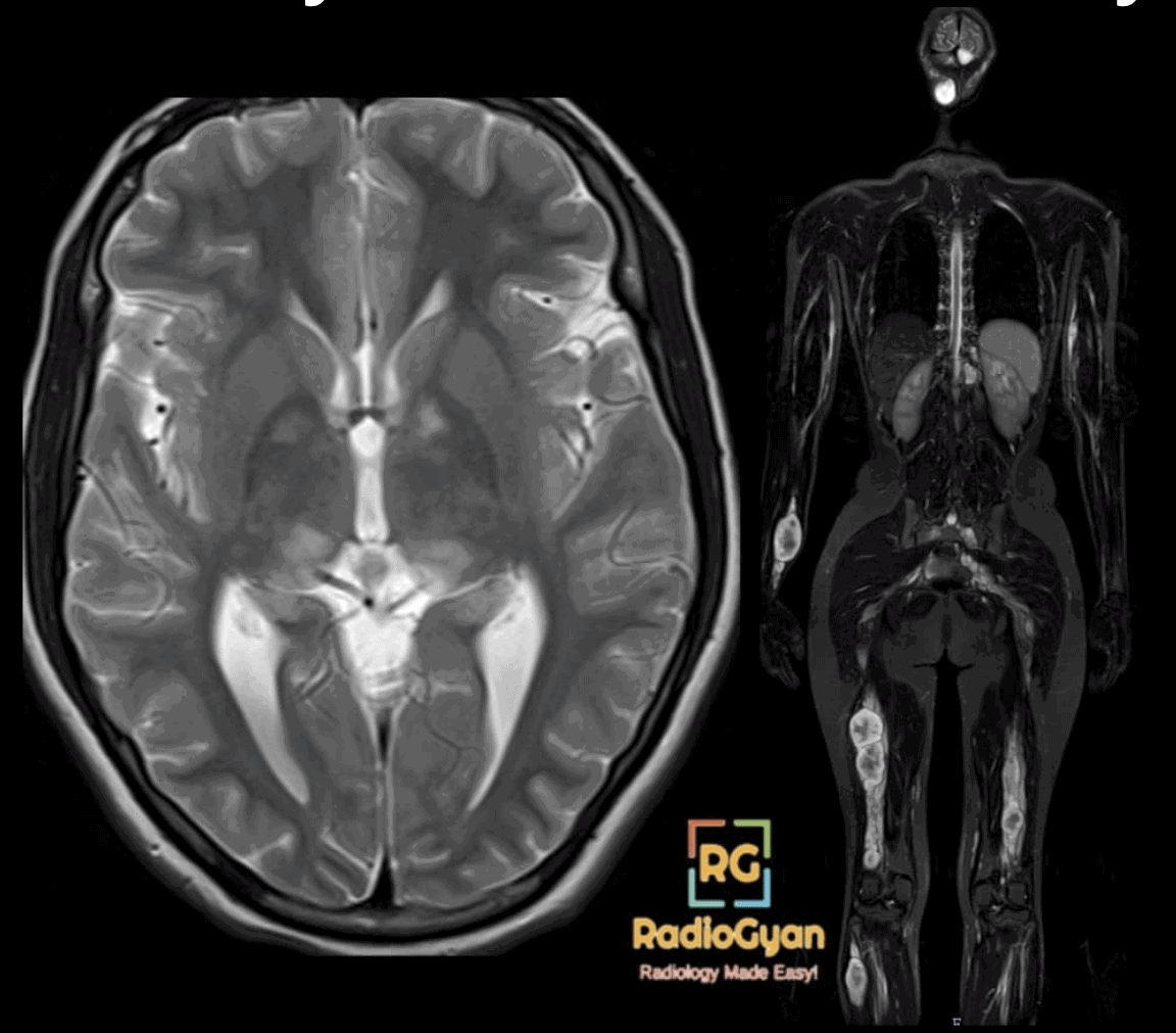
Neurofibromatosis is three separate genetic tumour syndromes. NF1 (chromosome 17) causes neurofibromas, optic pathway gliomas, FASI in the brain and skeletal dysplasias; NF2, renamed NF2-related schwannomatosis in 2022 (chromosome 22), causes bilateral vestibular schwannomas, meningiomas and ependymomas; and non-NF2 schwannomatosis causes multiple, often painful, schwannomas. Both sets of diagnostic criteria were rewritten, in 2021 and 2022 [1][2], so textbook lists based on the 1988 NIH and Manchester criteria are out of date.
This page is a working reference for the radiologist: the current criteria, the brain, skeletal and body findings of NF1 with the numbers behind them, how to tell a neurofibroma from a schwannoma, the imaging red flags for malignant transformation, the surveillance imaging you will be asked to report, and NF2-related schwannomatosis. For the shorter overviews see the NF1 and NF2 articles.
- Key facts for practice and radiology board exams
- NF1, NF2-related schwannomatosis and schwannomatosis at a glance
- NF1 diagnostic criteria: the 2021 revision
- NF1 brain MRI findings
- NF1 skull, spine and skeletal findings
- NF1 body imaging: plexiform neurofibromas, lung and associated tumours
- Neurofibroma imaging signs and neurofibroma versus schwannoma
- Atypical neurofibroma and MPNST: imaging red flags
- Surveillance imaging in NF1
- MEK inhibitors and volumetric MRI response
- NF2-related schwannomatosis (formerly NF2)
- Schwannomatosis: SMARCB1-, LZTR1- and 22q-related
- Differential diagnosis
- Frequently asked questions
- References
Key facts for practice and radiology board exams
- NF1 affects about 1 in 3,000 people [3]. NF2-related schwannomatosis affects about 1 in 25,000 to 33,000 births [4][5]. Memory aid: neurofibromatosis has 17 letters (chromosome 17); NF2 goes with chromosome 22.
- 2021 NF1 criteria: two of seven features, or one if a parent is affected. New: choroidal abnormalities as an alternative to Lisch nodules, and a pathogenic NF1 variant as a criterion [1].
- 2022 rename: NF2 is now NF2-related schwannomatosis; ependymoma replaced glioma in the criteria, and schwannomatosis is split into NF2-, SMARCB1-, LZTR1- and 22q-related types [2].
- FASI (focal areas of signal intensity, formerly UBOs) are vacuolar myelin change, not tumour [6]: no mass effect, no enhancement, and they regress with age [7]. Enhancement or mass effect means glioma until proven otherwise.
- Optic pathway glioma in NF1 involves the orbital optic nerve in 66 percent, keeps the shape of the optic pathway in 91 percent and is cystic in only 9 percent; sporadic OPG is chiasmatic in 91 percent and cystic in 66 percent [8]. Screening MRI in asymptomatic children is not recommended [9][3].
- Neurofibroma versus schwannoma: target sign 58 versus 15 percent, central enhancement 75 versus 8 percent; fascicular sign favours schwannoma, 63 versus 25 percent [10].
- MPNST lifetime risk in NF1 is 8 to 13 percent, median age at diagnosis 26 years [11]. Internal plexiform neurofibromas raise the risk about 20-fold [12].
- MPNST red flags: minimum ADC of 1.0 x 10⁻³ mm2/s or less (94 percent specificity at 100 percent sensitivity), SUVmax above 3.2 [13], growth, pain and loss of the target sign in a distinct nodular lesion [14].
- Adult surveillance (ERN GENTURIS 2023): whole-body MRI at least once at transition to adulthood, and annual breast MRI from 30 to 50 years in women [15].
- MEK inhibitors shrink plexiform neurofibromas: selumetinib partial response in 70 percent of children [16]; mirdametinib 41 percent in adults and 52 percent in children [17]. Response is read on volumetric MRI as a 20 percent volume change [18].
NF1, NF2-related schwannomatosis and schwannomatosis at a glance
| NF1 | NF2-related schwannomatosis (was NF2) | Other schwannomatosis | |
|---|---|---|---|
| Gene | NF1, chromosome 17q (neurofibromin) | NF2, chromosome 22q (merlin) | SMARCB1, LZTR1 or 22q-related; some unidentified [2] |
| Frequency | About 1 in 3,000 [3] | About 1 in 25,000 to 33,000 births [4][5] | Rarer |
| Defining tumours | Neurofibromas (cutaneous, nodular, plexiform), optic pathway glioma | Bilateral vestibular schwannomas, meningiomas, ependymomas (MISME) | Multiple non-vestibular schwannomas, often painful |
| Brain | FASI, optic pathway glioma, other low-grade gliomas, vasculopathy | Vestibular and other cranial nerve schwannomas, meningiomas | Usually none |
| Spine | Dural ectasia, dystrophic scoliosis, dumbbell neurofibromas | Intramedullary ependymomas, root schwannomas, meningiomas | Root and paraspinal schwannomas |
| Skin and eye | Cafe-au-lait macules, freckling, Lisch nodules, choroidal abnormalities | Juvenile cataract, retinal hamartoma, epiretinal membrane | None characteristic |
| Malignancy | MPNST, GIST, phaeochromocytoma, breast cancer, glioma | Tumours histologically benign; morbidity from location | MPNST risk higher in SMARCB1-related disease [19] |
Neurofibromatosis sits alongside tuberous sclerosis, von Hippel-Lindau syndrome and Sturge-Weber syndrome among the phakomatoses. NF1 is also a RASopathy, sharing its pathway with Noonan and Costello syndrome.
NF1 diagnostic criteria: the 2021 revision
The International Consensus Group on Neurofibromatosis Diagnostic Criteria replaced the NIH criteria in 2021 [1]. Two of the new items are directly relevant to imaging: optic pathway glioma and the distinctive osseous lesions.
| Criterion | 2021 definition | Imaging relevance |
|---|---|---|
| Cafe-au-lait macules | Six or more, over 5 mm before puberty or over 15 mm after puberty | None |
| Freckling | Axillary or inguinal | None |
| Neurofibromas | Two or more of any type, or one plexiform neurofibroma | MRI and ultrasound show deep and plexiform tumours |
| Optic pathway glioma | Present | MRI of orbits and chiasm |
| Ocular | Two or more Lisch nodules on slit lamp, or two or more choroidal abnormalities on optical coherence tomography or near-infrared reflectance imaging (new) | Ophthalmology |
| Distinctive osseous lesion | Sphenoid dysplasia, anterolateral bowing of the tibia, or pseudarthrosis of a long bone | Radiograph, CT; sphenoid dysplasia is not counted separately if an ipsilateral orbital plexiform neurofibroma is present |
| Genetics | Heterozygous pathogenic NF1 variant with a variant allele fraction of 50 percent in apparently normal tissue such as blood (new) | None |
What else changed. The revision defines mosaic NF1 (features limited to a body segment, or a lower variant allele fraction) and sets out parallel criteria for Legius syndrome, caused by SPRED1 variants: six or more bilateral cafe-au-lait macules with or without freckling, and none of the other NF1 features [1]. A child with only pigmentary findings may therefore have Legius syndrome, which carries no neurofibromas, optic gliomas or MPNST. FASI were never a diagnostic criterion and are still not one.
NF1 brain MRI findings
Focal areas of signal intensity (FASI)
FASI, previously called unidentified bright objects (UBOs), are T2 and FLAIR hyperintense foci with no mass effect and no enhancement. Histology shows vacuolar or spongiotic change in myelin rather than tumour or gliosis [6]. The globus pallidus and internal capsule are the commonest site [20]; the cerebellum, brainstem and thalamus are the other typical locations.
Their course is age-dependent. In a prospective cohort re-imaged after 8 years, T2 hyperintensities fell in number, size and intensity from childhood into adulthood, while lesions in the cortex and subcortical or deep white matter became more frequent with age and probably have a different substrate [7]. Quantitative follow-up shows a more uneven course than simple regression, with a fall between about 7 and 12 years and a rise again in adolescence in some children [20]. Childhood FASI, not their later disappearance, were the best predictor of adult cognitive difficulty [7].
| Feature | FASI | Low-grade glioma |
|---|---|---|
| Mass effect | None [6] | Present, expands the structure |
| Enhancement | None | Often present |
| Behaviour | Stable or regressing through childhood [7] | Grows on follow-up |
| Typical sites | Globus pallidus, cerebellar white matter, dentate, brainstem, thalamus [20] | Optic pathway, brainstem (tectum, medulla), cerebellum |
| What to do | Report as FASI; no dedicated follow-up | Short-interval MRI and neuro-oncology referral |
Optic pathway glioma
Optic pathway glioma (OPG), a pilocytic astrocytoma, develops in up to 15 percent of children with NF1 and makes up two thirds or more of NF1 CNS tumours [15]. In a cohort of 134 NF1 patients with OPG, 42.5 percent were symptomatic, diagnosed at a mean age of 7.6 years [21]. The imaging pattern differs from sporadic OPG enough to be a separate entity [8].
| Feature | NF1-associated OPG | Sporadic OPG |
|---|---|---|
| Orbital optic nerve involved | 66 percent | 32 percent |
| Chiasm involved | 62 percent | 91 percent |
| Extension beyond the optic pathway at diagnosis | 2 percent | 68 percent |
| Shape of the optic pathway preserved | 91 percent (fusiform, tortuous nerve) | 27 percent (globular mass) |
| Cystic component | 9 percent | 66 percent |
| Stable on follow-up | About half | 5 percent |
Screening. Routine MRI to look for OPG in an asymptomatic child with NF1 is not recommended; surveillance is by regular ophthalmological examination, with MRI when vision, fields, optic discs or puberty timing change [9][3]. The adult ERN GENTURIS guideline takes the same line and refers asymptomatic OPG to a specialist unit rather than scheduling repeat MRI [15]. An enlarged, J-shaped sella can accompany a chiasmatic glioma: see the J-shaped sella sign.
Other gliomas, vasculopathy and hydrocephalus
Brainstem gliomas in NF1 behave far more indolently than sporadic diffuse brainstem gliomas and are a distinct clinical entity [22]. In a paediatric series in which 80 of 181 NF1 children had brain or vascular imaging, vasculopathy was found in 18 percent of those imaged, a minimum population prevalence of 8 percent, almost all cerebrovascular (7 percent) and rarely peripheral (1 percent) [23]. Hydrocephalus affects an estimated 1 to 13 percent, from aqueductal webs, chiasmatic-hypothalamic tumours or thalamic mass effect; moyamoya and tumours can mimic it clinically [24].
NF1 skull, spine and skeletal findings
| Finding | Imaging appearance | Notes |
|---|---|---|
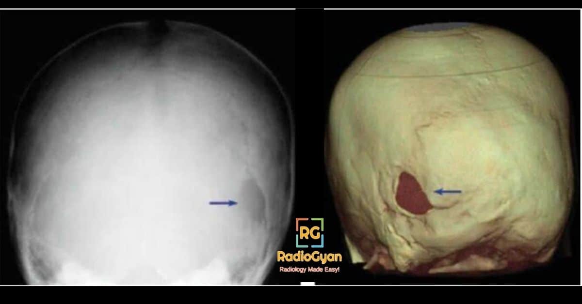
| Sphenoid wing dysplasia | Hypoplastic or absent greater wing, widened superior orbital fissure; empty or bare orbit on frontal radiograph; pulsatile exophthalmos, temporal lobe herniation into the orbit | Diagnostic criterion [1]; often with an orbital plexiform neurofibroma |
| Asterion (lambdoid) defect | Lucent defect of the posterior calvarium at the lambdoid suture | Can enlarge like a growing skull fracture; managed conservatively [25] |
| Dural ectasia and lateral meningocele | Posterior vertebral scalloping, widened canal and foramina; CSF-signal sac through the foramen | 75 percent of posteriorly scalloped vertebrae had dural ectasia on MRI [26]; see scalloped vertebra |
| Dystrophic kyphoscoliosis | Short-segment, sharply angulated curve with rib penciling, vertebral wedging and rotation, scalloping, spindled transverse processes | Rib penciling was the single radiographic predictor of progression [27] |
| Rib penciling | Thin, twisted ribbon ribs | Three or more penciled ribs: 87 percent progressed [27] |
| Anterolateral tibial bowing and pseudarthrosis | Anterolateral bowing, narrowed sclerotic medullary canal, then fracture non-union, usually distal tibia and fibula | Diagnostic criterion [1] |
| Dumbbell neurofibroma | Intradural-extradural tumour widening the neural foramen | Consider MPNST if large or painful |
| Focal overgrowth | Soft tissue and bone hypertrophy of a limb with a diffuse plexiform neurofibroma | Elephantiasis neuromatosa |
Dystrophic versus non-dystrophic scoliosis. Durrani analysed nine radiographic dystrophic features in 91 NF1 spinal deformities: rib penciling, vertebral rotation, posterior, anterior and lateral vertebral scalloping, vertebral wedging, spindling of the transverse processes, widened interpedicular distance and enlarged intervertebral foramina [27]. Curves change category over time (“modulation”): 81 percent of deformities diagnosed before 7 years acquired dystrophic features, against 25 percent diagnosed later. A curve that acquires three penciled ribs, or any three dystrophic features, progresses in 85 to 87 percent. MRI adds the cause of scalloping: posterior scalloping with dural ectasia, anterior or lateral scalloping with an adjacent neurofibroma [26].
NF1 body imaging: plexiform neurofibromas, lung and associated tumours
Plexiform neurofibromas are infiltrative tumours along multiple fascicles or branches of a nerve, appearing on MRI as lobulated, rope-like or “bag of worms” masses that are hyperintense on T2 and STIR, often with multiple target signs. Internal (deep) plexiform neurofibromas are largely invisible on examination, which is why whole-body MRI is used to count and measure them. People with internal plexiform neurofibromas were about 20 times more likely to develop MPNST than those without [12].
Lung. NF1-associated diffuse lung disease is characterised by upper lobe cysts and bullae with lower lobe reticulation and fibrosis, without honeycombing [28]. In 71 NF1 patients on CT, cysts (35 percent), nodules (32 percent) and paraseptal emphysema (30 percent) were the commonest findings, cysts occurred only in adults, and pulmonary nodules and metastases were more frequent in patients with MPNST [29]. Posterior mediastinal and intercostal neurofibromas, lateral thoracic meningoceles and chest wall deformity complete the chest picture.
| Tumour | Risk in NF1 | Imaging pearl |
|---|---|---|
| All cancers | Lifetime risk 59.6 percent versus 30.8 percent in the general population [30] | Avoid unnecessary CT; NF1 is treated as radiosensitive [15] |
| MPNST | Lifetime risk 8 to 13 percent; median age 26 years; 5-year survival 21 percent [11] | Arises in plexiform neurofibromas; see the red flags below |
| Breast cancer | Standardised incidence ratio 11.1 in women under 40 [30] | Annual breast MRI from 30 to 50 years [15] |
| Gastrointestinal stromal tumour | 6 of 95 adults (6.3 percent) on screening CT [31] | Often multiple, small bowel, KIT and PDGFRA wild-type |
| Phaeochromocytoma | 0.1 to 5.7 percent; 84 percent solitary adrenal, 9.6 percent bilateral, 6.1 percent ectopic [32] | Exclude before surgery or pregnancy, or with hypertension |
| CNS tumours | Optic pathway glioma up to 15 percent of children [15]; brainstem glioma usually indolent [22] | Screen with eye examination, not MRI, in children [3] |
Neurofibroma imaging signs and neurofibroma versus schwannoma
Both neurofibromas and schwannomas are benign nerve sheath tumours, so the old teaching that “neurofibromas arise from the nerve and schwannomas from the sheath” is wrong. The real difference is growth pattern: a schwannoma is a pure Schwann cell tumour that grows eccentrically and displaces the fascicles, so it can be shelled out; a neurofibroma contains Schwann cells, fibroblasts and mast cells, grows between the fascicles and cannot be separated from the nerve. That explains why the signs overlap.
| Sign | Definition | Seen in |
|---|---|---|
| Target sign | Central low and peripheral high T2 signal: a fibrocollagenous core with a myxoid rim [33] | Neurofibroma 58 percent, schwannoma 15 percent [10]; usually lost in MPNST |
| Fascicular sign | Multiple small ring-like low-signal foci in a high-signal mass on T2, reflecting fascicular bundles [33] | Schwannoma 63 percent, neurofibroma 25 percent [10] |
| Entering and exiting nerve sign | Fusiform mass tapering into the nerve at each pole [33] | Any nerve sheath tumour; confirms nerve origin |
| Split fat sign | Rim of fat around the poles of an intermuscular mass on T1 [33] | Any intermuscular nerve sheath tumour; see split fat sign |
| Bag of worms | Tortuous, multinodular expansion of a nerve and its branches | Plexiform neurofibroma |
| MRI feature | Neurofibroma (n=12) | Schwannoma (n=40) |
|---|---|---|
| Target sign on T2 | 58 percent | 15 percent |
| Central enhancement | 75 percent | 8 percent |
| Target sign plus central enhancement | 63 percent | 3 percent |
| Fascicular appearance | 25 percent | 63 percent |
| Thin T2 hyperintense rim | 8 percent | 58 percent |
| Diffuse enhancement | 13 percent | 67 percent |
| Central versus eccentric nerve position, cystic change, peripheral enhancement | Not significantly different | Not significantly different |
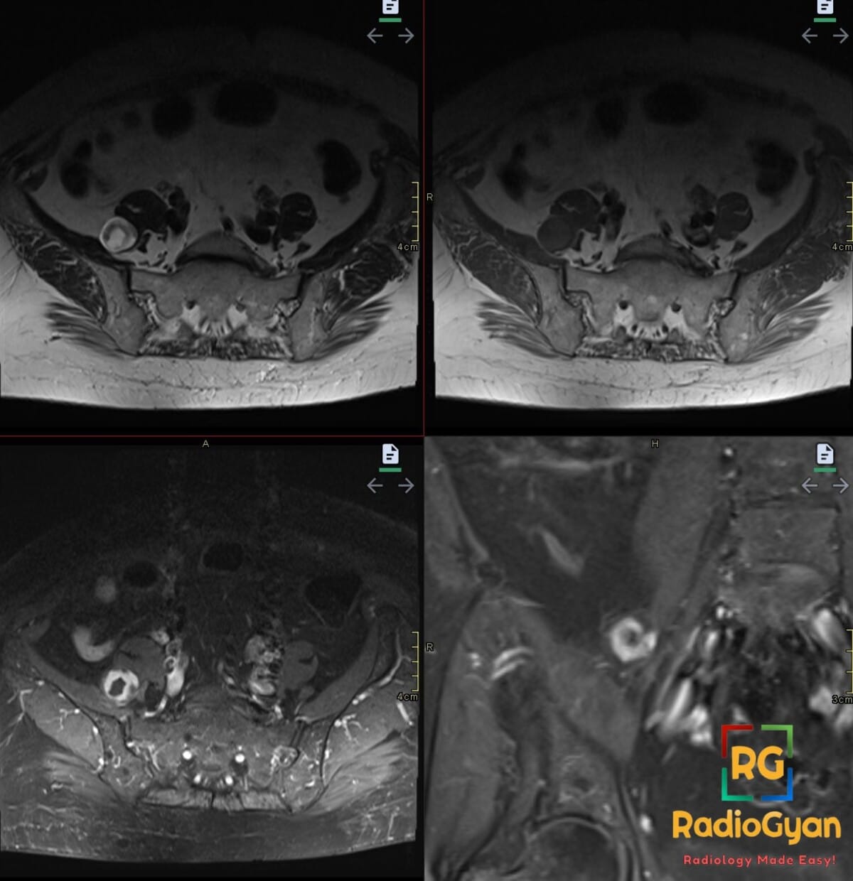
Ultrasound shows the same features: a fusiform hypoechoic mass in continuity with the nerve, a central echogenic area corresponding to the target sign, and posterior acoustic enhancement. Overlap between neurofibroma and schwannoma is as large on ultrasound as on MRI.
Atypical neurofibroma and MPNST: imaging red flags
Malignant transformation in NF1 usually happens inside a plexiform neurofibroma and passes through an intermediate stage. The 2017 consensus names it atypical neurofibromatous neoplasm of uncertain biologic potential (ANNUBP): at least two of cytological atypia, loss of neurofibroma architecture, hypercellularity and a mitotic count above 1 per 50 but below 3 per 10 high-power fields [34]. In 76 atypical neurofibromas from three centres, most appeared on MRI as distinct nodular lesions (well-defined nodules that stand out from the background plexiform tumour, typically without a target sign) and most were FDG-avid; none of the 57 completely resected lesions recurred, and 4 progressed to MPNST [14].
| Study | Discriminator | Result |
|---|---|---|
| Wasa, 61 PNST on MRI [35] | Two or more of: large diameter, peripheral enhancement, perilesional oedema-like zone, intratumoral cystic change | Sensitivity 61 percent, specificity 90 percent for MPNST |
| Demehri, 31 PNST [36] | Minimum ADC | 0.47 versus 1.08 x 10⁻³ mm2/s (AUC 0.89); ADC 1.0 or less plus diameter 4.2 cm or more detected every malignancy |
| Demehri [36] | Ill-defined margins; peritumoral oedema | 77 versus 32 percent; 66 versus 23 percent |
| Ahlawat, 55 NF1 PNST [13] | Mean diameter | 8.2 cm MPNST versus 4.3 cm benign |
| Ahlawat [13] | Minimum ADC 1.0 x 10⁻³ mm2/s or less | Specificity 94 percent at 100 percent sensitivity |
| Ahlawat [13] | FDG PET SUVmax above 3.2 | Specificity 83 percent at 100 percent sensitivity |
| Chirindel, 93 NF1 lesions [37] | FDG PET SULmax cut-off | 3.2 at 1 hour (AUC 0.973), 4.1 at 4 hours (AUC 0.978); normalising to liver raised specificity to 93 to 94 percent |
In practice: in an NF1 patient with new pain, neurological deficit or rapid growth of a known tumour, look for a nodule that has lost its target sign, ill-defined or infiltrative margins, perilesional oedema, necrosis or haemorrhage, low ADC, and FDG avidity, and state which lesion should be biopsied. Clinical symptoms matter: pain and motor weakness were common presentations of atypical neurofibromas [14].
Surveillance imaging in NF1
| Question | Recommendation | Source |
|---|---|---|
| Screening brain or orbit MRI for optic glioma in asymptomatic children | Not recommended; annual ophthalmological examination instead | [9][3] |
| Whole-body MRI | At least once at the transition from childhood to adulthood, to measure internal plexiform tumour burden as an MPNST risk marker; more often if indicated | [15] |
| Breast cancer screening in women | Annual breast MRI from 30 to 50 years; mammography only where MRI is unavailable; national screening after 50 | [15] |
| CT | Avoid where possible because of radiosensitivity | [15] |
| Plexiform neurofibroma response | Volumetric MRI; a 20 percent or greater volume change defines response or progression | [18] |
What to put in a whole-body MRI report: the number and location of plexiform neurofibromas, which are internal, volumes or three-plane measurements of index lesions for comparison, any distinct nodular lesion, any lesion without a target sign or with restricted diffusion, spinal and paraspinal disease, and incidental findings such as adrenal masses.
MEK inhibitors and volumetric MRI response
Plexiform neurofibromas that cannot be resected are now treated with MEK inhibitors, and the radiologist measures the response. In the SPRINT phase 2 trial, 35 of 50 children (70 percent) on selumetinib had a confirmed partial response, defined as a volume reduction of 20 percent or more, and 28 of these lasted a year or longer [16]. In the ReNeu trial of mirdametinib, the blinded confirmed response rate was 41 percent in 58 adults and 52 percent in 56 children, with median best volume reductions of 41 and 42 percent [17]. Mirdametinib was the first MEK inhibitor approved in the USA for both adults and children aged 2 years and older with symptomatic, inoperable NF1 plexiform neurofibromas [38].
Volumetric MRI, not linear measurement, is the REiNS-recommended method, with a 20 percent volume change as the threshold for response and progression [18]. Use the same sequence, typically fat-suppressed T2 or STIR in three planes, for every comparison.
NF2-related schwannomatosis (formerly NF2)
The 2022 consensus replaced the Manchester criteria and the name at the same time [2]. The mnemonic is still useful: MISME, multiple inherited schwannomas, meningiomas and ependymomas (illustrated mnemonic). Neurofibroma and glioma, part of the older criteria, are no longer in them.
| Diagnosis requires one of | Details |
|---|---|
| Bilateral vestibular schwannomas | Diagnostic on their own |
| Identical NF2 pathogenic variant in two anatomically distinct NF2-related tumours | Schwannoma, meningioma or ependymoma |
| Two major criteria, or one major plus two minor criteria | See below |
| Major criteria | Unilateral vestibular schwannoma; first-degree relative other than a sibling with NF2-related schwannomatosis; two or more meningiomas; NF2 pathogenic variant in unaffected tissue such as blood |
| Minor criteria | Ependymoma; non-vestibular schwannoma; single meningioma (two of the same tumour type count as two minor criteria); juvenile subcapsular or cortical cataract, retinal hamartoma or epiretinal membrane in a person under 40 years |
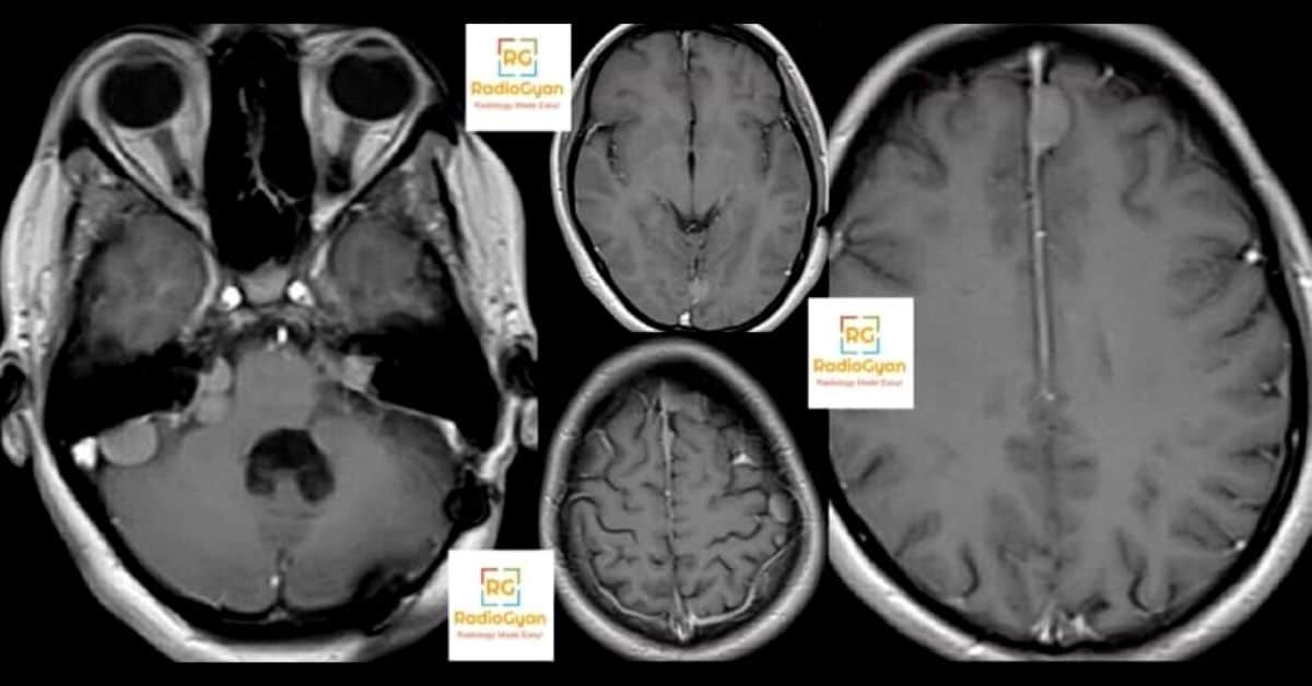
Imaging. Image the whole neuraxis: post-contrast brain MRI with thin sections through the internal auditory canals, and whole-spine MRI. Vestibular schwannomas enter and widen the internal auditory canal, meningiomas are dural-based with a broad base, and schwannomas of the trigeminal, oculomotor and lower cranial nerves and of spinal roots are common. In a 63-patient natural history study, 67.4 percent had spinal tumours and 49.2 percent meningiomas [39]. Ependymomas are intramedullary spinal cord tumours.
Children. Before hearing loss, children present with a meningioma, a non-vestibular schwannoma, a mononeuropathy, a cataract or seizures, and in a UK paediatric series every non-vestibular presentation was diagnosed late, by a mean of 3.2 years [40]. A meningioma or schwannoma in a child should prompt MRI of the internal auditory canals and spine and genetic referral.
Treatment response. Bevacizumab shrinks progressive vestibular schwannomas: in 31 patients, 55 percent had a radiographic response, defined as a volume reduction of 20 percent or more, and 57 percent a hearing response, with a median time to response of 3 months [41]. The original report showed a median pretreatment volumetric growth of 62 percent a year [42]. As in NF1, response is measured volumetrically.
Schwannomatosis: SMARCB1-, LZTR1- and 22q-related
Non-NF2 schwannomatosis produces multiple schwannomas of spinal and peripheral nerves, typically with chronic pain rather than a mass, and without the bilateral vestibular schwannomas, meningiomas and ependymomas of NF2-related disease [2]. The ERN GENTURIS guideline advises craniospinal MRI at symptomatic diagnosis and, in people with a confirmed variant and an affected relative, from 12 to 14 years; whole-body MRI can alternate with craniospinal MRI, ultrasound is useful for a painful limb without a palpable lump, and a rapidly growing tumour or loss of function should raise the possibility of MPNST, especially in SMARCB1-related disease [19].
Differential diagnosis
| Condition | Overlap with NF1 or NF2 | Distinguishing features |
|---|---|---|
| Legius syndrome (SPRED1) | Cafe-au-lait macules and freckling | No neurofibromas, Lisch nodules, optic glioma or skeletal dysplasia [1] |
| Mosaic (segmental) NF1 | Neurofibromas or pigmentary changes | Confined to one body segment [1] |
| Non-NF2 schwannomatosis | Multiple schwannomas | No bilateral vestibular schwannomas, pain-predominant [2] |
| Sporadic solitary neurofibroma or schwannoma | Target and fascicular signs | Single lesion, no other criteria |
| Glioma versus FASI | T2 hyperintense brain foci | Mass effect, enhancement or growth [6] |
| Other causes of posterior vertebral scalloping | Scalloping, widened canal | Dural ectasia of Marfan or Ehlers-Danlos syndrome, acromegaly, raised intraspinal pressure; no neurofibromas |
| Fibrous dysplasia (McCune-Albright) | Cafe-au-lait macules, skull base bone lesion | Ground-glass expansile bone, not sphenoid wing absence |
Frequently asked questions
References
- Legius E, Messiaen L, Wolkenstein P, Pancza P, Avery RA, Berman Y, et al. Revised diagnostic criteria for neurofibromatosis type 1 and Legius syndrome: an international consensus recommendation. Genet Med. 2021;23(8):1506-1513.
- Plotkin SR, Messiaen L, Legius E, Pancza P, Avery RA, Blakeley JO, et al. Updated diagnostic criteria and nomenclature for neurofibromatosis type 2 and schwannomatosis: An international consensus recommendation. Genet Med. 2022;24(9):1967-1977.
- Miller DT, Freedenberg D, Schorry E, Ullrich NJ, Viskochil D, Korf BR. Health Supervision for Children With Neurofibromatosis Type 1. Pediatrics. 2019;143(5):e20190660.
- Asthagiri AR, Parry DM, Butman JA, Kim HJ, Tsilou ET, Zhuang Z, et al. Neurofibromatosis type 2. Lancet. 2009;373(9679):1974-1986.
- Evans DG. Neurofibromatosis type 2. Handb Clin Neurol. 2015;132:87-96.
- DiPaolo DP, Zimmerman RA, Rorke LB, Zackai EH, Bilaniuk LT, Yachnis AT. Neurofibromatosis type 1: pathologic substrate of high-signal-intensity foci in the brain. Radiology. 1995;195(3):721-724.
- Hyman SL, Gill DS, Shores EA, Steinberg A, Joy P, Gibikote SV, et al. Natural history of cognitive deficits and their relationship to MRI T2-hyperintensities in NF1. Neurology. 2003;60(7):1139-1145.
- Kornreich L, Blaser S, Schwarz M, Shuper A, Vishne TH, Cohen IJ, et al. Optic pathway glioma: correlation of imaging findings with the presence of neurofibromatosis. AJNR Am J Neuroradiol. 2001;22(10):1963-1969.
- Listernick R, Louis DN, Packer RJ, Gutmann DH. Optic pathway gliomas in children with neurofibromatosis 1: consensus statement from the NF1 Optic Pathway Glioma Task Force. Ann Neurol. 1997;41(2):143-149.
- Jee WH, Oh SN, McCauley T, Ryu KN, Suh JS, Lee JH, et al. Extraaxial neurofibromas versus neurilemmomas: discrimination with MRI. AJR Am J Roentgenol. 2004;183(3):629-633.
- Evans DG, Baser ME, McGaughran J, Sharif S, Howard E, Moran A. Malignant peripheral nerve sheath tumours in neurofibromatosis 1. J Med Genet. 2002;39(5):311-314.
- Tucker T, Wolkenstein P, Revuz J, Zeller J, Friedman JM. Association between benign and malignant peripheral nerve sheath tumors in NF1. Neurology. 2005;65(2):205-211.
- Ahlawat S, Blakeley JO, Rodriguez FJ, Fayad LM. Imaging biomarkers for malignant peripheral nerve sheath tumors in neurofibromatosis type 1. Neurology. 2019;93(11):e1076-e1084.
- Higham CS, Dombi E, Rogiers A, Bhaumik S, Pans S, Connor SEJ, et al. The characteristics of 76 atypical neurofibromas as precursors to neurofibromatosis 1 associated malignant peripheral nerve sheath tumors. Neuro Oncol. 2018;20(6):818-825.
- Carton C, Evans DG, Blanco I, Friedrich RE, Ferner RE, Farschtschi S, et al. ERN GENTURIS tumour surveillance guidelines for individuals with neurofibromatosis type 1. EClinicalMedicine. 2023;56:101818.
- Gross AM, Wolters PL, Dombi E, Baldwin A, Whitcomb P, Fisher MJ, et al. Selumetinib in Children with Inoperable Plexiform Neurofibromas. N Engl J Med. 2020;382(15):1430-1442.
- Moertel CL, Hirbe AC, Shuhaiber HH, Bielamowicz K, Sidhu A, Viskochil D, et al. ReNeu: A Pivotal, Phase IIb Trial of Mirdametinib in Adults and Children With Symptomatic Neurofibromatosis Type 1-Associated Plexiform Neurofibroma. J Clin Oncol. 2025;43(6):716-729.
- Dombi E, Ardern-Holmes SL, Babovic-Vuksanovic D, Barker FG, Connor S, Evans DG, et al. Recommendations for imaging tumor response in neurofibromatosis clinical trials. Neurology. 2013;81(21 Suppl 1):S33-S40.
- Evans DG, Mostaccioli S, Pang D, Fadzil O Connor M, Pittara M, Champollion N, et al. ERN GENTURIS clinical practice guidelines for the diagnosis, treatment, management and surveillance of people with schwannomatosis. Eur J Hum Genet. 2022;30(7):812-817.
- Kraut MA, Gerring JP, Cooper KL, Thompson RE, Denckla MB, Kaufmann WE. Longitudinal evolution of unidentified bright objects in children with neurofibromatosis-1. Am J Med Genet A. 2004;129A(2):113-119.
- Friedrich RE, Nuding MA. Optic Pathway Glioma and Cerebral Focal Abnormal Signal Intensity in Patients with Neurofibromatosis Type 1: Characteristics, Treatment Choices and Follow-up in 134 Affected Individuals and a Brief Review of the Literature. Anticancer Res. 2016;36(8):4095-4121.
- Molloy PT, Bilaniuk LT, Vaughan SN, Needle MN, Liu GT, Zackai EH, et al. Brainstem tumors in patients with neurofibromatosis type 1: a distinct clinical entity. Neurology. 1995;45(10):1897-1902.
- Kaas B, Huisman TA, Tekes A, Bergner A, Blakeley JO, Jordan LC. Spectrum and prevalence of vasculopathy in pediatric neurofibromatosis type 1. J Child Neurol. 2013;28(5):561-569.
- Roth J, Constantini S. Neurofibromatosis Type 1 Related Hydrocephalus. Neurol India. 2021;69(Supplement):S372-S375.
- Solanki C, Ramachandran S, Devi BI, Sharma R. Calvarial defects in the region of the lambdoid suture in neurofibromatosis type-1 patients. J Pediatr Neurosci. 2015;10(1):22-24.
- Tsirikos AI, Ramachandran M, Lee J, Saifuddin A. Assessment of vertebral scalloping in neurofibromatosis type 1 with plain radiography and MRI. Clin Radiol. 2004;59(11):1009-1017.
- Durrani AA, Crawford AH, Chouhdry SN, Saifuddin A, Morley TR. Modulation of spinal deformities in patients with neurofibromatosis type 1. Spine (Phila Pa 1976). 2000;25(1):69-75.
- Zamora AC, Collard HR, Wolters PJ, Webb WR, King TE. Neurofibromatosis-associated lung disease: a case series and literature review. Eur Respir J. 2007;29(1):210-214.
- Avanesov M, Well L, Laqmani A, Derlin T, Riccardi VM, Adam G, et al. Structural alteration of lung parenchyma in patients with NF1: a phenotyping study using multidetector computed tomography (MDCT). Orphanet J Rare Dis. 2021;16(1):29.
- Uusitalo E, Rantanen M, Kallionpää RA, Pöyhönen M, Leppävirta J, Ylä-Outinen H, et al. Distinctive Cancer Associations in Patients With Neurofibromatosis Type 1. J Clin Oncol. 2016;34(17):1978-1986.
- Nishida T, Tsujimoto M, Takahashi T, Hirota S, Blay JY, Wataya-Kaneda M. Gastrointestinal stromal tumors in Japanese patients with neurofibromatosis type I. J Gastroenterol. 2016;51(6):571-578.
- Walther MM, Herring J, Enquist E, Keiser HR, Linehan WM. von Recklinghausen’s disease and pheochromocytomas. J Urol. 1999;162(5):1582-1586.
- Kakkar C, Shetty CM, Koteshwara P, Bajpai S. Telltale signs of peripheral neurogenic tumors on magnetic resonance imaging. Indian J Radiol Imaging. 2015;25(4):453-458.
- Miettinen MM, Antonescu CR, Fletcher CDM, Kim A, Lazar AJ, Quezado MM, et al. Histopathologic evaluation of atypical neurofibromatous tumors and their transformation into malignant peripheral nerve sheath tumor in patients with neurofibromatosis 1-a consensus overview. Hum Pathol. 2017;67:1-10.
- Wasa J, Nishida Y, Tsukushi S, Shido Y, Sugiura H, Nakashima H, et al. MRI features in the differentiation of malignant peripheral nerve sheath tumors and neurofibromas. AJR Am J Roentgenol. 2010;194(6):1568-1574.
- Demehri S, Belzberg A, Blakeley J, Fayad LM. Conventional and functional MR imaging of peripheral nerve sheath tumors: initial experience. AJNR Am J Neuroradiol. 2014;35(8):1615-1620.
- Chirindel A, Chaudhry M, Blakeley JO, Wahl R. 18F-FDG PET/CT qualitative and quantitative evaluation in neurofibromatosis type 1 patients for detection of malignant transformation: comparison of early to delayed imaging with and without liver activity normalization. J Nucl Med. 2015;56(3):379-385.
- Hoy SM. Mirdametinib: First Approval. Drugs. 2025;85(7):977-984.
- Parry DM, Eldridge R, Kaiser-Kupfer MI, Bouzas EA, Pikus A, Patronas N. Neurofibromatosis 2 (NF2): clinical characteristics of 63 affected individuals and clinical evidence for heterogeneity. Am J Med Genet. 1994;52(4):450-461.
- Anand G, Vasallo G, Spanou M, Thomas S, Pike M, Kariyawasam DS, et al. Diagnosis of sporadic neurofibromatosis type 2 in the paediatric population. Arch Dis Child. 2018;103(5):463-469.
- Plotkin SR, Merker VL, Halpin C, Jennings D, McKenna MJ, Harris GJ, et al. Bevacizumab for progressive vestibular schwannoma in neurofibromatosis type 2: a retrospective review of 31 patients. Otol Neurotol. 2012;33(6):1046-1052.
- Plotkin SR, Stemmer-Rachamimov AO, Barker FG 2nd, Halpin C, Padera TP, Tyrrell A, et al. Hearing improvement after bevacizumab in patients with neurofibromatosis type 2. N Engl J Med. 2009;361(4):358-367.
Further reading: Osborn’s Brain, the standard neuroradiology text for the phakomatoses.


Improve Content And Help Your Colleagues!
Found an error in the post? Please let us know using our contact page and we will update it with due credits!
If you wish to contribute radiological images for this case (or any other article on the website), you can submit them here and you will be duly credited: Submit radiology case
Sir you are best, great contribution for residents.
Thank you for your kind words, Asim :).
Many thanks, It is very simple and easy way to give information.
Glad you liked it Dr. Elshamsy.
Do check out more simplified radiology videos here: https://www.youtube.com/channel/UCrq_N7nwDk_Wo_AGhNakrMA?sub_confirmation=1