


Breast microcalcifications are tiny calcium deposits (usually under 1 mm) seen on mammography. Most are benign, but calcifications are the earliest and often the only mammographic sign of ductal carcinoma in situ (DCIS), accounting for roughly 85–95% of screen-detected DCIS. Accurate characterisation using the ACR BI-RADS 5th edition lexicon — assessing morphology and distribution — is what separates a “leave alone” from a “biopsy now”.
This page distils the current BI-RADS 5th edition approach to mammographic calcifications: the nine typically benign descriptors, the four suspicious morphologies (with their probability of malignancy), the five distribution patterns, and how these translate into a BI-RADS category and management. A comprehensive video lecture by a breast-imaging subspecialist is embedded below.
How to analyse breast calcifications: the two questions
Every group of calcifications is described by two axes, and a third step is mandatory:
- Morphology — the shape of the individual particles (typically benign vs suspicious). Morphology is the stronger predictor of malignancy.
- Distribution — how the particles are arranged in the breast (diffuse to segmental). Distribution modifies the level of suspicion.
- Compare with priors — stability over 2–3 years or a classically benign appearance can downgrade findings; new, increasing, or linear/segmental calcifications are upgraded. Assess on magnification views (craniocaudal and 90° mediolateral) before finalising.
Typically benign calcifications (BI-RADS 2)
Nine morphologies are labelled typically benign. When characteristic, they need no further workup. In BI-RADS 5th edition the old “eggshell” and “lucent-centred” terms were consolidated into a single descriptor, rim.
| Descriptor | Mammographic appearance | Underlying cause |
|---|---|---|
| Skin (dermal) | Lucent-centred, polygonal; confirmed as superficial on a tangential view | Calcium in dermis (sebaceous glands) |
| Vascular | Parallel “tram-track” or discontinuous linear pairs along a tubular structure | Arterial wall (medial) calcification |
| Coarse / popcorn-like | Large (over 2–3 mm), dense, may coalesce, sometimes with a mass | Involuting (hyalinising) fibroadenoma |
| Large rod-like | Smooth rods, over 0.5 mm wide, following ducts toward the nipple; usually bilateral, women over 60 | Secretory disease / plasma-cell mastitis (duct ectasia) |
| Round / punctate | Round, under 1 mm (punctate if under 0.5 mm) | Acini of the terminal ductal lobular unit |
| Rim | Thin calcium around a lucent centre (“eggshell”) | Wall of an oil cyst (fat necrosis) or simple cyst |
| Dystrophic | Irregular, coarse, over 1 mm, tend to coalesce | Post-surgery, post-radiation, or trauma (fat necrosis) |
| Milk of calcium | Sediment in a cyst: “teacup” curvilinear on the 90° lateral, faint smudge on the CC view | Layering calcium within microcysts |
| Suture | Linear or tubular, may show knots | Calcification on retained suture material |
Caveat: grouped round or punctate calcifications that are new, or seen for the first time with no priors, are placed in BI-RADS 3 (probably benign) and followed at 6 months rather than dismissed.
Suspicious morphology (BI-RADS 4–5)
BI-RADS 5th edition collapsed the previous “intermediate concern” and “higher probability of malignancy” groups into a single suspicious morphology category with four descriptors, listed below in order of increasing probability of malignancy (PPV). Any suspicious morphology warrants tissue sampling.
| Descriptor | Appearance | Approx. PPV (malignancy) | Usual BI-RADS category |
|---|---|---|---|
| Amorphous | So small/hazy that a shape cannot be assigned (“powdery”, “cloud-like”) | ~20% | 4B |
| Coarse heterogeneous | Irregular, conspicuous, generally 0.5–1 mm, may coalesce | ~15% | 4B |
| Fine pleomorphic | Varying shapes and sizes, under 0.5 mm (“crushed stone”); no fine-linear forms | ~29% | 4B |
| Fine linear / fine-linear branching | Thin (under 0.5 mm), linear, discontinuous, may branch (“casting”) | ~70% | 4C (5 if segmental/linear and new) |
PPVs vary between series. Bent et al. (AJR 2010) reported fine linear/branching 70%, fine pleomorphic 28%, coarse heterogeneous 20%, amorphous 20% and typically benign 0%. Park et al. (AJR 2019), applying the 5th edition, found amorphous 15.9%, coarse heterogeneous 31.7%, fine pleomorphic 58.2% and fine linear/branching 90.6% — confirming that fine-linear branching morphology carries the highest risk.
Distribution descriptors
Distribution is assessed after morphology and shifts the level of suspicion. The five descriptors run from almost-always-benign (diffuse) to highly suspicious (segmental).
| Distribution | Definition | Significance |
|---|---|---|
| Diffuse (scattered) | Random throughout the breast, typically bilateral | Almost always benign |
| Regional | Scattered over a large volume (over 2 cm), not in a ductal pattern | Lower suspicion; depends on morphology (~26%) |
| Grouped (clustered) | At least 5 calcifications within 1 cm, in an area up to 2 cm | Needs evaluation; risk rises with small size + suspicious shape (~31%) |
| Linear | Arrayed in a line, may branch — suggests deposition within a duct | Suspicious unless classically vascular/rod-like (~60%) |
| Segmental | Deposits in the ducts and branches of a single lobe (triangular, apex at nipple) | Highly suspicious for multifocal/extensive DCIS (~62%) |
BI-RADS assessment categories and management
| Category | Meaning | Malignancy risk | Management |
|---|---|---|---|
| 0 | Incomplete | — | Magnification views (CC + 90° lateral) and/or prior films |
| 2 | Benign | 0% | Routine screening |
| 3 | Probably benign | Under 2% | Short-interval (6-month) follow-up |
| 4A | Low suspicion | Over 2% to 10% | Image-guided biopsy |
| 4B | Moderate suspicion | Over 10% to 50% | Biopsy; radiologic-pathologic concordance essential |
| 4C | High suspicion | Over 50% to 95% | Biopsy; a benign result is discordant |
| 5 | Highly suggestive of malignancy | 95% or more | Biopsy; a benign result must prompt re-biopsy |
| 6 | Known biopsy-proven cancer | — | Definitive treatment |
Bent et al. validated this subdivision in a digital mammography cohort: PPV 13% for 4A, 36% for 4B, 79% for 4C and 100% for category 5.
Workup of suspicious calcifications
- Magnification views (spot magnification, CC and true 90° lateral) better resolve particle morphology and reveal milk-of-calcium layering.
- Stereotactic or tomosynthesis-guided vacuum-assisted biopsy is the standard for calcifications with no ultrasound or MRI correlate.
- Specimen radiography confirms the targeted calcifications were retrieved; a clip is deployed for localisation.
- Radiologic-pathologic concordance is mandatory. A benign result on a 4C or 5 lesion is discordant and requires repeat (often surgical) biopsy.
- Upgrade rates: atypical ductal hyperplasia or DCIS on core biopsy may be upgraded at excision, so histology guides surgical planning.
What changed in BI-RADS 5th edition
- “Eggshell” and “lucent-centred” were merged into the single typically benign descriptor rim.
- The suspicious morphologies “intermediate concern” and “higher probability of malignancy” were merged into one suspicious morphology group (amorphous, coarse heterogeneous, fine pleomorphic, fine linear/branching).
- Suspicious calcifications are stratified with the 4A / 4B / 4C subdivisions, aligning descriptor PPV with the assigned category — a change validated by later 4th-vs-5th-edition comparisons.
Watch: breast microcalcifications lecture
A detailed lecture on the importance of microcalcifications on mammography, with histopathological correlation:
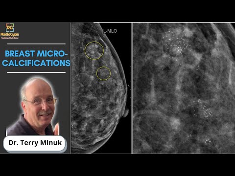
Timestamps:
0:50 Introduction
1:13 Normal breast anatomy with histopathological correlation
6:00 The origin of calcifications
7:23 Ductal calcifications
19:19 Lobular calcifications
40:55 Miscellaneous calcifications including benign and malignant processes
48:00 Take-home points, Q and A
Speaker: Dr. Terry Minuk, Clinical Associate Professor, Department of Radiology, Faculty of Health Sciences, McMaster University; Radiologist and Head of Breast Imaging, Hamilton Health Sciences.

Frequently asked questions
Are breast microcalcifications a sign of cancer?
Most are benign. However, calcifications are the commonest mammographic sign of DCIS (early, non-invasive breast cancer) and account for 85–95% of screen-detected DCIS. The morphology and distribution decide whether follow-up or biopsy is needed.
Which calcification morphology is the most suspicious?
Fine linear or fine-linear branching (“casting”) calcifications carry the highest probability of malignancy, roughly 70% in the ACR atlas and up to 90% in some series. Amorphous, coarse heterogeneous and fine pleomorphic forms are also suspicious but lower risk.
What BI-RADS category are suspicious calcifications given?
Amorphous, coarse heterogeneous and fine pleomorphic calcifications are usually BI-RADS 4B; fine linear or fine-linear branching calcifications are usually 4C, and BI-RADS 5 when new and in a linear or segmental distribution. All suspicious calcifications need biopsy.
How are suspicious microcalcifications biopsied?
Because calcifications are usually invisible on ultrasound, the standard is stereotactic or tomosynthesis-guided vacuum-assisted biopsy, followed by specimen radiography to confirm retrieval and clip placement, with mandatory radiologic-pathologic concordance.
Do benign-looking calcifications need follow-up?
Classically benign calcifications (vascular, popcorn, rim, milk of calcium) are BI-RADS 2 and need only routine screening. New grouped round or punctate calcifications without priors are BI-RADS 3 and are followed at 6 months.
References
- D’Orsi CJ, Sickles EA, Mendelson EB, Morris EA, et al. ACR BI-RADS Atlas, Breast Imaging Reporting and Data System, 5th edition. American College of Radiology; 2013.
- Rao AA, Feneis J, Lalonde C, Ojeda-Fournier H. A Pictorial Review of Changes in the BI-RADS Fifth Edition. RadioGraphics. 2016;36(3):623–639. PMID: 27082663.
- Bent CK, Bassett LW, D’Orsi CJ, Sayre JW. The positive predictive value of BI-RADS microcalcification descriptors and final assessment categories. AJR Am J Roentgenol. 2010;194(5):1378–1383. PMID: 20410428.
- Burnside ES, Ochsner JE, Fowler KJ, et al. Use of microcalcification descriptors in BI-RADS 4th edition to stratify risk of malignancy. Radiology. 2007;242(2):388–395. PMID: 17255409.
- Park GE, Kim SH, Lee JM, Kang BJ, Chae BJ. Comparison of Positive Predictive Values of Categorization of Suspicious Calcifications Using the 4th and 5th Editions of BI-RADS. AJR Am J Roentgenol. 2019;213(3):710–715. PMID: 31063419.
- Malignancy Risk Stratification of Suspicious Breast Microcalcifications Detected on Mammograms. Indian J Radiol Imaging. 2026. PMID: 41523350.
More radiology video content:




Improve Content And Help Your Colleagues!
Found an error in the post? Please let us know using our contact page and we will update it with due credits!
If you wish to contribute radiological images for this case (or any other article on the website), you can submit them here and you will be duly credited: Submit radiology case