
Quiz
Which of the following is a feature of this pathology?
- Spiral fracture pattern
- Transverse fracture pattern
- Oblique fracture pattern
- Stable, rounded bump or protrusion on the bone
Pathophysiology
- Common in children and distal radius is the most common site
- Rare in adults and occurs in flat bones (ribs)
- Fall on an outstretched arm causing forced dorsiflexion is most common mechanism of injury
- These are incomplete fractures caused by axial loading along the long axis of the bone causing trabecular compression near the metaphysis resulting buckling of the cortex.
- Pediatric bones are porous and vascular compared to adult skeleton and able to tolerate the deformation. Pores retard the progression of fracture line but increase the likelihood of compression.
Key Imaging Features
Radiograph
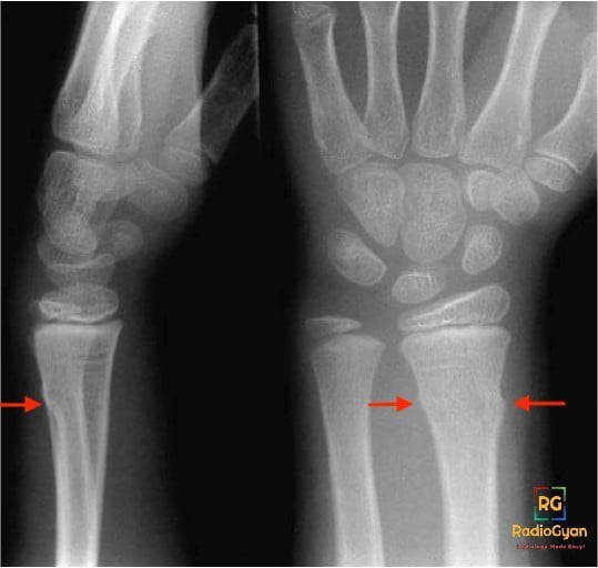
- Buckling of one or both of the cortexes of a long bone
- May associated with angulation of the bone.
- Complete fracture line is not visualized
- Callus formation and soft tissue swelling
- An adjacent bone may undergo plastic deformation. For eg-the ulna in fractures of the distal radius
- Comparison with contralateral limb’s radiograph in case of suspicious plastic deformation
Imaging Recommendation:
Radiograph in two orthogonal views.
Top 3 Differential Diagnosis:
- Greenstick fracture- mid diaphyseal location with complete break in single cortex with angulation of the bone
- Salter harris fracture- epiphyseal plate fractures, depending on the type-epiphyseal displacement with / without fracture of metaphysis
- Bowing fracture- bowing of the long tubular bones without visible fracture line or cortical break
Clinical Features:
- Symptoms – pain and edema at the fracture site, tenderness and restricted range of motion
- Age/Sex predilection 7-12 year age group, no sex predilection
- Risk factors -fall on outstretched hand, weightlifting, gymnastics, genetic predisposition to low bone density,
Etymology and synonyms:
The name “torus” has Latin roots that means “swelling” or “protuberance’’. The cortical buckling in fracture resembles the convex portion of the upper part of the base of a Greek column. Here is an image from https://buffaloah.com/ depicting a Torus.

Even though the terms buckle and torus fractures are used interchangeably, depending on the force involved in causing fracture, these fractures are described as
- Buckle fracture- single cortex is buckled/compressed
- Torus fracture – both cortices are buckled/compressed
- Greenstick fracture – complete break in a single cortex
- Complete fracture – complete break in both cortices
Treatment:
- Rest, immobilization with a splint or cast,
- Physical therapy to increase range of motion and strength
References:
Single best review article
Other references
- Davidson JS, Brown DJ, Barnes SN, Bruce CE. Simple treatment for torus fractures of the distal radius. J Bone Joint Surg Br. 2001 Nov;83(8):1173-5. doi:10.1302/0301-620x.83b8.11451.PMID: 11764434.
- Imaging in Pediatric Skeletal Trauma: Techniques and Applications by Karl J. Johnson (Editor), E. Bache (Editor), A.L. Baert (Foreword). ISBN-10 : 3540661964
Done by – Dr Gauri / edited by Mansi.

Improve Content And Help Your Colleagues!
Found an error in the post? Please let us know using our contact page and we will update it with due credits!
If you wish to contribute radiological images for this case (or any other article on the website), you can submit them here and you will be duly credited: Submit radiology case