


A boxer’s fracture is a fracture of the neck of the fifth metacarpal, almost always caused by punching something solid with a clenched fist. It is the most common metacarpal fracture and one of the most common fractures presenting to any emergency department. On radiographs you see a transverse or slightly comminuted fracture line through the metacarpal neck with the head tipped volarly, giving an apex-dorsal deformity and a lost knuckle contour.
The fracture itself is easy. The reporting decisions around it are where most of the value sits, and they are the three things this article is built around: which view you measure the angulation on, how much angulation actually matters (far more than most people quote), and the two findings that override the angle entirely – rotational malalignment and an open wound over the knuckle. Get those three right and the rest of the management follows.
| At a glance | |
|---|---|
| Definition | Fracture of the fifth metacarpal neck, typically transverse and impacted, with volar angulation of the distal fragment |
| Also called | Brawler’s fracture, scrapper’s fracture, street fighter’s fracture, bar-room fracture, punch fracture, subcapital fifth metacarpal fracture |
| Typical patient | Young adult male, punch injury to a wall, door or another person |
| Mechanism | Axial load along the clenched fifth metacarpal, or a direct blow to the dorsum of the hand |
| First-line imaging | PA, oblique and lateral radiographs of the hand |
| View to measure on | True lateral or 45-degree pronated oblique. Never the PA |
| Commonly quoted acceptable angulation | About 40 degrees, but randomised data support accepting up to 70 degrees in the fifth metacarpal |
| Findings that force surgery | Rotational malalignment (scissoring), open fracture or fight bite, neurovascular injury, intra-articular extension |
| Default treatment | Buddy taping or a soft wrap with early mobilisation. Reduction and casting are not required in most cases |
| Union | Radiological union at roughly 7 to 8 weeks; return to work is usually much earlier |
| Main long-term issue | Cosmetic loss of the knuckle prominence, which rarely affects function |
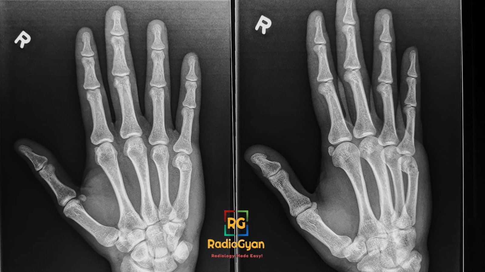
Quiz
A 24-year-old man has the radiographs above after punching another man in the mouth during a fight. Which single finding would most change your management?
- Volar angulation of the metacarpal head measured at 35 degrees
- A 4 mm laceration over the fifth metacarpophalangeal joint
- Mild impaction at the fracture site
- Loss of the normal knuckle contour on clinical examination
Click here for the answer
Answer: 2. A small laceration over the knuckle after a punch to the mouth is a fight bite (clenched-fist injury) until proven otherwise. The tooth enters the extensor tendon and the metacarpophalangeal joint capsule with the finger flexed; when the hand opens, the wound slides away from the joint breach and looks trivial. It carries a high rate of septic arthritis and osteomyelitis and needs urgent exploration, washout and antibiotics. The other three findings are all expected features of an ordinary boxer’s fracture and none of them, on its own, changes the plan: 35 degrees of volar angulation is well within the accepted range, impaction is the norm, and the lost knuckle contour is a cosmetic consequence rather than a functional one.
What is a boxer’s fracture?
The fifth metacarpal neck is the narrow, thin-cortexed segment just proximal to the metacarpal head. When a clenched fist strikes a solid object, the force runs proximally along the shaft and the neck buckles because it is the weakest point in that column. The metacarpal head then rotates volarly, pulled by the intrinsic interosseous muscles that cross palmar to the axis of the metacarpophalangeal joint, and the dorsal cortex is comminuted or impacted. That combination gives the classic apex-dorsal angulation.
The fifth ray tolerates this deformity better than any other because of the mobility of the fifth carpometacarpal joint, which allows roughly 20 to 30 degrees of flexion-extension and lets the hand compensate for a malunited neck. The index and middle carpometacarpal joints are essentially rigid, which is exactly why the acceptable angulation for the second and third metacarpals is a fraction of what is acceptable for the fifth.

Why the name is a misnomer, and what else it is called
Trained boxers rarely sustain this fracture. A boxer strikes with the fist in neutral alignment through the second and third metacarpals, which are the strong, immobile radial rays, and does so wearing wraps and gloves. It is the untrained striker who throws a hooking or roundhouse blow, lands on the ulnar border of the hand, and fractures the fourth or fifth metacarpal. The eponym therefore describes the wrong sport and the wrong metacarpal, which makes it a textbook example of Stigler’s law of eponymy: no scientific discovery is named after its original discoverer.
Dr. Indra Neil Mekala made exactly this point in our radiology learning group: a trained professional strikes with the fist in a neutral position, whereas an inexperienced street fighter delivers a roundhouse blow that exposes the fourth and fifth metacarpals. In current radiological and orthopaedic usage, however, “boxer’s fracture” unambiguously means the fifth metacarpal neck, and that is how it should be reported.
| Term | What it actually refers to | Comment |
|---|---|---|
| Boxer’s fracture | Fracture of the fifth metacarpal neck | The standard usage in radiology reports and in the orthopaedic literature, despite the misnomer |
| Brawler’s / scrapper’s / street fighter’s fracture | Fracture of the fourth or fifth metacarpal neck from an untrained punch | Etymologically the more accurate label for what we call a boxer’s fracture |
| Bar-room fracture | Same injury, named for the setting | Purely colloquial; not used in reporting |
| Punch fracture | Any metacarpal fracture from a punch | Non-specific; describes mechanism rather than site |
| True boxer’s injury | Second or third metacarpal neck or head fracture, or a Bennett fracture of the thumb | The injury pattern actually seen in trained fighters |
| Fight bite (clenched-fist injury) | Wound over the metacarpophalangeal joint from striking teeth, with or without a fracture | A different diagnosis that frequently coexists. Always excluded explicitly |
X-ray of a boxer’s fracture: which views and what to look for
Plain radiographs are diagnostic and are all that most of these fractures need. Request three views of the hand, not two, because the amount of angulation is the number that drives management and it can only be assessed on a lateral or a properly pronated oblique.
| View | What it is good for | What it will mislead you about |
|---|---|---|
| PA (dorsopalmar) | Detecting the fracture, comminution, intra-articular extension, shortening, and screening the other metacarpals and the carpus | Angulation. Volar tilt is out of plane on the PA and is almost invisible; a badly angulated fracture can look near-anatomical |
| 45-degree pronated oblique | The workhorse view for the fourth and fifth metacarpal necks, which overlap on the true lateral | It slightly overestimates volar angulation compared with CT, so compare against the normal side rather than against a textbook number |
| True lateral | The reference plane for angulation and for detecting dorsal comminution | The metacarpals superimpose, so the fifth metacarpal neck can be hard to isolate |
| Brewerton view (MCP joints flexed 65 degrees, dorsum on the plate, tube angled 15 degrees ulnar) | Metacarpal head fractures and collateral-recess erosions | Not a routine trauma view; use it only for a suspected head fracture |
| CT | Occult fracture, intra-articular head fracture, carpometacarpal fracture-dislocation, suspected associated hamate fracture, preoperative planning | Not required for a straightforward neck fracture |
Reporting checklist
- Which metacarpal, and which part. Neck, shaft, base or head. Head fractures are intra-articular and are managed differently.
- Fracture pattern. Transverse, oblique, spiral or comminuted. Spiral and long oblique patterns shorten and rotate; transverse neck fractures angulate.
- Volar angulation in degrees, and say which view you measured it on.
- Dorsal comminution. A comminuted dorsal cortex means the fracture will lose any reduction you achieve, which is an argument against attempting one.
- Shortening, compared with the adjacent metacarpal.
- Intra-articular extension into the metacarpophalangeal joint.
- The other metacarpals, the carpus and the carpometacarpal joints. Multiple metacarpal fractures behave differently from an isolated one, and a fifth carpometacarpal fracture-dislocation is a classic miss on the PA view.
- Soft tissues. Look specifically for gas, a radio-opaque foreign body or a tooth fragment over the metacarpophalangeal joint. That finding turns a routine fracture into a surgical emergency.
How to measure volar angulation (and the baseline everyone forgets)
The fifth metacarpal neck is already angled volarly in a normal hand, so an angle measured off the fracture film is not the same thing as the deformity caused by the injury. Lowdon measured a normal head-shaft angle of about 26 degrees on the oblique view, and roughly 15 degrees is quoted on the true lateral. Whatever number you measure, the clinically meaningful figure is the excess over that physiological baseline, and the cleanest way to establish the baseline is the patient’s own uninjured hand.
| Method | How it is drawn | Performance |
|---|---|---|
| Metacarpal neck-shaft centre (MNSC) angle | Angle between the mid-medullary axis of the shaft and a line through the centre of the head and the centre of the neck | The most reliable method. On 45-degree pronated oblique radiographs, Lee et al. found inter-observer ICCs of 0.88 to 0.93, better than the shaft-articular-surface method and better than sagittal CT |
| Shaft-articular surface (SAS) angle | Angle between the shaft axis and a line along the articular surface of the metacarpal head | Consistently reads a few degrees higher than MNSC and is less reproducible |
| Dorsal tangent line | Angle between tangents drawn along the dorsal cortices of the proximal and distal fragments | Proposed as a more reproducible alternative to intersecting central axes, particularly for short distal fragments |
| Comparison with the contralateral hand | Measure the same angle on the uninjured side and subtract | The only way to separate injury-related angulation from the patient’s own anatomy. Worth doing when the measured angle sits near a decision threshold |
Two practical consequences. First, a measured angle carries an error of several degrees between observers, so a report should not imply precision it does not have: approximately 45 degrees is honest, 47 degrees is not. Second, because there is no consensus on the measurement method or the surgical cut-off, a number quoted without the method and the view attached is not actionable. State both.
How much angulation is acceptable?
This is the question the fracture is really about, and the honest answer is that the traditional teaching numbers are considerably more conservative than the evidence requires. The commonly quoted limits below reflect standard hand-surgery teaching and are useful as a framework, particularly for the radial metacarpals where they are not in dispute.
| Metacarpal | Commonly quoted acceptable volar angulation | Why the limit is what it is |
|---|---|---|
| Second (index) | About 10 to 15 degrees | The second carpometacarpal joint is essentially immobile, so there is no compensation for a malunion |
| Third (middle) | About 15 to 20 degrees | Also a rigid carpometacarpal joint |
| Fourth (ring) | About 30 to 40 degrees | The fourth carpometacarpal joint allows roughly 15 degrees of motion |
| Fifth (little) | About 40 degrees by classical teaching, and up to 70 degrees on randomised evidence | The fifth carpometacarpal joint allows 20 to 30 degrees of flexion-extension, which absorbs a large malunion. This is the range where the literature and the textbooks disagree |
The randomised data are consistent and they point one way. Statius Muller and colleagues randomised boxer’s fractures angulated up to 70 degrees to a pressure bandage with immediate mobilisation or to three weeks in an ulnar gutter cast, and found no difference in range of motion, pain, satisfaction, return to work or need for physiotherapy. A meta-analysis of comparative studies found that conservative management leaves a worse radiographic angle than surgery but produces equivalent QuickDASH scores, equivalent grip strength and an earlier return to work, regardless of the palmar angulation. And a Bayesian network meta-analysis of six randomised trials ranked conservative treatment first for the lowest total complication rate, ahead of plate fixation and antegrade intramedullary nailing, with transverse K-wire pinning last.
The practical position, which the 2024 current-concepts review states plainly, is that 70 degrees is the acceptable angulation for conservative management in most studies. Severe angulation beyond that causes two problems worth naming in a report: loss of the knuckle prominence with a compensatory prominence of the metacarpal head in the palm, which can be uncomfortable when gripping, and pseudoclawing – an extension lag at the metacarpophalangeal joint with compensatory hyperextension, seen when the deformity is large enough to slacken the extensor mechanism.
Rotation is the finding that actually forces surgery
Angulation is negotiable. Rotation is not. A malrotated metacarpal makes the finger cross over its neighbour when the hand closes – scissoring – and that is a functional problem that does not remodel and does not compensate. Seitz and Froimson quantified the amplification: 10 degrees of rotational malalignment at the metacarpal produces about 2 cm of overlap at the fingertip.
- Rotation is a clinical diagnosis, not a radiographic one. Ask the patient to make a fist, or, if too painful, flex the fingers passively and look at the plane of the nail plates. All four fingers should converge towards the scaphoid tubercle. Any digit that crosses another is malrotated.
- What the radiograph can show is an abrupt change in the calibre of the medullary canal or in the cortical thickness across the fracture, which suggests the two fragments are being viewed in different planes. It is a soft sign and its absence excludes nothing.
- Which patterns rotate. Spiral and long oblique shaft fractures rotate and shorten. Transverse neck fractures usually do not, which is why the classic boxer’s fracture is so often managed without surgery.
- Shortening matters through the extensor mechanism. In a cadaveric study, each 2 mm of metacarpal shortening produced roughly 7 degrees of extensor lag at the metacarpophalangeal joint. Shortening beyond about 5 mm is generally regarded as an indication for fixation.
When does a boxer’s fracture need surgery?
| Category | Finding | Why it matters |
|---|---|---|
| Absolute | Rotational malalignment / scissoring | Does not remodel; 10 degrees at the metacarpal equals about 2 cm of fingertip overlap |
| Absolute | Open fracture, including any wound over the knuckle after a punch to the mouth | Needs urgent debridement, washout and antibiotics. A splint is not treatment for an open fracture |
| Absolute | Neurovascular injury | Requires exploration |
| Absolute | Displaced intra-articular fracture of the metacarpal head | Articular incongruity leads to stiffness and post-traumatic arthritis |
| Relative | Volar angulation beyond about 70 degrees | Loss of knuckle contour, palmar prominence of the head, pseudoclawing |
| Relative | Shortening beyond about 5 mm | Approximately 7 degrees of extensor lag per 2 mm of shortening |
| Relative | Multiple metacarpal fractures | The adjacent intact metacarpals normally act as an internal splint. Lose them and the construct is unstable |
| Relative | Fracture-dislocation of the fifth carpometacarpal joint | Unstable; frequently missed on the PA view alone |
Treatment: what the randomised evidence actually shows
For a closed, non-rotated boxer’s fracture, the default is buddy taping or a soft wrap with early mobilisation. Formal reduction under block, a moulded ulnar gutter cast and serial radiographs have all been tested against the simpler option and none of them has come out ahead. The evidence spans more than twenty years and several countries and is unusually consistent.
| Study | Design and population | Comparison | Result |
|---|---|---|---|
| Statius Muller et al., Arch Orthop Trauma Surg 2003 | RCT, 40 patients, angulation up to 70 degrees | Pressure bandage for 1 week with immediate mobilisation vs 3 weeks in an ulnar gutter cast | No difference in range of motion, satisfaction, pain, return to work or physiotherapy need. Reduction below 70 degrees was of no value |
| van Aaken et al., Arch Orthop Trauma Surg 2016 | Prospective multicentre randomised non-inferiority trial | Soft wrap and buddy taping vs reduction and casting | Soft wrap and buddy taping non-inferior |
| Dunn et al., Orthopedics 2016 | Systematic review of RCTs of cast vs soft wrap without reduction | Cast immobilisation vs soft wrap | Cast immobilisation is not superior in most cases; reduction and casting are not necessary |
| Zong et al., Medicine 2016 | Bayesian network meta-analysis, 6 RCTs, 288 patients | Conservative vs plate fixation vs antegrade intramedullary nailing vs transverse K-wire pinning | Conservative treatment ranked best for lowest total complications (SUCRA 94.1%). Transverse pinning ranked worst (15.7%) |
| Pellatt et al., Ann Emerg Med 2019 | RCT, adults 18 to 70, uncomplicated fractures, 2 Australian hospitals | Buddy taping vs plaster immobilisation | Buddy taping gave equivalent functional outcomes |
| Martinez-Catalan et al., J Hand Surg Am 2020 | Prospective RCT, angulation below 70 degrees, no rotational deformity | Buddy taping vs closed reduction and cast | No functional advantage to reduction and casting |
| Chong et al., J Hand Surg Asian Pac Vol 2020 | Meta-analysis of comparative studies | Conservative vs operative | Conservative leaves a worse radiographic angle but gives equivalent QuickDASH, grip strength and total active motion, with fewer days off work and no surgical complications |
| Zawam et al., Eur J Trauma Emerg Surg 2024 | RCT, 90 active adults, palmar angulation 30 to 70 degrees | Ulnar gutter slab vs percutaneous transverse pinning | Comparable union (7.8 vs 7.4 weeks), QuickDASH and total active motion. All patients returned to pre-injury work |
One historical technique deserves a caveat. The Jahss manoeuvre – flexing the metacarpophalangeal and proximal interphalangeal joints to 90 degrees and pushing the head dorsally through the proximal phalanx – is still described for reduction, but immobilising the hand in that position has caused pressure necrosis over the dorsum of the proximal interphalangeal joint and stiffness. If a reduction is performed, the hand should be immobilised in the intrinsic-plus (safe) position, not in the 90-90 position.
Where fixation is genuinely required, the usual options are antegrade intramedullary K-wires (bouquet nailing), intramedullary headless compression screws, transverse pinning to the adjacent intact metacarpal, and plate fixation for comminution or multiple metacarpal fractures. The network meta-analysis above is a useful reminder that transverse pinning carried the highest complication burden of the operative options.
Associated injuries and the things that catch people out
| Entity | How it presents | How not to miss it |
|---|---|---|
| Fight bite (clenched-fist injury) | A small, innocuous-looking laceration over the dorsum of a metacarpophalangeal joint after a punch to the mouth, sometimes with a fracture | The wound is over the joint with the finger flexed and moves away from the breach when the hand opens, so it looks trivial. Look for gas, a tooth fragment or a foreign body on the radiograph. Organisms include Streptococcus anginosus, Staphylococcus aureus, Eikenella corrodens and anaerobes such as Fusobacterium and Prevotella, so co-amoxiclav is the usual empirical cover |
| Fifth carpometacarpal fracture-dislocation | Same punch mechanism, pain at the base rather than the neck of the metacarpal | The PA view can look near-normal. Check the parallel carpometacarpal joint lines and look at the lateral or a 30-degree pronated oblique. CT if in doubt |
| Hamate fracture | Ulnar-sided hand pain after a punch or a fall | Overlapping structures obscure the hamate on the PA view; carpal tunnel view or CT |
| Metacarpal head fracture | Intra-articular fracture distal to the neck | A different injury with a different threshold for surgery. Brewerton view or CT |
| Multiple metacarpal fractures | Higher-energy injury, often a crush | The adjacent intact metacarpals are what make an isolated fracture stable. Always look at all five |
| Intentional and recurrent injury | A boxer’s fracture is frequently the result of a deliberate act | A recognised marker for injury recidivism. Worth a brief social and safeguarding thought, particularly in adolescents and in repeat attenders |
Healing, complications and what to tell the patient
| Outcome | Expected course |
|---|---|
| Radiological union | Around 7 to 8 weeks in randomised series, whether treated conservatively or with pinning |
| Return to work and activity | Usually well before radiological union; earlier with conservative treatment than after surgery |
| Loss of knuckle prominence | Common and permanent with a significant malunion. Cosmetic rather than functional in almost all patients |
| Palmar prominence of the metacarpal head | Can be uncomfortable on gripping when angulation is severe |
| Pseudoclawing | Extension lag at the metacarpophalangeal joint with compensatory hyperextension; associated with severe angulation |
| Extensor lag from shortening | Roughly 7 degrees per 2 mm of metacarpal shortening |
| Stiffness | The main risk of prolonged immobilisation, which is the argument for early mobilisation |
| Infection | Uncommon in closed fractures; the dominant risk in fight bites and after pin fixation |
| Non-union | Rare in the metacarpal neck |
Frequently asked questions
References
- Statius Muller MG, Poolman RW, van Hoogstraten MJ, Steller EP. Immediate mobilization gives good results in boxer’s fractures with volar angulation up to 70 degrees: a prospective randomized trial comparing immediate mobilization with cast immobilization. Arch Orthop Trauma Surg. 2003;123(10):534-537. PMID: 14639483
- van Aaken J, Fusetti C, Luchina S, et al. Fifth metacarpal neck fractures treated with soft wrap/buddy taping compared to reduction and casting: results of a prospective, multicenter, randomized trial. Arch Orthop Trauma Surg. 2016;136(1):135-142. PMID: 26559192
- Dunn JC, Kusnezov N, Orr JD, Pallis M, Mitchell JS. The boxer’s fracture: splint immobilization is not necessary. Orthopedics. 2016;39(3):188-192. PMID: 27018606
- Zong SL, Zhao G, Su LX, et al. Treatments for the fifth metacarpal neck fractures: a network meta-analysis of randomized controlled trials. Medicine (Baltimore). 2016;95(11):e3059. PMID: 26986129
- Pellatt R, Fomin I, Pienaar C, et al. Is buddy taping as effective as plaster immobilization for adults with an uncomplicated neck of fifth metacarpal fracture? A randomized controlled trial. Ann Emerg Med. 2019;74(1):88-97. PMID: 30853124
- Martinez-Catalan N, Pajares S, Llanos L, Mahillo I, Calvo E. A prospective randomized trial comparing the functional results of buddy taping versus closed reduction and cast immobilization in patients with fifth metacarpal neck fractures. J Hand Surg Am. 2020;45(12):1134-1140. PMID: 32718787
- Chong HH, Hau MY, Shah R, Singh H. Management of little finger metacarpal fractures: a meta-analysis of the current evidence. J Hand Surg Asian Pac Vol. 2020;25(3):281-290. PMID: 32723052
- Zawam SH, Abdelrazek BH, Elmofty A, Morsy A, Abousayed M. Conservative treatment versus transverse pinning in fifth metacarpal neck fractures in active adults: a randomized controlled trial. Eur J Trauma Emerg Surg. 2024;50(2):531-542. PMID: 38151577
- Sain A, Arif S, Manyar H, et al. Current concepts in the management of boxer’s fracture. Georgian Med News. 2024;(347):122-124. PMID: 38609127
- Lee JK, Cho WM, Lee HI, et al. The inter- and intra-observer reliability of volar angulation measurements in a fifth metacarpal neck fracture. Arch Orthop Trauma Surg. 2022;142(7):1705-1713. PMID: 35316389
- Lowdon IM. Fractures of the metacarpal neck of the little finger. Injury. 1986;17(3):189-192. PMID: 3818059
- Strauch RJ, Rosenwasser MP, Lunt JG. Metacarpal shaft fractures: the effect of shortening on the extensor tendon mechanism. J Hand Surg Am. 1998;23(3):519-523. PMID: 9620194
- Seitz WH Jr, Froimson AI. Management of malunited fractures of the metacarpal and phalangeal shafts. Hand Clin. 1988;4(3):529-536. PMID: 3049643
- Talan DA, Abrahamian FM, Moran GJ, et al. Clinical presentation and bacteriologic analysis of infected human bites in patients presenting to emergency departments. Clin Infect Dis. 2003;37(11):1481-1489. PMID: 14614671
- Carreno A, Ansari MT, Malhotra R. Management of metacarpal fractures. J Clin Orthop Trauma. 2020;11(4):554-561. PMID: 32684692
- Hussain MH, Ghaffar A, Choudry Q, Iqbal Z, Khan MN. Management of fifth metacarpal neck fracture (boxer’s fracture): a literature review. Cureus. 2020;12(7):e9442. PMID: 32864266
Co-Authors: Dr. Mansi Sarmalkar and Dr. Gauri Parvathy.

Improve Content And Help Your Colleagues!
Found an error in the post? Please let us know using our contact page and we will update it with due credits!
If you wish to contribute radiological images for this case (or any other article on the website), you can submit them here and you will be duly credited: Submit radiology case