


CT appearance of Pulmonary Langerhans Cell Histiocytosis (LCH) and approach to imaging of cystic lung diseases
Pulmonary LCH is a rare disease found almost exclusively in cigarette smokers.
Radiologic findings of pulmonary LCH include
- Nodules, usually smaller than 1 to 5 mm, centrilobular, and peribronchiolar, may be cavitary, may be seen in association with cysts.
- Thick-walled or thin-walled cysts. – Progression over time from nodules to thick-walled cysts to thin-walled cysts.
- Upper lobe predominance in size and number of nodules or cysts, costophrenic angles spared.
- It can be associated with lymphadenopathy.
Pulmonary LCH vs lymphangioleiomyomatosis radiology
| Imaging features of Pulmonary Langerhans Cell Histiocytosis | Imaging features of Lymphangioleiomyomatosis |
|---|---|
| – Thick-walled, or thin-walled cysts – Nodules, usually smaller than 1 to 5 mm, centrilobular and peribronchiolar, may be cavitary, may be seen in association with cysts – Progression over time from nodules to thick-walled cysts to thin-walled cysts – Upper lobe predominance in size and number of nodules or cysts, costophrenic angles spared – Fine reticular opacities – Ground-glass opacity – Mosaic perfusion or air trapping – Pulmonary hypertension | – Thin-walled lung cysts, usually round in shape – Diffuse distribution, costophrenic angles involved – Mild septal thickening or ground-glass opacity – Lymph node enlargement – Small nodules in patients with TSC – Pleural effusion – Pneumothorax |
Common causes of cystic lung disease
Common (not truly cystic lung diseases but often confused as cysts)
- Emphysema (e.g., bullae) – Thin imperceptible walls.
- Cystic bronchiectasis
- Honeycombing
Less Common
- Pulmonary Langerhans Cell Histiocytosis
- Lymphangioleiomyomatosis (isolated LAM or associated with the TSC)
- Lymphocytic interstitial pneumonia (LIP) – Associated with Sjogren’s, AIDS, SLE
- Cystic metastases
- Pneumatoceles –
- Birt-Hogg-Dube syndrome = Lung cysts and renal oncocytomas.
Approach to imaging of cystic lung diseases
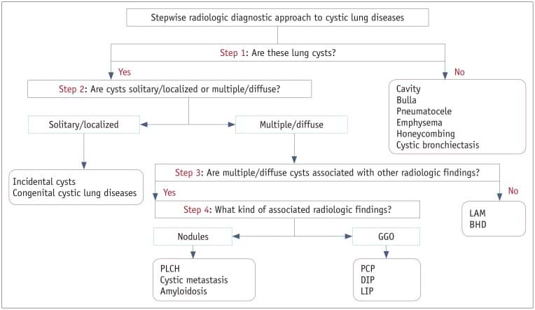
Reference: A Stepwise Diagnostic Approach to Cystic Lung Diseases for Radiologists
Watch the video for the detailed case discussion and PACS based image stack.
Reference and further reading:

To attend live, join our Telegram group to get regular updates for these webinars:
More Radiology videos:


Improve Content And Help Your Colleagues!
Found an error in the post? Please let us know using our contact page and we will update it with due credits!
If you wish to contribute radiological images for this case (or any other article on the website), you can submit them here and you will be duly credited: Submit radiology case