
Topics covered:
- Normal myelination.
- Hypoxic Ishemic Encephalopathy.
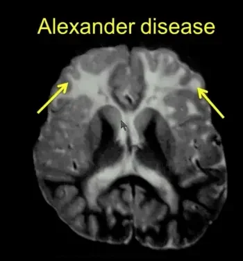
- Dysmelinating and demyelinating disorders.
- Approach to dysmyelinating disorders depending on
- Involvement of grey matter / white matter or both
- Central/ Peripheral involvement.
- Enhancement pattern
- Symmetrical involvement.
- Head size.
- Systemic involvement.
- Important disorders are covered with typical imaging findings.
- Check out the Atlas of normal myelination in neonates on our anatomy page.
- Go through this excellent Imaging flowchart from the AJR article titled here.
Click here to to read an excellent presentation on Normal Myelination by Fang Yu 1 M.D., Michael Wang 1 M.D., Kiran Sargar 2 M.D., Yutaka Sato 3 M.D., Achint K. Singh 1 M.D. 1: University of Texas Health Science Center San Antonio.
Resources:
- Normal MR Imaging Anatomy of Brain.
- AJR : Inborn Errors of Metabolism: Combining Clinical and Radiologic Clues to Solve the Mystery
- RSNA : Leukodystrophy in Children A Pictorial Review of MR Imaging Features.
- Barkovich Pediatric Neuroimaging

Improve Content And Help Your Colleagues!
Found an error in the post? Please let us know using our contact page and we will update it with due credits!
If you wish to contribute radiological images for this case (or any other article on the website), you can submit them here and you will be duly credited: Submit radiology case
White matter diseases lecture is excellent
Glad you liked it Tarun.
We share more webinars over telegram: https://t.me/radiogyan