Lithium Nephropathy / Lithium induced renal disease
Radiology features
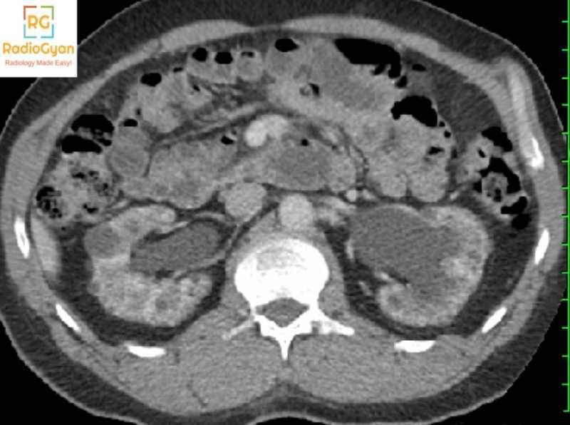
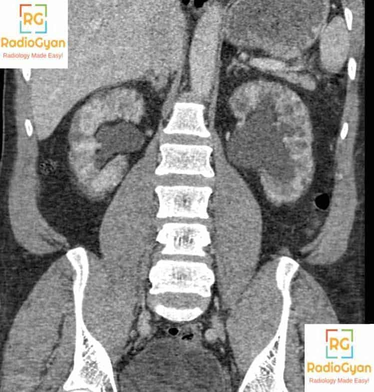
- Multiple innumerable micro cysts randomly distributed in BOTH cortex and medulla or predominantly cortex are characteristic radiology findings in cases of lithium induced renal disease/lithium nephropathy.
- Cysts are usually 1-2mm in diameter.
- Cysts arise from distal tubular structures and collecting ducts.
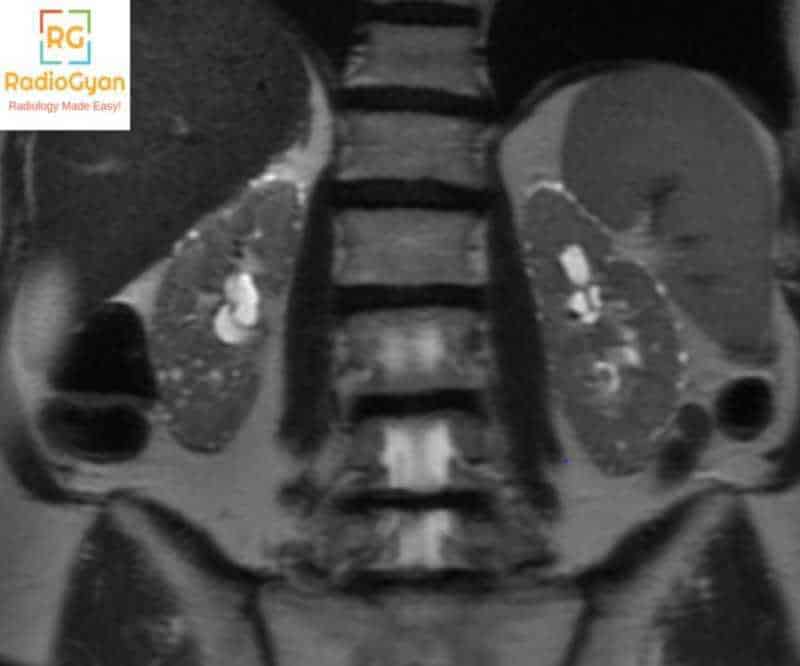
- Best seen on T2W MRI images.
T2W MRI images in another patient shows tiny subcentimeter sized in both kidneys.
- Cysts can also bee seen on USG : Lithium nephropathy sonographic findings
- Differentials diagnosis with imaging features:
- Autosomal polycystic kidney (ADPKD)
- Nephromegaly with large cysts.
- Cysts are often of varying sizes and few of them are complicated.
- Glomerulocystic kidney disease:
- Patients are usually children or young adult.
- Cysts arise from Bowman space so ONLY in CORTEX of the kidney.
- Medullary cystic kidney disease:
- Cysts are present in the medulla and corticomedullary junction but spare the cortex.
- Acquired cystic kidney disease
- Seen in patients on long term dialysis.
- Cysts are NOT uniform in size although they affect both cortex and medulla.
- Autosomal polycystic kidney (ADPKD)
Clinical features and pathology of lithium nephropathy:
- Lithium is used to treat unipolar major depression and bipolar affective disorders.
- Lithium toxicity spectrum includes:
- Acute intoxication.
- Nephrogenic diabetes insipidus / Polyuria-polydipsia syndrome: Harmless and reversible.
- Chronic renal disease (10-20 years): Chronic focal interstitial nephritis
- Approx. 30-60% of patients on long term lithium therapy can have cysts.
- Patients with chronic interstitial nephritis and typical cysts are at risk of developing end-stage renal disease.
References and further reading:
- RadCases
- Genitourinary Imaging: Case Review
- Genitourinary Imaging: The Requisites (Requisites in Radiology)
- Lithium-induced Nephropathy Radiology
- Chronic Lithium Nephropathy: MR Imaging for Diagnosis
- Lithium Nephropathy USG
More than 400 interesting Radiology Cases: Radiology Spotters Cases


