
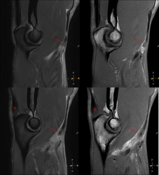
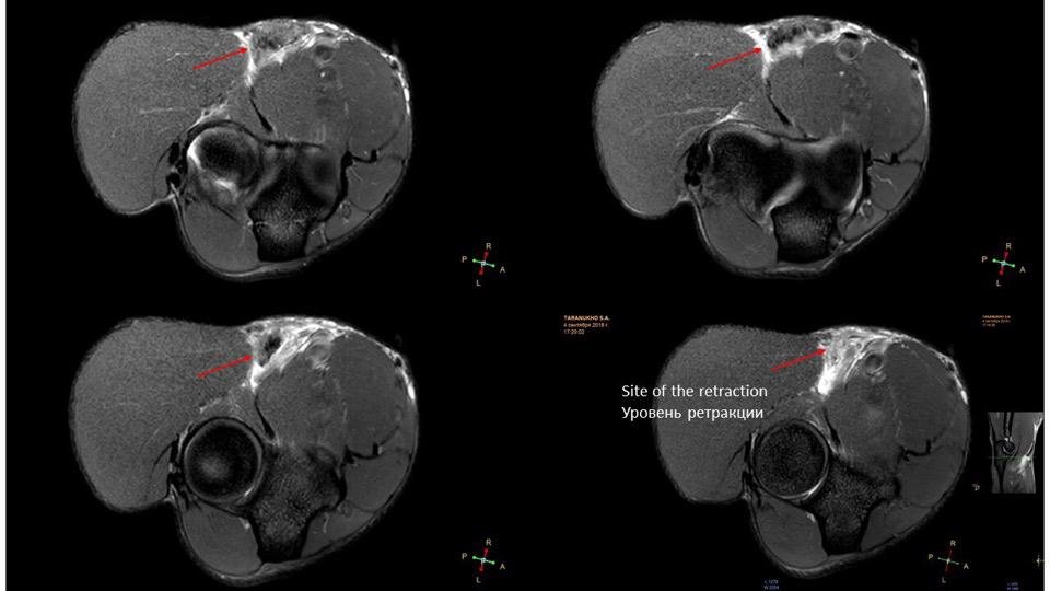
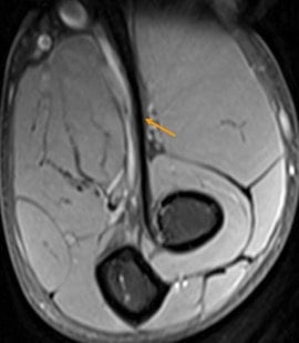
Elbow. Male, 31 years, acute pain and swelling after work out. Full-thickness tear of the distal biceps tendon. MR images showing retracted biceps tendon fibers (arrow) surrounded by edema. Note the thickened distal triceps tendon (asterisk) as a result of chronic overuse.
Biceps brachii distal tendon rupture:
Clinical Features:
- Most commonly in the dominant arm of males after the fourth decade.
- Most tears occur 1–2 cm above the radial tuberosity as this is a hypoperfused zone.
- Complete tears are evident clinically however, differentiation between complete and partial tears is difficult.
| Complete Tear | Partial Tear |
| Single traumatic event involving large force acting against resistance from an elbow flexed to 90° | Minor trauma or not even associated with a traumatic event when the underlying tendon is degenerated) |
| Usually acute | Usually chronic presentation |
| Hook test does not elicit pain. “Popeye sign” | Hook test elicits pain |
| Discontinuity with or without retraction | At least some fibers are intact |
| Surgical management | Conservative to start with. |

Ultrasound:
- Dynamic imaging can be used to evaluate continuity of the tendon or the abnormal movement of a disconnected proximal tendon.
- Partial tears show changed caliber with reduced echogenicity which is often difficult to evaluate.
- Posterior acoustic shadowing at the distal biceps has proven to be highly sensitive for a full thickness tear.
- Anisotropy at distal tendon end can mimic complete rupture.
Tendon retraction of less than 8 cm correlates with an intact aponeurosis, whereas a retraction of more than that indicates torn aponeurosis.
MRI
- Fluid-signal filled gap on STIR sequence, increased intratendinous signal intensity, and edema in the biceps muscle belly and surrounding soft tissues are characteristic for acute full thickness rupture.
- Partial tears show altered signal and caliber of the distal biceps tendon.
- FABS view: Flexed ABducted Supinated view is superior to routine imaging for evaluation of the distal biceps tendon.
Associated pathological conditions:
- Enthesophyte formation at the radial tuberosity which also can be a contributing factor.
- Bicipitoradial bursitis.
Differentials:
- Brachialis muscle sprain.
Further reading:
- RadCases MSK
- USG of Biceps tendon rupture
- Radiographics: Distal Biceps tendon pathology
- RadSource.
- Radiology Assistant
Case courtesy: musculoskeletal_rad_cases
More radiology content at RadioGyan : Radiology resources

Improve Content And Help Your Colleagues!
Found an error in the post? Please let us know using our contact page and we will update it with due credits!
If you wish to contribute radiological images for this case (or any other article on the website), you can submit them here and you will be duly credited: Submit radiology case