


Key facts for practice and radiology board exams
ASPECTS is a 10-point score read on non-contrast CT. The middle cerebral artery (MCA) territory is divided into 10 regions and one point is deducted for each region showing early ischemic change, so a normal scan scores 10. [1] A low score no longer excludes a patient from thrombectomy: the 2026 AHA/ASA guideline recommends it down to ASPECTS 3 within 6 hours. [5]
- 10 is normal, 0 means every region is involved. Pexman described a sharp
increase in dependence and death at an ASPECTS of 7 or less, [2] but later work points to a
linear relationship between score and outcome, and no clear cut-off for a low score can be
given. [4] - Narrow the window before you score. Interobserver agreement improved with a
window width of 8 HU and a center level of 32 HU rather than a standard brain window. [3] - It is an anterior circulation score only. The posterior circulation has its
own 10-point version, pc-ASPECTS, with a different region map. [15]
How is the ASPECTS score calculated?
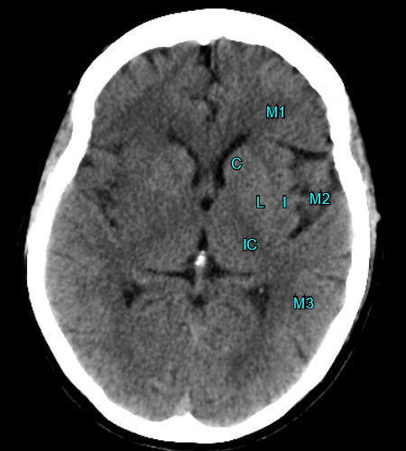
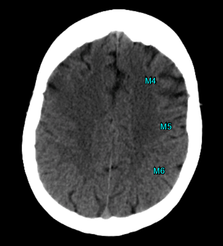
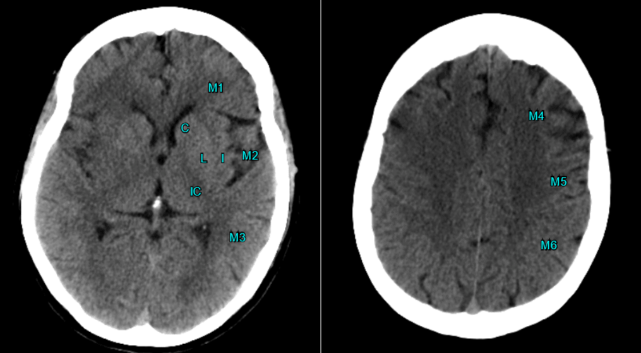
Start at 10 and subtract one point for each of the 10 MCA regions showing hypoattenuation or loss of grey-white differentiation. Seven regions sit at the ganglionic level, at or below the caudate head, and three sit above it at the supraganglionic level. Every region is worth one point, whatever its volume. [2]
| Region | Level | What it covers |
|---|---|---|
| C | Ganglionic | Caudate head |
| L | Ganglionic | Lentiform nucleus |
| IC | Ganglionic | Internal capsule |
| I | Ganglionic | Insular ribbon |
| M1 | Ganglionic | Anterior MCA cortex |
| M2 | Ganglionic | MCA cortex lateral to the insular ribbon |
| M3 | Ganglionic | Posterior MCA cortex |
| M4 | Supraganglionic | Anterior MCA territory, immediately superior to M1 |
| M5 | Supraganglionic | Lateral MCA territory, immediately superior to M2 |
| M6 | Supraganglionic | Posterior MCA territory, immediately superior to M3 |
- Window settings matter more than reading speed. A narrowed window (width 8 HU,
center level 32 HU) raised agreement between readers at every level of experience. [3] - Subtract only for acute change. Old infarcts, leukoaraiosis and motion
artifact are the three recurring traps, and the score has no way of flagging them. - Know which hemisphere is symptomatic. The good agreement reported in the
original validation (kappa 0.71 to 0.89) was obtained with knowledge of the affected
hemisphere. [1] - Report the number, not just a category. The treatment thresholds have moved
since 2019, and a recorded score survives the next revision.
Does a low ASPECTS score rule out thrombectomy?
No. This is the single biggest change in stroke CT reporting in the last decade. Under the 2019 guideline, thrombectomy within 6 hours required an ASPECTS of 6 or more, so a score of 5 was an exclusion. Six randomized trials of thrombectomy in large infarcts have since reversed that, and the 2026 AHA/ASA guideline now reads as follows. [5]
| Presentation | ASPECTS | Recommendation |
|---|---|---|
| ICA or M1 occlusion, within 6 hours, NIHSS 6 or more, prestroke mRS 0 to 1 | 3 to 10 | Class 1, recommended |
| 6 to 24 hours, age under 80, NIHSS 6 or more, prestroke mRS 0 to 1, no significant mass effect | 3 to 5 | Class 1, recommended |
| Within 6 hours, age under 80, NIHSS 6 or more, prestroke mRS 0 to 1, no significant mass effect | 0 to 2 | Class 2a, reasonable |
| Within 6 hours, NIHSS 6 or more, prestroke mRS 2 | 6 or more | Class 2a, reasonable |
| Basilar artery occlusion, within 24 hours, NIHSS 10 or more, baseline mRS 0 to 1 | pc-ASPECTS 6 or more | Class 1, recommended |
Five of the six large-core trials were positive and one was not. Pooling all six with central imaging adjudication, 1,886 patients, thrombectomy gave an adjusted generalized odds ratio of 1.63 (95% CI 1.42 to 1.88) for a better modified Rankin Scale (mRS) outcome, with lower mortality (adjusted risk ratio 0.82). The benefit held across ASPECTS strata out to 24 hours, the one exception being a core of 150 mL or more presenting beyond 6 hours. [6]
| Trial | ASPECTS eligible | Result |
|---|---|---|
| RESCUE-Japan LIMIT [7] | 3 to 5 | mRS 0 to 3 at 90 days in 31.0% vs 12.7% (relative risk 2.43) |
| SELECT2 [8] | 3 to 5, or core 50 mL or more | mRS shift, generalized odds ratio 1.51 (1.20 to 1.89); stopped early for efficacy |
| ANGEL-ASPECT [9] | 3 to 5, or core 70 to 100 mL | Generalized odds ratio 1.37 (1.11 to 1.69) |
| TENSION [10] | 3 to 5 | Adjusted common odds ratio 2.58 (1.60 to 4.15), with lower mortality |
| TESLA [11] | 2 to 5 | No significant benefit: adjusted difference 0.63 (95% CrI -0.09 to 1.34) |
| LASTE [12] | 0 to 5 | Generalized odds ratio 1.63 (1.29 to 2.06); death 36.1% vs 55.5% |
Two cautions survive the change. Symptomatic hemorrhage was more common with thrombectomy in the lowest-scoring patients, [12] and the one trial that selected on non-contrast CT alone down to ASPECTS 2 did not meet its success threshold. [11] A low ASPECTS is now a reason to discuss the case, not a reason to close it.
How reproducible is the score you just read?
Less reproducible than the decisions built on it. When 15 raters scored the same scans, interrater agreement on the full 10-point score was only slight to moderate (kappa 0.129 to 0.315), and dichotomizing at 0 to 5 versus 6 to 10 did not reach substantial agreement either (kappa 0.561). The authors concluded that agreement may be insufficient for ASPECTS to be used on its own as a treatment criterion. [13] Agreement tracks experience: against an expert consensus, a neuroradiology fellow reached kappa 0.486 to 0.678 where a senior resident reached 0.198 to 0.491. [3]
Automated scoring is now at least as consistent as the reader. Across 11 studies and 1,976 patients, automated ASPECTS agreed with the reference standard at an intraclass correlation of 0.72 against 0.62 for expert readers, the best-performing package reaching 0.86. [14] Treat the software output as a second reader rather than an answer, and look hardest at the regions where machine and human disagree most, the cortical M regions.
pc-ASPECTS: the posterior circulation score
pc-ASPECTS applies the same idea to the posterior circulation, also out of 10. Two points each are deducted for early ischemic change in the midbrain or pons, and one point each for the left or right thalamus, cerebellum and posterior cerebral artery territory. Note the last of these: the region is the PCA territory, not the occipital lobe alone. In basilar artery occlusion the score carried prognosis on CT angiography source images, where 52% of patients scoring 8 or more had a favorable outcome against 4% of those below 8, but the same score read on non-contrast CT did not predict independence. [15] The 2026 guideline uses pc-ASPECTS 6 or more as its threshold for basilar thrombectomy. [5]
Frequently asked questions
What does ASPECTS stand for?
ASPECTS stands for Alberta Stroke Program Early CT Score. It was developed at the University of Calgary and first validated in 203 patients treated with intravenous alteplase within 3 hours of onset.
Does a low ASPECTS score rule out thrombectomy?
No. Not under the current guideline. The 2026 AHA/ASA guideline recommends thrombectomy for an ASPECTS of 3 to 10 within 6 hours as a Class 1 recommendation, and for 3 to 5 between 6 and 24 hours in selected patients under 80 without significant mass effect. An ASPECTS of 0 to 2 within 6 hours is a Class 2a recommendation. The older rule, ASPECTS 6 or more, is superseded.
What window settings should I use to read ASPECTS?
A narrowed window. Interobserver agreement was better with a window width of 8 HU and a center level of 32 HU than with a standard brain window, at every level of reader experience. The figure of a 30 HU level with an 80 HU width, which circulates widely, matches no published comparison.
What is pc-ASPECTS?
pc-ASPECTS is the posterior circulation version, also scored out of 10. Two points each are deducted for the midbrain and pons, and one point each for the left and right thalamus, cerebellum and posterior cerebral artery territory. It is the score the 2026 guideline uses for basilar artery occlusion, at a threshold of 6 or more.
What is DWI-ASPECTS?
DWI-ASPECTS is the same 10-region template read on diffusion-weighted imaging instead of non-contrast CT. It detects early ischemic change more sensitively, but the published thresholds for predicting poor outcome vary between studies and no single cut-off is established. Two of the large-core thrombectomy trials enrolled more than 80% of their patients on MRI rather than CT.
References
- Barber PA, Demchuk AM, Zhang J, Buchan AM. Validity and reliability of a quantitative computed tomography score in predicting outcome of hyperacute stroke before thrombolytic therapy. Lancet. 2000;355(9216):1670-4. PMID: 10905241.
- Pexman JH, Barber PA, Hill MD, Sevick RJ, Demchuk AM, Hudon ME, et al. Use of the Alberta Stroke Program Early CT Score (ASPECTS) for assessing CT scans in patients with acute stroke. AJNR Am J Neuroradiol. 2001;22(8):1534-42. PMID: 11559501.
- Kobkitsuksakul C, Tritanon O, Suraratdecha V. Interobserver agreement between senior radiology resident, neuroradiology fellow, and experienced neuroradiologist in the rating of Alberta Stroke Program Early Computed Tomography Score (ASPECTS). Diagn Interv Radiol. 2018;24(2):104-7. PMID: 29467112.
- Schroder J, Thomalla G. A critical review of Alberta Stroke Program Early CT Score for evaluation of acute stroke imaging. Front Neurol. 2016;7:245. PMID: 28127292.
- Prabhakaran S, Gonzalez NR, Zachrison KS, Adeoye O, Alexandrov AW, Ansari SA, et al. 2026 guideline for the early management of patients with acute ischemic stroke: a guideline from the American Heart Association/American Stroke Association. Stroke. 2026;57(8):e316-e436. PMID: 41582814.
- Sarraj A, Thomalla G, Yoshimura S, Huo X, Costalat V, Zaidat OO, et al. Endovascular thrombectomy for patients with large-core ischaemic stroke presenting up to 24 h after onset (ATLAS): a systematic review and individual patient data meta-analysis with central imaging adjudication. Lancet. 2026;407(10543):2015-26. PMID: 42107392.
- Yoshimura S, Sakai N, Yamagami H, Uchida K, Beppu M, Toyoda K, et al. Endovascular therapy for acute stroke with a large ischemic region. N Engl J Med. 2022;386(14):1303-13. PMID: 35138767.
- Sarraj A, Hassan AE, Abraham MG, Ortega-Gutierrez S, Kasner SE, Hussain MS, et al. Trial of endovascular thrombectomy for large ischemic strokes. N Engl J Med. 2023;388(14):1259-71. PMID: 36762865.
- Huo X, Ma G, Tong X, Zhang X, Pan Y, Nguyen TN, et al. Trial of endovascular therapy for acute ischemic stroke with large infarct. N Engl J Med. 2023;388(14):1272-83. PMID: 36762852.
- Bendszus M, Fiehler J, Subtil F, Bonekamp S, Aamodt AH, Fuentes B, et al. Endovascular thrombectomy for acute ischaemic stroke with established large infarct: multicentre, open-label, randomised trial. Lancet. 2023;402(10414):1753-63. PMID: 37837989.
- Writing Committee for the TESLA Investigators, Yoo AJ, Zaidat OO, Sheth SA, Rai AT, Ortega-Gutierrez S, et al. Thrombectomy for stroke with large infarct on noncontrast CT: the TESLA randomized clinical trial. JAMA. 2024;332(16):1355-66. PMID: 39374319.
- Costalat V, Jovin TG, Albucher JF, Cognard C, Henon H, Nouri N, et al. Trial of thrombectomy for stroke with a large infarct of unrestricted size. N Engl J Med. 2024;390(18):1677-89. PMID: 38718358.
- Farzin B, Fahed R, Guilbert F, Poppe AY, Daneault N, Durocher AP, et al. Early CT changes in patients admitted for thrombectomy: intrarater and interrater agreement. Neurology. 2016;87(3):249-56. PMID: 27316243.
- Azzam AY, Hadadi I, Al-Shahrani LM, Shanqeeti UA, Alqurqush NA, Alsehli MA, et al. Human readers versus AI-based systems in ASPECTS scoring for acute ischemic stroke: a systematic review and meta-analysis with region-specific guidance. ASIDE Intern Med. 2025;1(4):1-9. PMID: 41049459.
- Puetz V, Sylaja PN, Coutts SB, Hill MD, Dzialowski I, Mueller P, et al. Extent of hypoattenuation on CT angiography source images predicts functional outcome in patients with basilar artery occlusion. Stroke. 2008;39(9):2485-90. PMID: 18617663.
Check out other useful radiology calculators:

Improve Content And Help Your Colleagues!
Found an error in the post? Please let us know using our contact page and we will update it with due credits!
If you wish to contribute radiological images for this case (or any other article on the website), you can submit them here and you will be duly credited: Submit radiology case
About the Author
Dr. Amar Udare, MD, DNB
Dr. Udare holds an MBBS and MD degree, and his expertise lies in the field of radiology. He has authored multiple peer-reviewed publications, contributing significantly to the medical field. His works can be accessed on PubMed and Google Scholar.
In addition to his academic and professional achievements, Dr. Udare is an avid reader and enjoys exploring the latest advancements in medical technology. His commitment to making complex medical knowledge accessible to patients and the general public aligns with our mission at RadioGyan.com.
For any further questions or clarifications, feel free to reach out to Dr. Udare via the contact form.