


Updated for 2026: revised to reflect current ultrasound criteria (the echogenic intrauterine mass now outweighs endometrial thickness), the Gutenberg color-Doppler grading table, the modern concept of enhanced myometrial vascularity (EMV) and how it differs from a true uterine AVM, and the shift toward expectant, medical and hysteroscopic management. All references are PubMed-indexed.
Retained products of conception (RPOC) is placental and/or fetal (trophoblastic) tissue that persists in the uterus after a delivery, miscarriage, or termination of pregnancy. It is a common cause of secondary postpartum haemorrhage and abnormal uterine bleeding, and transvaginal ultrasound with color Doppler is the first-line imaging test. This article covers the definition and full form of RPOC, the ultrasound and MRI findings, the Gutenberg vascularity grading, key differentials (including uterine AVM and EMV), and current management.
- RPOC full form: Retained Products Of Conception.
- Best single sign: an echogenic intrauterine mass on grayscale ultrasound.
- Grading: color-Doppler vascularity is scored Type 0 to Type 3 (Gutenberg), which stratifies bleeding risk.
- Main mimic: enhanced myometrial vascularity (EMV), historically mislabelled “acquired uterine AVM”.
Quiz
There is increased risk of retained products of conception in case of:
- Advanced maternal age.
- Vacuum assisted delivery.
- Placenta accreta.
- Past history of Cesarean section.
Pathophysiology
Persistence of intrauterine tissues formed after conception beyond the termination of pregnancy is known as retained products of conception. Microscopically it is diagnosed based on the presence of chorionic villi.
Key Imaging Features
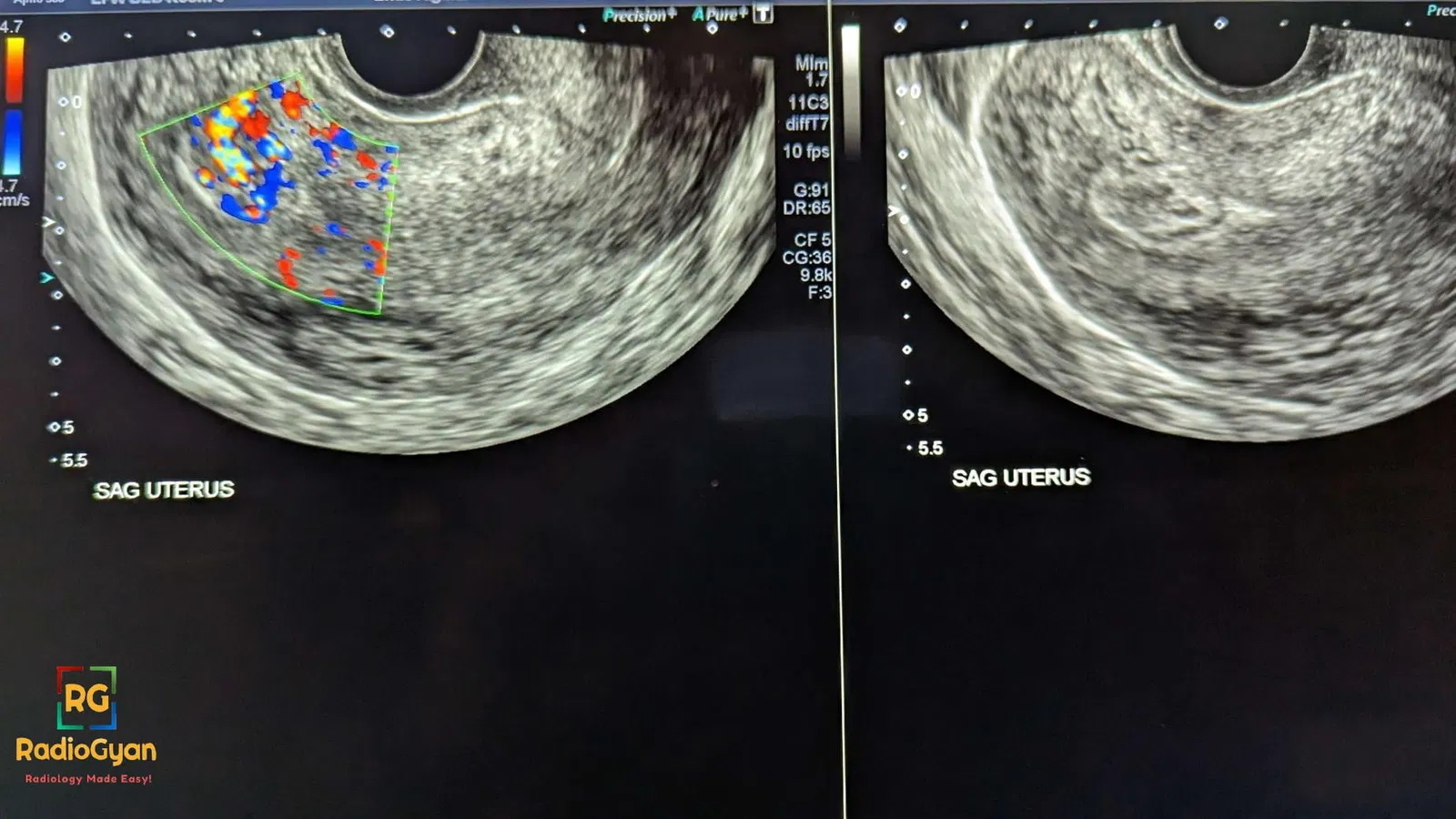
- Ultrasound ( gray scale):
- Increased endometrial echo complex. (8 to 13 mm)
- Intrauterine mass.
- Doppler:
- Vascularity seen from myometrium extending towards endometrium. ( not isolated to myometrium)
- Absence of doppler has low negative predictive value ( RPOC can be avascular)
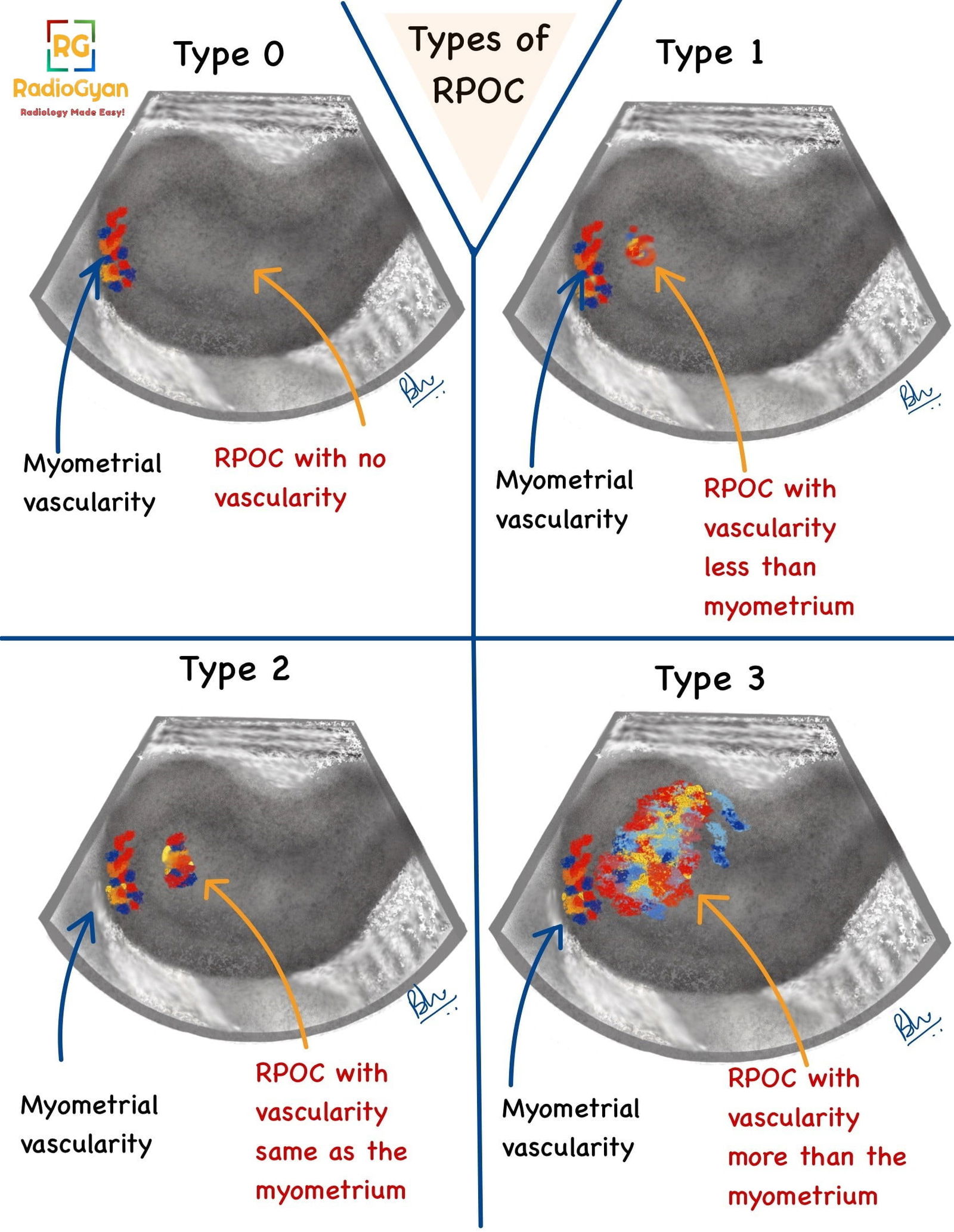
- The degree of vascularity of the intrauterine component can be compared with the myometrial vascularity in the same image section and graded as type 0, 1, 2, or 3.Refer Gutenberg classification for more details. A schematic representation is as follows:
- MRI/CT:
- Intracavitary uterine soft tissue.
- Variable myometrial thinning, junctional zone obliteration.
- Post contrast enhancement within the endometrial cavity.
- T1, T2 characteristics majorly depend on the extent of necrosis and hemorrhage within the RPOC.
Imaging Recommendation:
Grayscale and doppler ultrasound as first line modality. MRI is suggested for complicated cases.
Updated ultrasound criteria (what actually makes the diagnosis)
- Echogenic intrauterine mass is the most sensitive and specific grayscale sign of RPOC. A discrete, vascular endometrial mass is far more reliable than thickening alone.
- Endometrial thickness (ET) is a weaker criterion. An ET of ≥15 mm is often used to raise suspicion, but published cut-offs vary widely and specificity is limited, so a thick but featureless endometrium alone should not be over-called.
- Color Doppler is used mainly to stratify bleeding risk (see grading below), not to confirm the diagnosis: absent flow does not exclude RPOC, because avascular (Type 0) RPOC is well recognised.
- Spectral Doppler (peak systolic velocity, resistive index) overlaps substantially between benign and high-risk lesions and is not reliable on its own.
Top Differential Diagnosis:
- Blood clots- No vascularity.
- Uterine arteriovenous malformation – Vascularity in the myometrium.
- Uterine lesions like endometrial polyp or submucosal fibroid.
- Subinvolution – rare entity.
- Invasive hydatidiform mole.
Clinical Features:
- Symptoms :
- Bleeding:
- Primary(within first 24 hours) postpartum hemorrhage.
- Secondary (bleeding after more than 24 hours upto 6 weeks) postpartum hemorrhage.
- Lower abdominal pain.
- Fever.
- Bleeding:
- Age predilection: More common with advanced maternal age.
- Risk factors if any :
- Placenta accreta.
- History of RPOC in previous pregnancy.
- Advanced maternal age.
- Instrumental delivery.
- Second trimester delivery/miscarriage/abortion.
- Arrest during delivery/ Prolonged labor > 14 hours.
- Prolonged oxytocin use.
- Nulliparity.
- Past uterine surgery.
- Congenital uterine anomaly.
Classification System:
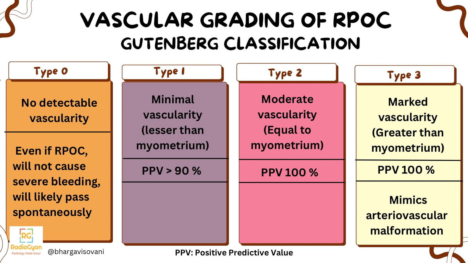
The Gutenberg classification categorises RPOC by its color-Doppler vascularity, because the degree of flow, compared with adjacent myometrium in the same image, predicts the risk of bleeding at treatment. It defines four grades:
| Type | Grayscale appearance | Color-Doppler vascularity | Bleeding risk / implication |
|---|---|---|---|
| Type 0 | Hyperechoic mass | Avascular (no flow) | Lowest risk; frequently resolves without intervention |
| Type 1 | Mixed echogenicity mass | Minimal or no flow | Low risk |
| Type 2 | Intracavitary mass | Marked vascularity confined to the endometrial cavity | Higher bleeding risk |
| Type 3 | Intracavitary mass | Marked vascularity extending into adjacent myometrium (enhanced myometrial vascularity) | Highest bleeding risk |
EMV vs uterine AVM vs RPOC: how to tell them apart
A common diagnostic trap is a hypervascular area in the uterus after pregnancy loss. This is usually enhanced myometrial vascularity (EMV): a cluster of tortuous, high-velocity myometrial vessels that develops with RPOC and was historically (and incorrectly) reported as an “acquired arteriovenous malformation (AVM)”. Current evidence shows EMV almost always coexists with retained tissue and is typically self-limiting, so the label matters because it changes management.
| Feature | RPOC | EMV (pregnancy-related) | True congenital uterine AVM |
|---|---|---|---|
| Context | After delivery, miscarriage or termination | After miscarriage or termination; often with RPOC | No temporal link to pregnancy; may be congenital |
| Grayscale | Echogenic intracavitary mass | Serpiginous myometrial vessels, heterogeneous endometrium | Tubular anechoic myometrial spaces |
| Doppler | Variable flow within the mass (Type 0 to 3) | High-velocity turbulent myometrial flow (high PSV) | High-velocity, low-resistance arterialised flow |
| Natural history | Resolves or is evacuated | Usually resolves spontaneously over weeks to months | Persists; may need embolization |
| First-line management | Expectant / medical / hysteroscopy | Expectant with serial ultrasound if PSV low and bleeding mild | Uterine artery embolization if symptomatic |
Practical point: in a stable patient with mild bleeding, a low peak systolic velocity and imaging suggestive of EMV, expectant management with serial ultrasound is safe and avoids unnecessary embolization or hysterectomy. Rising bleeding or a very high PSV warrants escalation.
Treatment:
Management is individualised to symptoms, bleeding, lesion vascularity and the desire to preserve fertility. The trend is away from routine blind curettage toward more conservative and targeted options.
- Expectant management: reasonable for small, low-grade (Type 0 to 1) RPOC and for EMV with low PSV; many resolve spontaneously. Serial ultrasound confirms resolution.
- Medical: misoprostol. Because RPOC can take time to pass, follow-up ultrasound should be appropriately delayed to avoid over-diagnosing persistent tissue and over-treating.
- Surgical: hysteroscopic resection: increasingly the preferred surgical route. Direct visualisation allows targeted removal of retained tissue and lowers the risk of intrauterine adhesions (Asherman syndrome) compared with blind dilatation and curettage.
- Surgical: dilatation and curettage (D&C): still widely used; ultrasound guidance improves completeness and safety, including when marked vascularity (EMV) is present.
- Uterine artery embolization: reserved for a true AVM or uncontrolled haemorrhage; it is not required for most cases of pregnancy-related EMV.
References:
- Sellmyer MA, et al. Physiologic, histologic, and imaging features of retained products of conception. RadioGraphics. 2013;33(3):781–796. doi:10.1148/rg.333125177 (PMID: 23674774).
- Incognito GG, et al. Ultrasound assessment of retained products of conception (RPOC): insights from the current literature. J Clin Med. 2025;14(16):5864. doi:10.3390/jcm14165864 (PMID: 40869690).
- Grewal K, et al. Natural history of pregnancy-related enhanced myometrial vascularity following miscarriage. Ultrasound Obstet Gynecol. 2020;55(5):676–682. doi:10.1002/uog.21872 (PMID: 31503383).
- Groszmann YS, et al. Diagnosis and management of patients with enhanced myometrial vascularity associated with retained products of conception. Ultrasound Obstet Gynecol. 2018;52(3):396–399. doi:10.1002/uog.18954 (PMID: 29124818).
- Xholli A, et al. Clinical and ultrasonographic characteristics of pregnancy-related enhanced myometrial vascularity: prospective cohort study. Ultrasound Obstet Gynecol. 2024;63(5):672–682. doi:10.1002/uog.27537 (PMID: 37984401).
- Shahulhameed MS, et al. Role of hysteroscopy in the management of uterine vascular malformations with a focus on enhanced myometrial vascularity. Gynecol Minim Invasive Ther. 2024;13(4):209–214. doi:10.4103/gmit.gmit_29_24 (PMID: 39660239).
Case co-authored by TeamGyan Member Dr. Bhargavi Sovani
Frequently asked questions
What is the full form of RPOC?
RPOC stands for Retained Products Of Conception: placental or fetal (trophoblastic) tissue that remains in the uterus after a delivery, miscarriage, or termination of pregnancy.
What is the best imaging test for RPOC?
Transvaginal ultrasound with color Doppler is first-line. The most reliable sign is an echogenic intrauterine mass; MRI is reserved for complicated or equivocal cases.
How is RPOC graded on ultrasound?
The Gutenberg classification scores color-Doppler vascularity from Type 0 (avascular) to Type 3 (marked vascularity extending into the myometrium). Higher grades carry a higher risk of bleeding at treatment.
What is the difference between RPOC and a uterine AVM?
Most hypervascular areas seen after pregnancy loss are enhanced myometrial vascularity (EMV) associated with RPOC, not a true arteriovenous malformation. EMV usually resolves spontaneously and is managed expectantly, whereas a true congenital AVM has no link to a recent pregnancy and may require uterine artery embolization.
How is RPOC treated?
Options are expectant management, medical treatment with misoprostol, or surgery. Hysteroscopic resection is increasingly preferred over blind dilatation and curettage because it targets the tissue and lowers the risk of intrauterine adhesions.

Improve Content And Help Your Colleagues!
Found an error in the post? Please let us know using our contact page and we will update it with due credits!
If you wish to contribute radiological images for this case (or any other article on the website), you can submit them here and you will be duly credited: Submit radiology case