
What genital tract pathologies are associated with long term Tamoxifen use?
Long term Tamoxifen use in patients with breast cancer can cause endometrial polyps, endometrial hyperplasia, cystic endometrial atrophy, adenomyosis, fibroids, ovarian cysts, endometrial carcinoma.
What is the most common endometrial pathology associated with Tamoxifen use?
Endometrial polyps are the most common endometrial pathology reported in association with tamoxifen exposure.
- Tamoxifen is a selective estrogen receptor modulator (SERM) used for chemoprophylaxis of breast cancer.
- 50 percent of patients on Tamoxifen develop an endometrial lesion within 6 – 36 months. Severity is dependant on the duration of treatment.
- Abnormal endometrial thickness criteria ⁉️ – Answer in the comments. Check your answer in the video below.
- Endometrial thickness ranges from 9-13mm in patients with Tamoxifen use.
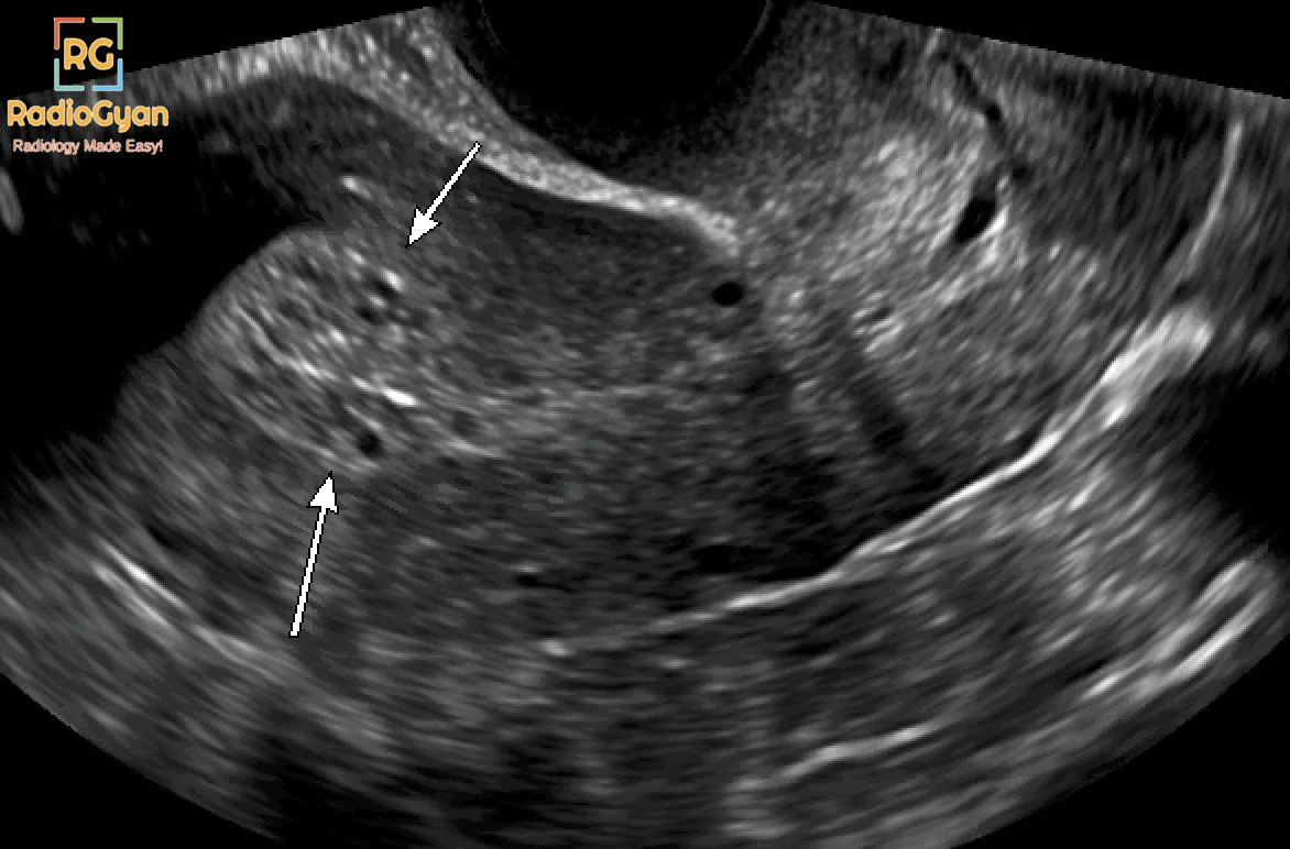
Imaging of tamoxifen-associated endometrial changes:
- Ultrasound is the imaging modality of choice.
- Typical findings: Thickened endometrium with multiple cystic spaces giving a “Swiss cheese’ pattern”.
- Polyps will be seen as echogenic lesions with a vascular stalk.
- Hysterosonography can be used as a problem solving tool.
How to distinguish endometrial polyps and submucosal fibroids on ultrasound and hyseterosonography?
Polyps appear as smoothly marginated oblong echogenic lesions with a narrow attachment to the endometrium but may be broad-based.
Submucosal fibroids appear as round structures arising from the myometrium with wide attachment to the myometrium.
Role of MRI in Tamoxifen associated endometrial changes:
MRI can be used in equivocal cases, however, it is not used routinely. A study by Ascher et al. suggested that MR imaging may help identify which patients should undergo a sampling procedure and those who can be followed up with MR imaging.
Management of Patients with Abnormal Bleeding on Tamoxifen
👉 Asymptomatic patients with ONLY imaging findings: No further assessment.
👉 Postmenopausal women with any bleeding, spotting, staining, or bloody vaginal discharge: Rule out endometrial cancer (Transvaginal ultrasound and biopsy).
👉 Premenopausal women with amenorrhea/oligomenorrhea and no risk factors for endometrial cancer: Expectant management.
👉 Premenopausal women with heavy bleeding: Endometrial biopsy.
👉 Endometrial polyps in patients on Tamoxifen: Resection suggested.
Detailed video with PACS based case images:

References:
- The effect of tamoxifen on the genital tract.
- Tamoxifen and endometrial cancer. Is screening necessary? A review of the literature.
- Tamoxifen-induced Uterine Abnormalities: The Role of Imaging.
- Abnormal uterine bleeding and uterine pathology in patients on tamoxifen therapy
For more Genitourinary radiology cases, check this link:

Improve Content And Help Your Colleagues!
Found an error in the post? Please let us know using our contact page and we will update it with due credits!
If you wish to contribute radiological images for this case (or any other article on the website), you can submit them here and you will be duly credited: Submit radiology case