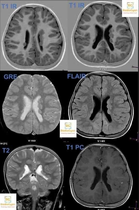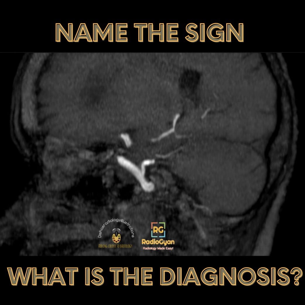
Quiz
Which of the following is not a type of Persistent Fetal Carotid-Vertebrobasilar Anastomoses?
- Persistent trigeminal artery
- Persistent otic (acoustic) artery
- Fetal origin of PCA artery
- Persistent Hypoglossal artery
Pathophysiology
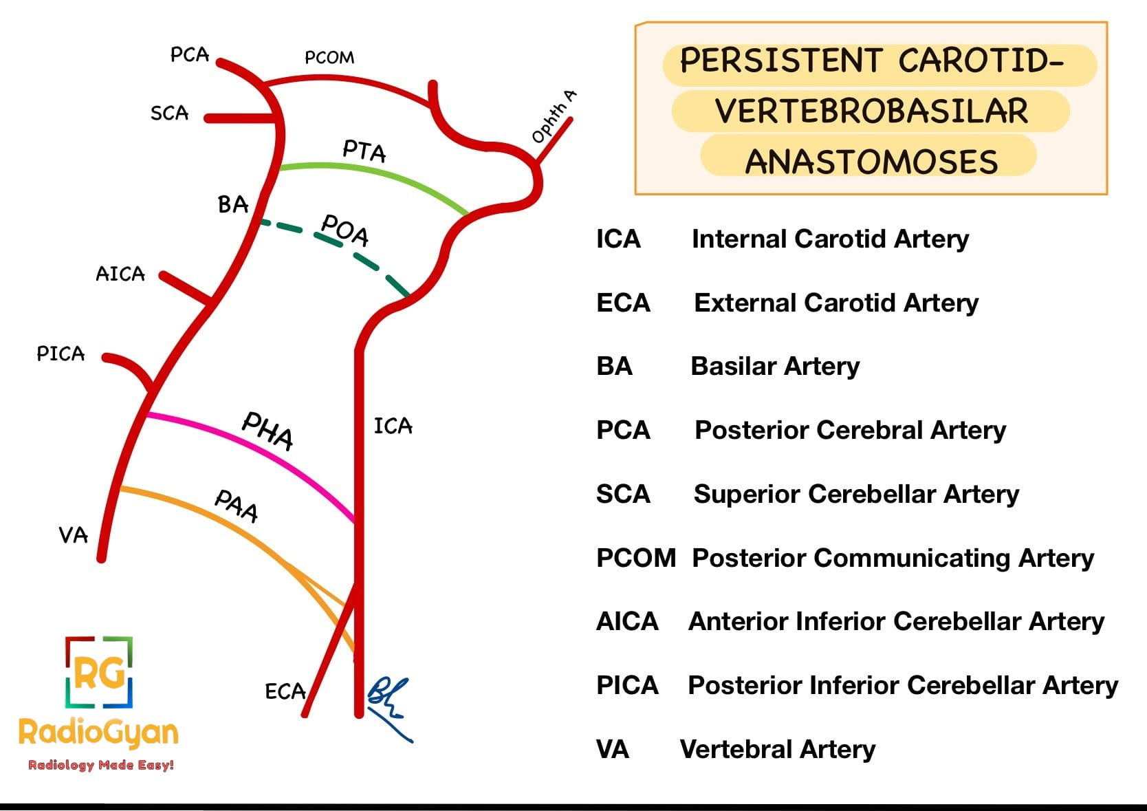
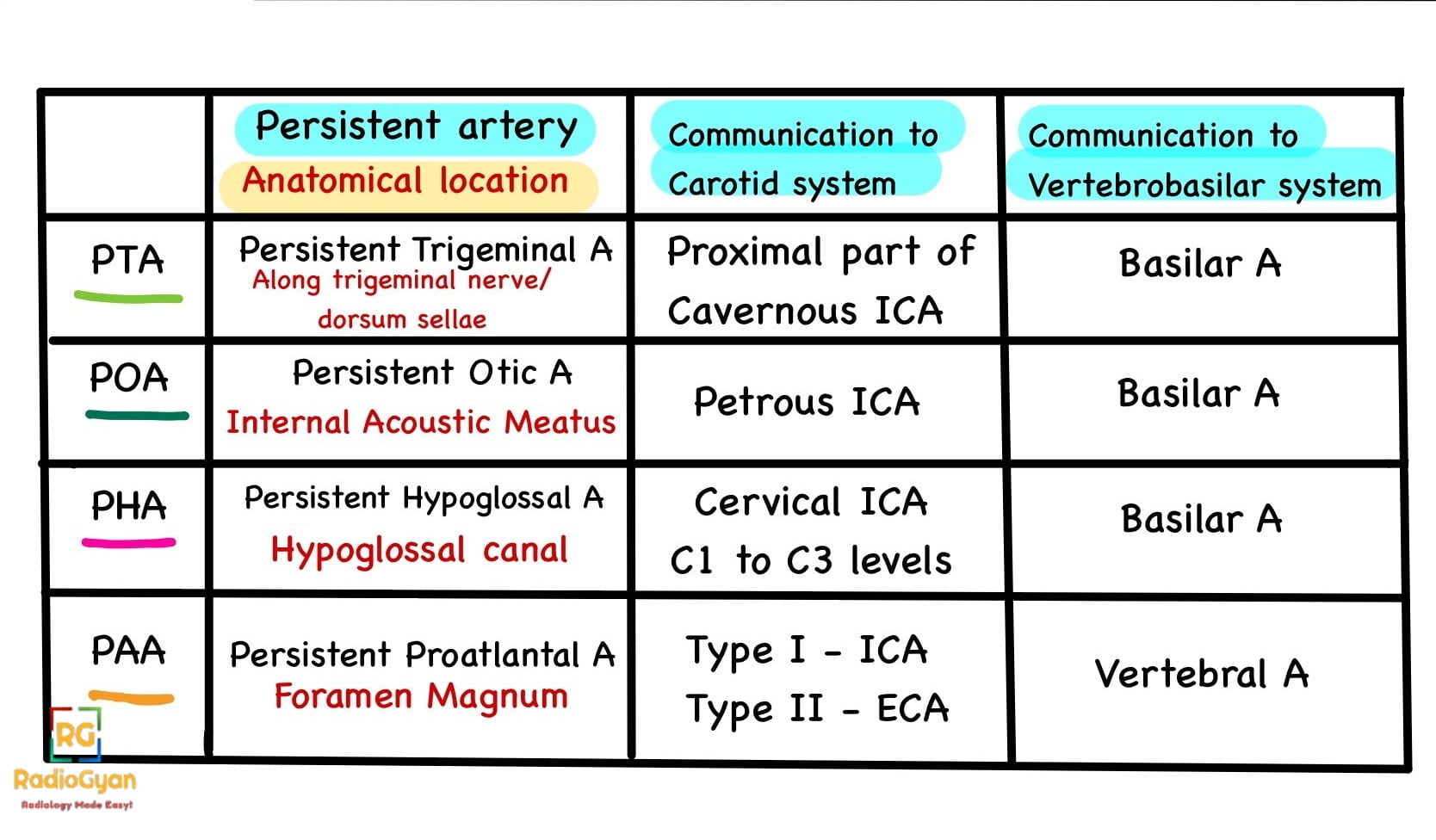
- In embryonic development, there are four channels between the carotid and vertebrobasilar arteries, namely, the trigeminal, otic, hypoglossal, and proatlantal intersegmental arteries which eventually regress.
- Persistent primitive trigeminal artery (PPTA) is the most common of the persistent carotid-basilar connections. The PPTA connects the cavernous segment of the internal carotid artery (ICA) to the basilar artery (BA) between the origins of the anterior inferior cerebellar artery (AICA) and superior cerebellar artery (SCA).
- It is commonly seen unilaterally and often associated with hypoplastic vertebral, posterior communicating (PCom) and caudal basilar arteries.
Key Imaging Features
- CT/ MR angiography– Anomalous vessel is seen arising from the cavernous segment of the internal carotid artery extending toward the basilar artery.
- Tau sign- On sagittal images, the combination of the vertical and horizontal segments of the ICA and the proximal portion of the trigeminal artery resembles the greek letter ‘Tau’.
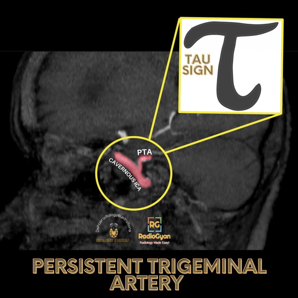
Imaging Recommendation: Angiography-CT/MRI
Top 3 Differential Diagnosis:
- Persistent carotid-vertebrobasilar anastomoses (PCBA) include
- Persistent otic artery– first artery to regress and rarest anomaly, arising from the petrous part of the ICA within the carotid canal, and joins the basilar artery, inferiorly.
- Persistent hypoglossal artery– a second most common type of PCBA arising from the posterior aspect of cervical ICA and courses along the hypoglossal canal to anastomose with basilar artery.
- Persistent proatlantal artery– Type I arises from the cervical ICA at the C2-3 level and runs posterosuperior and joins the vertebral artery before coursing upward through the foramen magnum. Type II arises from the ECA and follows a similar course as type I.
- A simple schematic diagram of these persistent carotid- vertebrobasilar anastomoses is as follows:
Clinical Features:
- Symptoms- Usually asymptomatic. PPTA is often associated with intracranial aneurysms, brain arteriovenous malformation, trigeminal artery-cavernous fistula, Moyamoya and disease and acute and chronic large vessel occlusion, which can produce symptoms.
- No specific age/sex predilection or risk factors are known.
Classification System:
Salas classification
- Medial (sphenoidal) type- PPTA directly perforate the central portion of the dorsum sellae and forms an anastomosis with the basilar artery
- Lateral (petrosal) type- PPTA courses along the lateral portion of the dorsum sellae and then forms an anastomosis with the basilar artery
Saltzman classification
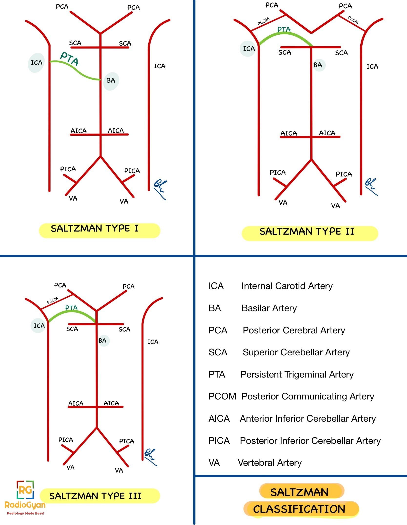
Most commonly used classification. An easy line diagram to depict this classification is as follows:
| Type | Description |
| I | PPTA enters the BA between the SCA and AICA; the PPTA supplies the BA, SCAs, and posterior cerebral arteries (PCAs); the posterior communicating arteries (PcomAs), vertebral artery (VA) and proximal BA may be absent or hypoplastic. |
| II | PPTA supplies the BA and SCAs, the proximal BA is well formed, and PcomAs are present and, with the BA, contribute to the distal posterior circulation |
| I + II | PPTA supplies the BA and bilateral SCA as well as the opposite or ipsilateral PCA, while the other PCA is supplied by a patent PcomA |
| III | PPTA terminates as a cerebellar artery [ types IIIa, b, and c PPTAs terminate as the SCA, AICA, and posterior inferior cerebellar artery (PICA), respectively] |
Etymology and synonyms:
The anastomosis between the cavernous segment of ICA and the vertebro-basillar artery has a close relationship to the Gasserian ganglion and the fifth cranial nerve, giving the name- ‘persistent trigeminal artery’.
Treatment:
- No treatment in incidentally detected cases.
- Treatment of the associated complication depends on the symptoms.
References:
Single best review article:
Other references:
- Luh GY, Dean BL, Tomsick TA, Wallace RC. The persistent fetal carotid-vertebrobasilar anastomoses. AJR Am J Roentgenol. 1999;172(5):1427-1432. doi:10.2214/ajr.172.5.10227532
- Aguiar, Guilherme & Conti, Mario & Veiga, José Carlos & Jory, M & Souza, Rodrigo. (2011). Basilar Artery Aneurysm at a Persistent Trigeminal Artery Junction: A Case Report and Literature Review. Interventional neuroradiology : journal of peritherapeutic neuroradiology, surgical procedures and related neurosciences. 17. 343-6. 10.1177/159101991101700310.
- Osborn’s Brain: Imaging, Pathology, and Anatomy by Anne G. Osborn MD FACR (Author), Gary L. Hedlund DO (Author), Karen L. Salzman MD (Author)-Elsevier publication– 2nd edition. ISBN-10 : 9780323477765
Case co-authored by TeamGyan Member Dr Mansi Sarmalkar , edited by Dr. Bhargavi Sovani


