
Pathophysiology of the Canal of Nuck
- Extension of the parietal peritoneum follows the round ligament as it passes to the inguinal canal.
- Usually gets obliterated.
- This evagination of the parietal peritoneum = the canal of Nuck = female counterpart of processus vaginalis in men.
- If it is:
- Completely patent: Avenue for an indirect inguinal hernia and hydrocele (Canal of Nuck hernia).
- Obliterated at the level of both the superficial and deep inguinal rings: Fluid may be trapped in between both ends of the canal, creating a Canal of Nuck cyst.
- Obliterated at lower end – Infantile hydrocele / encysted hydrocele.
- Hydrocele vs cyst canal of Nuck : The terms hydrocele and cyst of Canal of Nuck are used interchangeably but these are separate pathologies.
- Symptoms: It usually presents as a painless swelling in the inguinal region. Infection can cause pain.
Imaging appearances:
- Canal of Nuck cyst is an inguinal cyst in females.
- US : Cystic lesion in the inguinal canal with posterior acoustic attenuation.
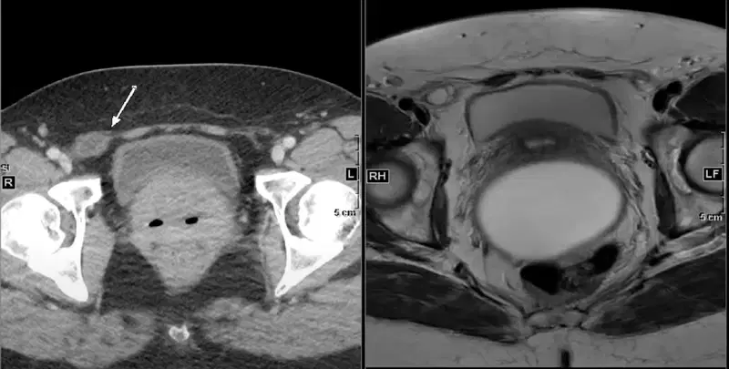
- CT : Hypodense lesion with our without internal septations.
- MRI: T1 isointense, T2 hyperintense, typical appearance of a cystic lesion.
- Can show peripheral enhancement.
Differentials for Canal of Nuck cyst
- Inguinal hernia containing fluid in patients with ascites.
- Soft tissue tumor.
- Endometriosis cyst
- Cyst
- Abscess
- Lymphadenopathy
Management:
Surgical excision or conservative management depending on patient presentation.
Detailed video with PACS based case images:


Improve Content And Help Your Colleagues!
Found an error in the post? Please let us know using our contact page and we will update it with due credits!
If you wish to contribute radiological images for this case (or any other article on the website), you can submit them here and you will be duly credited: Submit radiology case