


Dilatation and dilation mean the same thing: a hollow structure that is wider than it should be, or the act of widening it. Standard medical dictionaries list them as synonyms, and English has only one verb for both, to dilate. The two words differ in style, not in meaning.
Where radiologists actually go wrong is not the spelling. It is using dilated when the right word is distended, ectatic or aneurysmal, and writing prominent instead of giving a number. This page covers what dilatation means, what the evidence and the style manuals say about each form, how the neighbouring terms differ, and the measurement thresholds that decide when a duct, vessel, bowel loop or ventricle is genuinely dilated.
What does dilatation mean?
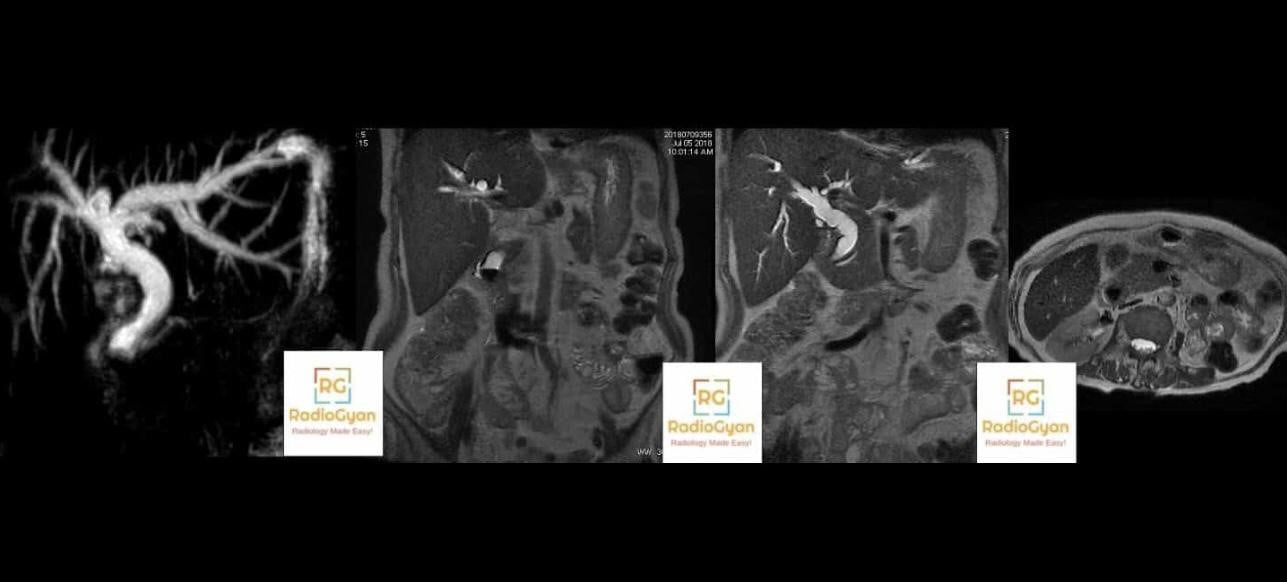
Dilatation is enlargement of the lumen or cavity of a hollow structure beyond its normal calibre a duct, blood vessel, cardiac chamber, bowel loop, renal collecting system or cerebral ventricle. It is a descriptive finding, not a diagnosis: the report should say what is dilated, by how much, and what appears to be causing it.
The word comes from the Latin verb dilatare (to spread wide) through the noun dilatatio, which gives dilatation directly. Dilation is the same idea rebuilt in English by adding the live suffix -tion to the verb dilate [2].
- The verb is always dilate. A duct is dilated. It is never dilatated that form was recorded once in the seventeenth century and is obsolete [1].
- Dilatation belongs to hollow structures. A solid organ is enlarged (hepatomegaly, splenomegaly), not dilated.
- Dilatation is not the same as obstruction. Ducts stay dilated for weeks after a stone passes, and the common bile duct widens with age and after cholecystectomy.
Dilatation vs dilation: is there any real difference?
Three respectable positions exist, and they do not agree.
| Position | What it holds | Source |
|---|---|---|
| They are interchangeable | Medical dictionaries define each in terms of the other. There is one verb, to dilate, and no difference in meaning. | Standard medical dictionaries |
| Dilation is the act, dilatation is the condition | The AMA Manual of Style, 11th edition: dilate is the verb; dilation means the act of dilating; dilatation means the condition of being stretched or expanded. Hence treated with dilation and curettage, but cervical dilatation was 3 cm. | AMA Manual of Style (11th ed), crediting ACOG |
| The distinction is invented | Bloom and colleagues traced both words through the Oxford English Dictionary: dilatation is recorded from 1386 and dilation is the later, etymologically irregular formation. Their verdict on the action-versus-condition split was that it seems arbitrary and without historical evidence or support. | Bloom et al, J Urol 1992 [1] |
For a radiology report, only one of these matters in practice: both forms are understood, so the failure mode is inconsistency, not choice. Mixing the two within a single report or paper looks careless.
Which one is winning?
Mahroo and colleagues measured this directly across PubMed, Google Scholar and the Google Books corpora [2]:
- The first PubMed-indexed use of dilatation dates to 1809, almost a century before dilation in 1907.
- Across all of PubMed, dilatation still led by roughly 1.9 to 1, but that ratio had fallen to about 1.3 to 1 in the five years to 2013.
- In Google Scholar, which reaches beyond medicine, the ratio had already dropped below parity to 0.83 dilation had overtaken.
- In the Google Books English corpus, dilation passed dilatation in 1996 two years earlier in the American English corpus, while in British English the two were still level at the end of the series.
So dilation is gradually displacing the older, etymologically better-founded dilatation, and the shift is running faster in American English and faster outside medicine than inside it.
Even current guidelines disagree
This is not a settled question at guideline level, which is the strongest argument that neither form is wrong:
- The 2024 Fleischner Society glossary of terms for thoracic imaging uses dilatation throughout. It defines bronchiectasis as a clinical condition of irreversible bronchial dilatation, and recommends the plain term bronchial dilatation whenever chronicity has not been established [3].
- The 2022 ACC/AHA aortic disease guideline uses dilation throughout, and goes further: it states that in clinical practice the term dilation is preferred to ectasia for mild aortic enlargement, because radiologists have applied ectatic inconsistently to mildly enlarged aortas, to tortuous aortas, and even to aortas of 4.5 to 5.4 cm, which overlaps with what most experts would call an aneurysm [4].
Two international bodies, two years apart, two spellings. Use whichever your department or target journal uses, and be consistent.
Dilated, distended, ectatic, prominent or aneurysmal?
These are the distinctions that actually change how a report is read.
| Term | What it means | Use it when | Pitfall |
|---|---|---|---|
| Dilated / dilatation | Lumen wider than the accepted normal calibre for that structure. | You can name the structure and, ideally, quote a measurement. | Implies pathology. Do not use it for a normally full viscus. |
| Distended | Stretched by its contents gas, fluid, urine, contrast. | Bladder, stomach, gallbladder, bowel filled with content. | A distended bladder or stomach is often physiological. Distension describes state, not cause. |
| Ectatic / ectasia | Mild, usually diffuse widening, often with tortuosity or an abnormal course. | Diffuse coronary widening, tubular ectasia of the rete testis, dural ectasia. | Applied inconsistently to the aorta. The 2022 ACC/AHA guideline prefers dilation there [4]. |
| Dolichoectatic | Elongated as well as widened. | Vertebrobasilar dolichoectasia. | Needs both features, not just a wide vessel. |
| Aneurysmal | Focal enlargement to at least 1.5 times the expected normal diameter of that artery. | Once the threshold or the guideline definition for that segment is met. | Not interchangeable with dilated. Committing to aneurysm triggers surveillance pathways. |
| Prominent | Nothing measurable. | Ideally never. | Hedging word. Replace it with the measurement. |
| Enlarged | Increased overall size of a solid organ or cardiac chamber. | Liver, spleen, kidney, prostate, cardiac chambers. | Do not write dilated liver. Chambers may be dilated when the reference is a volume. |
How dilated is dilated? Reference thresholds
The word only means something once it is anchored to a number. These are the thresholds most often used to call a structure dilated. They vary with modality, measurement convention (inner-edge versus outer-edge), age and body size, so treat them as decision points rather than absolutes. For a wider list see our normal radiology measurements reference.
| Structure | Threshold commonly used for dilatation | Source |
|---|---|---|
| Common bile duct (gallbladder in situ) | Endoscopic ultrasound in 647 patients showed the CBD widens significantly after age 70, but even in the oldest group it did not exceed 7.6 mm. Anything wider warrants investigation. Cholecystectomy was the one other factor that independently widened it. | Benjaminov et al, Surg Endosc 2013 [5] |
| Main pancreatic duct | Segmental or diffuse dilatation greater than 5 mm defines main-duct IPMN. 5 to 9 mm is a worrisome feature; 10 mm or more is a high-risk stigma. | Kyoto IPMN guidelines 2024 [6] |
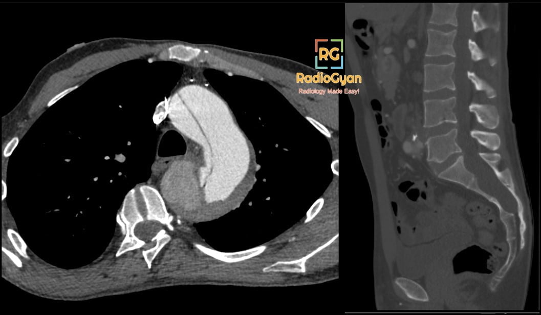
| Ascending aorta and aortic root | A diameter of 4.0 cm or more is described as dilated. The conventional definition of an arterial aneurysm is at least 1.5 times the expected normal diameter, which works for the abdominal and descending aorta but underperforms at the root. | 2022 ACC/AHA aortic disease guideline [4] |
| Abdominal aorta | Greater than 3.0 cm on ultrasound, measured outer edge to outer edge, defines an abdominal aortic aneurysm. | 2022 ACC/AHA aortic disease guideline [4] |
| Main pulmonary artery | 30 mm or more, or a pulmonary artery to aorta ratio above 0.9, suggests pulmonary hypertension on CT, particularly combined with RVOT wall thickening and septal deviation. | 2022 ESC/ERS pulmonary hypertension guidelines [7] |
| Bronchus | Dilatation of the bronchus relative to its accompanying pulmonary artery (the signet-ring sign, broncho-arterial ratio above 1), lack of tapering, or bronchi visible within 1 cm of the pleura. Call it bronchial dilatation unless irreversibility is established, in which case it is bronchiectasis. | Fleischner glossary 2024 [3]; Aliberti et al, Lancet Respir Med 2022 [8] |
| Small bowel | Loops above 2.5 to 3 cm in calibre on CT are taken as dilated, with a transition point and collapsed distal bowel confirming obstruction. | Silva et al, RadioGraphics 2009 [9] |
| Colon | Working thresholds of 6 cm for the colon and 9 cm for the caecum, above which perforation risk rises in acute colonic pseudo-obstruction and toxic megacolon. | Widely used surgical working figures |
| Renal collecting system (postnatal) | Anteroposterior renal pelvic diameter 10 mm or more places a postnatal kidney in UTD P1 or higher; the classification also grades calyceal dilatation, parenchymal thickness and ureteric involvement. | UTD classification, Nguyen et al 2014 [10] |
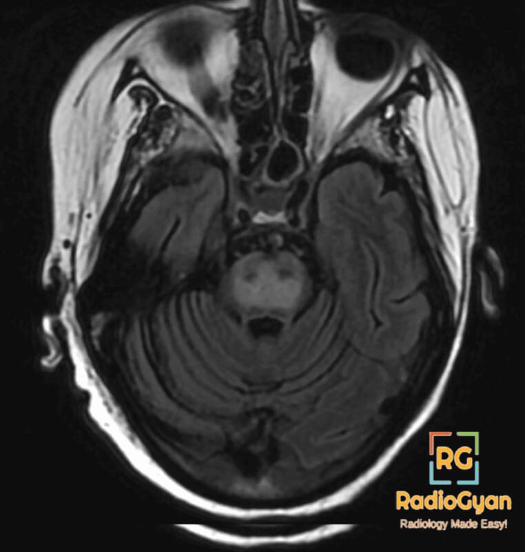
| Lateral ventricles | Evans index (maximum width of the frontal horns divided by maximum inner skull width) above 0.30 defines ventriculomegaly in the normal pressure hydrocephalus work-up. | iNPH guidelines, 3rd edition [11] |
| Cardiac chambers | Chamber dilatation is defined by volumes indexed to body surface area, not by eyeballing a four-chamber view. | ASE/EACVI chamber quantification 2015 [12] |
Which spelling is conventional where?
| Context | Usual form | Comment |
|---|---|---|
| Dilatation and curettage | Both | The procedure is traditionally D and C as dilatation; American usage has largely shifted to dilation and curettage. The abbreviation is unaffected. |
| Obstetrics: the cervix in labour | Dilatation for the measured state | The AMA style example is cervical dilatation was 3 cm, while the obstetrician dilates the cervix. |
| Ophthalmology: pupils | Dilation | Dilated fundus examination and pupillary dilation dominate current usage. |
| Balloon and endoscopic procedures | Both | Balloon dilatation of a stricture and balloon dilation are both standard; American journals prefer the shorter form. |
| Vessels: physiological widening | Vasodilation | Vasodilatation survives in older physiology texts but is now uncommon. |
| Thoracic imaging | Dilatation | The Fleischner Society glossary uses it consistently [3]. |
| Aortic imaging and cardiology | Dilation | The 2022 ACC/AHA guideline uses it consistently [4]. |
| Physics and geometry | Dilation only | Time dilation, and dilation as a geometric transformation. Dilatation is never used here. |
Five rules for your reports
- Pick one form and keep it. Consistency within a report, a template library and a department matters far more than which form you pick.
- Never write dilatated. It is not a word in current English.
- Match the word to the structure. Lumens dilate, content-filled viscera distend, solid organs enlarge.
- Give the number. CBD 11 mm is worth more to the referrer than prominent CBD, and it is what the next reader will compare against.
- Say what it implies. Dilatation is a finding: pair it with the level, the transition point or the cut-off, and the likely cause.
The same discipline applies whenever a threshold decides management see our aortic dissection and double duct sign articles, or the radiology calculators for the measurements themselves.
Frequently asked questions
References
- Bloom DA, Mory RN, Hinman F Jr. Dilation vs. dilatation: a brief history. J Urol. 1992;147(6):1682. PMID: 1593720
- Mahroo OA, Shalchi Z, Hammond CJ. Dilatation and dilation: trends in use on both sides of the Atlantic. Br J Ophthalmol. 2014;98(6):845-846. PMID: 24568871
- Bankier AA, MacMahon H, Colby T, et al. Fleischner Society: glossary of terms for thoracic imaging. Radiology. 2024;310(2):e232558. PMID: 38411514
- Isselbacher EM, Preventza O, Hamilton Black J 3rd, et al. 2022 ACC/AHA guideline for the diagnosis and management of aortic disease. Circulation. 2022;146(24):e334-e482. PMID: 36322642
- Benjaminov F, Leichtman G, Naftali T, et al. Effects of age and cholecystectomy on common bile duct diameter as measured by endoscopic ultrasonography. Surg Endosc. 2013;27(1):303-307. PMID: 22903627
- Ohtsuka T, Fernandez-del Castillo C, Furukawa T, et al. International evidence-based Kyoto guidelines for the management of intraductal papillary mucinous neoplasm of the pancreas. Pancreatology. 2024;24(2):255-270. PMID: 38182527
- Humbert M, Kovacs G, Hoeper MM, et al. 2022 ESC/ERS guidelines for the diagnosis and treatment of pulmonary hypertension. Eur Heart J. 2022;43(38):3618-3731. PMID: 36017548
- Aliberti S, Goeminne PC, O Donnell AE, et al. Criteria and definitions for the radiological and clinical diagnosis of bronchiectasis in adults for use in clinical trials: international consensus recommendations. Lancet Respir Med. 2022;10(3):298-306. PMID: 34570994
- Silva AC, Pimenta M, Guimaraes LS. Small bowel obstruction: what to look for. RadioGraphics. 2009;29(2):423-439. PMID: 19325057
- Nguyen HT, Benson CB, Bromley B, et al. Multidisciplinary consensus on the classification of prenatal and postnatal urinary tract dilation (UTD classification system). J Pediatr Urol. 2014;10(6):982-998. PMID: 25435247
- Nakajima M, Yamada S, Miyajima M, et al. Guidelines for management of idiopathic normal pressure hydrocephalus (third edition). Neurol Med Chir (Tokyo). 2021;61(2):63-97. PMID: 33455998
- Lang RM, Badano LP, Mor-Avi V, et al. Recommendations for cardiac chamber quantification by echocardiography in adults: an update from the American Society of Echocardiography and the European Association of Cardiovascular Imaging. Eur Heart J Cardiovasc Imaging. 2015;16(3):233-270. PMID: 25712077

Improve Content And Help Your Colleagues!
Found an error in the post? Please let us know using our contact page and we will update it with due credits!
If you wish to contribute radiological images for this case (or any other article on the website), you can submit them here and you will be duly credited: Submit radiology case
Good stuff!
I always used to have this confusion. Thanks for sharing :).
Cody.
“Another differentiation is that dilation usually refers to physiological processes such as dilation of pupils, vessels and cervix vis-a-vis dilatation which refers to surgical procedures such as dilatation and curettage, balloon dilation valvuloplasty.”
I think you used the wrong word! Is it cervical dilatation or dilation there?
Hey Ilya,
My understanding is that if it physiological, it would be cervical dilation. If it is done surgically, it would be dilatation.