


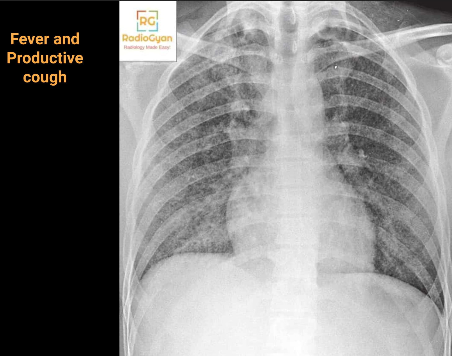
Miliary tuberculosis is haematogenous tuberculosis, and its imaging signature is a pattern rather than a lesion: innumerable 1 to 2 mm nodules of uniform size, distributed randomly through both lungs from apex to base, without cavitation. The single most useful thing to know about it is that the chest radiograph is frequently normal early in the disease, because the nodules are not yet large or profuse enough to be seen, while high-resolution CT already shows them [4][12].
This article is a working reference for reporting a suspected miliary pattern: what the 2024 Fleischner Society glossary now means by miliary and by micronodule [3], what the radiograph and CT actually show and in what proportion of patients, how to separate a random distribution from centrilobular and perilymphatic nodules, the differential diagnosis of a miliary pattern, what changes in HIV and other immunocompromised hosts, why a brain MRI belongs in the workup, and the one sentence in your report that changes which treatment regimen the patient is eligible for. Clinical anchors are the 2026 comprehensive review in Infection and Drug Resistance [1], the Indian Journal of Medical Research review by Sharma and colleagues [2], and WHO consolidated guidelines module 4 [13].
- Key facts for practice and radiology exams
- What the word miliary actually means
- Chest X-ray findings in miliary tuberculosis
- CT and HRCT findings
- Random, centrilobular or perilymphatic: the decisive step
- Differential diagnosis of a miliary pattern
- Miliary tuberculosis in HIV and other immunocompromised hosts
- Beyond the chest: what else to image
- Confirming the diagnosis
- Treatment, and the sentence in your report that changes it
- How to report a miliary pattern
- Frequently asked questions
- References
Key facts for practice and radiology exams
- Miliary means 1 to 2 mm, uniform and random. The 2024 Fleischner glossary defines the miliary pattern as profuse, diffuse, randomly distributed, well-defined micronodules of uniform size measuring 1 to 2 mm, resembling millet seeds [3].
- Micronodule no longer means under 3 mm. The same 2024 glossary raised the micronodule threshold from less than 3 mm to less than 6 mm in average diameter, to align with incidental nodule management thresholds. Miliary is now a much narrower term than micronodular [3].
- A normal chest radiograph does not exclude it. Nodules must reach a critical size and profusion to become radiographically visible, and the pattern evolves over days to weeks. HRCT shows them first [2][4][12].
- Ground-glass opacity is the second finding, not a distractor. In a 25-patient HRCT series, 23 patients had ground-glass opacity alongside the nodules; the patients with dyspnoea had the largest areas, and the two with impending ARDS had extensive ground-glass [5].
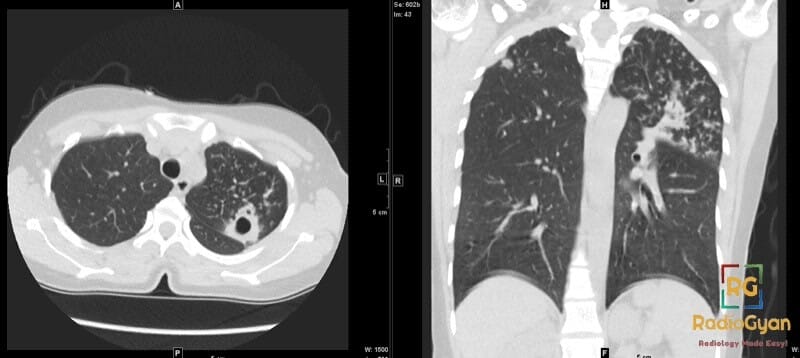
- There is no cavity. Cavitation, an upper-lobe or superior-segment focus and tree-in-bud clusters belong to post-primary disease with bronchogenic spread, not to miliary disease, although the two can coexist.
- The brain is involved far more often than it is symptomatic. More than 60 percent of miliary tuberculosis patients who had brain imaging in one series had cerebral lesions, and 5 of 21 had no neurological symptoms [9]; a 282-patient series reported central nervous system involvement in 87.6 percent, with contrast-enhanced MRI at 96.1 percent sensitivity against 34.7 percent for CT [8].
- Mortality is high and it is a diagnostic-delay disease. Miliary tuberculosis accounts for roughly 1 to 2 percent of tuberculosis in immunocompetent adults but carries a mortality above 30 percent, and untreated it is essentially uniformly fatal within a year [1][2].
- Reporting the pattern changes the regimen. A miliary pattern excludes the patient from the WHO 4-month rifapentine-based regimen and, in children, from the non-severe disease category that permits shortened treatment [13].
What the word miliary actually means
Miliary comes from milium, the millet seed, and the term is descriptive of size and profusion rather than of aetiology. The 2024 Fleischner Society glossary places it as a subcategory of the nodular pattern and defines it as profuse, diffuse and randomly distributed well-defined micronodules of uniform size, with diameters of 1 to 2 mm [3]. Every element of that sentence does work: profuse excludes a handful of nodules, diffuse excludes zonal predominance, randomly distributed excludes sarcoidosis and bronchiolitis, and uniform size excludes haematogenous metastases.
The 2024 glossary also changed the parent term. A micronodule was previously any nodule under 3 mm; it is now defined as less than 6 mm in average diameter, a threshold chosen to separate micronodules from larger, potentially actionable incidental nodules [3]. The practical consequence for reporting is that “diffuse micronodules” and “miliary” are no longer interchangeable. If you mean 1 to 2 mm random nodules, write miliary; if you mean 4 mm nodules, they are micronodules but they are not a miliary pattern, and the differential is different.
Pathologically, the nodules are discrete caseating granulomas seeded through the pulmonary capillary bed after lymphohaematogenous dissemination of Mycobacterium tuberculosis from a primary focus or from reactivation. Because the seeding is via blood rather than via airways, the granulomas land anywhere in the secondary pulmonary lobule, which is exactly what produces the random distribution on CT.
Chest X-ray findings in miliary tuberculosis
On a frontal radiograph the pattern is innumerable tiny, discrete, sharply or poorly defined nodules of near-identical size spread evenly through both lungs, from the apices to the costophrenic angles. They are at the edge of radiographic resolution, so the first impression is often a diffuse granular or fine reticulonodular haze rather than countable nodules. Look specifically in the retrocardiac lung and behind the diaphragm, where superimposed nodules summate and the pattern declares itself, and window a digital radiograph brightly before dismissing it.
Supporting features are as informative as the nodules. Lung volumes are normal. There is usually no consolidation, no cavity and no pleural effusion, and the absence of cavitation is what separates the appearance from post-primary tuberculosis. Overlapping nodules can generate curvilinear densities and a spurious reticular pattern, and in some patients the profusion is visibly asymmetric [2].
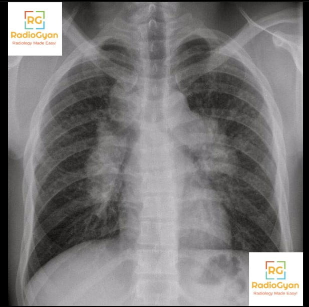
The important negative is that the radiograph can be entirely normal. Haematogenous seeding precedes radiographic visibility, and in patients who present with tuberculosis-related acute respiratory distress syndrome the radiograph may be indistinguishable from ARDS of any other cause, with the miliary pattern emerging only later or only on CT [2]. Cryptic miliary tuberculosis, classically in elderly and immunosuppressed patients presenting as pyrexia of unknown origin or unexplained wasting that mimics metastatic carcinoma, was for decades a post-mortem diagnosis for exactly this reason; HRCT is what moved it to an antemortem one [2].
CT and HRCT findings
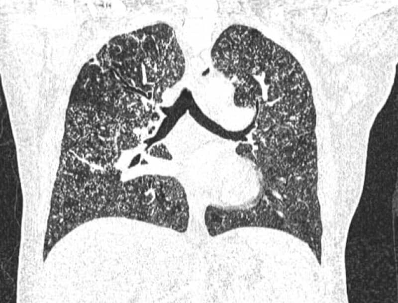
CT is the more sensitive test and the one that settles the pattern. Across the two classic HRCT series, nodules measured 1 to 2 mm in most patients, with ranges extending to 1 to 4 mm and, in one 25-patient series, to 5 mm; margins were sharply defined in the majority and poorly defined in a minority, and in every patient the distribution was random both across the lungs and within the secondary pulmonary lobule [4][5]. In one of those series, a patient with numerous nodules on HRCT had no miliary nodules at all on the chest radiograph [4].
| Finding | How often | What it means |
|---|---|---|
| Random 1 to 2 mm micronodules | 24 of 25 patients; 10 of 11 in a second series [4][5] | The defining feature. Nodules touch pleura, fissures and vessels indiscriminately |
| Ground-glass opacity | 23 of 25 patients [5] | Variable extent. Large areas correlate with dyspnoea, extensive areas with impending ARDS |
| Intralobular reticulation and interlobular septal thickening | 10 of 25 patients [5] | Reflects interstitial oedema and lymphatic congestion, not fibrosis |
| Nodules not visible on the chest radiograph | 1 of 10 patients with CT nodules [4] | The reason a normal radiograph cannot exclude the diagnosis |
| Mediastinal or hilar lymphadenopathy, often with necrotic centres | Common, particularly in HIV [7] | Supports tuberculosis over sarcoidosis when nodes show low-attenuation centres and rim enhancement |
| Cavitation | Absent in pure miliary disease | If present, there is coexisting post-primary disease with bronchogenic spread |
Thin sections matter. A 5 mm reconstruction blurs 1 to 2 mm nodules into ground-glass and can hide the pattern entirely, so review lung windows on 1 mm or thinner reconstructions, ideally with a maximum-intensity-projection slab, which makes profuse micronodules and their relationship to the pleura far easier to judge. If the first CT is equivocal in a patient with a strong clinical suspicion, a repeat scan a few days later can show the pattern that was not yet established [12].
Random, centrilobular or perilymphatic: the decisive step
Almost all of the diagnostic work in a micronodular lung is done by locating the nodules relative to the secondary pulmonary lobule, and the algorithm is short [6][11]. Start at the pleural surface and the fissures. If nodules sit directly on them, the distribution is either random or perilymphatic; if the subpleural few millimetres are spared, it is centrilobular.
| Distribution | Relationship to the lobule | Pleura and fissures | Zonal pattern | Typical causes |
|---|---|---|---|---|
| Random | Anywhere in the lobule, haematogenous | Nodules touch both | Uniform, apex to base | Miliary tuberculosis, haematogenous metastases, disseminated fungal and non-tuberculous mycobacterial infection, varicella |
| Perilymphatic | Along lymphatics: subpleural, fissural, interlobular septa and bronchovascular bundles | Nodules line the fissures in beaded rows | Upper and posterior zone predominance | Sarcoidosis, silicosis and coal workers pneumoconiosis, lymphangitic carcinomatosis |
| Centrilobular | Centred on the lobular core structures | Subpleural few millimetres spared | Often patchy or lobar, following airways | Bronchogenic spread of tuberculosis, infectious bronchiolitis, hypersensitivity pneumonitis, respiratory bronchiolitis |
Two practical caveats. First, perilymphatic and random are the pair that get confused, not perilymphatic and centrilobular, because both touch the pleura; the discriminator is that perilymphatic nodules line up along the fissures and septa in beaded rows and cluster in the upper posterior zones, whereas random nodules are scattered with no such order and no zonal gradient. Second, the tree-in-bud pattern is definitionally centrilobular: micronodules connected to branching linear opacities [3]. Tree-in-bud in a patient with tuberculosis means endobronchial spread from a cavity, and it is not part of miliary disease, so the older habit of listing tree-in-bud among miliary findings confuses two different routes of dissemination.
Differential diagnosis of a miliary pattern
Once the distribution is confirmed as random, the list is short. Size uniformity and the clinical setting do most of the remaining work [6][11].
| Diagnosis | What separates it from miliary tuberculosis |
|---|---|
| Haematogenous metastases | Nodules vary in size because they were seeded at different times; lower zone predominance is common as perfusion is greater there. Classic primaries are thyroid, renal cell carcinoma, melanoma and choriocarcinoma. Look for a known primary and for larger nodules elsewhere |
| Disseminated fungal infection (histoplasmosis, coccidioidomycosis, cryptococcosis) | Radiologically can be identical. Separated by geography, exposure history, serology and antigen testing; healed histoplasmosis leaves calcified nodules plus calcified nodes and splenic calcification |
| Disseminated non-tuberculous mycobacterial infection | Overlaps completely on imaging in the immunocompromised; culture and speciation separate them. Non-disseminated non-tuberculous mycobacterial lung disease is far more often nodular bronchiectatic and centrilobular |
| Varicella pneumonia and its residua | Acute varicella gives ill-defined nodules with surrounding ground-glass in an acutely unwell adult with the rash; healed disease leaves scattered small calcified nodules that are stable over years |
| Sarcoidosis | Perilymphatic, not random: beaded fissures, subpleural and bronchovascular nodules, upper and posterior zone predominance, symmetric hilar lymphadenopathy that is usually not necrotic |
| Silicosis and coal workers pneumoconiosis | Perilymphatic with an upper and posterior predominance, occupational exposure history, eggshell nodal calcification and progressive massive fibrosis in complicated disease |
| Hypersensitivity pneumonitis | Centrilobular ground-glass nodules rather than discrete dense nodules, with mosaic attenuation and air trapping on expiration, and an antigen exposure |
| Pulmonary alveolar microlithiasis | Micronodules are calcific and very dense, giving a sandstorm appearance with subpleural cystic lucencies and pleural calcification; the radiograph looks far worse than the patient |
Miliary tuberculosis in HIV and other immunocompromised hosts
Haematogenous dissemination is a failure of containment, so the epidemiology tracks immunosuppression. The 2026 review identifies HIV co-infection, anti-tumour necrosis factor alpha agents and chronic malnutrition as the principal risk factors [1]; solid organ transplantation, chronic corticosteroid use, end-stage renal disease, diabetes, alcohol use disorder and the extremes of age complete the list. The global context is that in 2024 an estimated 10.7 million people fell ill with tuberculosis and 1.23 million died, with India accounting for about a quarter of cases [14], so this is a common disease presenting in a rare way rather than a rare disease.
The miliary nodules themselves are the same in HIV-positive and HIV-negative patients. In a 29-patient CT comparison, every patient in both groups had randomly distributed small nodules and micronodules; what differed was everything around them [7].
| Feature | More common in HIV-seropositive patients | Comment |
|---|---|---|
| Interlobular septal thickening | Yes (p = 0.017) | Reflects greater interstitial and lymphatic involvement |
| Necrotic lymph nodes | Yes (p = 0.005) | Low-attenuation centres with rim enhancement; strongly favours mycobacterial disease over sarcoidosis |
| Extrathoracic involvement | Yes (p = 0.040) | Abdominal nodes, splenic and hepatic microabscesses, adrenal involvement |
| Large nodules | No, less common (p = 0.031) | Larger nodules were relatively more frequent in the seronegative group |
The practical inference is that necrotic nodes, septal thickening and extrathoracic disease alongside a miliary pattern should prompt a comment on the likely immune status, and that the abdomen deserves a look on any CT that includes it. Immune reconstitution inflammatory syndrome adds a second trap: after antiretroviral therapy is started, nodes can enlarge, pulmonary infiltrates can worsen and new central nervous system lesions can appear on effective antitubercular treatment, which is a restored immune response rather than treatment failure [2].
Beyond the chest: what else to image
Miliary tuberculosis is disseminated disease by definition, and more than 60 percent of patients have extrapulmonary complications: central nervous system involvement in 10 to 30 percent, abdominal involvement in 15 to 30 percent and skeletal involvement in up to 17 percent [1]. Hepatosplenomegaly and low-attenuation splenic and hepatic microabscesses are the commonest abdominal findings; necrotic abdominal and retroperitoneal nodes, ileocaecal wall thickening, ascites and adrenal enlargement follow.
The brain is the organ worth arguing about. Series that imaged the brain systematically, rather than only when neurological signs appeared, found far more disease than expected. The commonest lesions are intracranial tuberculomas. In a French cohort of 34 patients who underwent brain imaging, 21 had cerebral involvement, 5 of them without any neurological symptoms, and 9 patients with a normal cerebrospinal fluid nonetheless had an abnormal MRI [9]. In a Chinese two-hospital series of 282 patients assessed for central nervous system involvement, 87.6 percent had it, and contrast-enhanced MRI was substantially more sensitive (96.1 percent) than cerebrospinal fluid examination (71.9 percent) or CT (34.7 percent) [8]. Even seven patients with no symptoms and no signs all showed brain lesions on contrast-enhanced MRI in an early Indian series, most under 3 mm and visible only after contrast [10].
That is a strong argument for contrast-enhanced brain MRI, not CT, in a patient with a miliary pattern, and it is a recommendation with a therapeutic consequence rather than an academic one, because central nervous system involvement extends treatment and adds corticosteroids [1]. Fundoscopy is the cheap adjunct: choroid tubercles, bilateral pale grey-white or yellowish lesions smaller than a quarter of the optic disc and within 2 cm of the optic nerve, are regarded as pathognomonic of miliary tuberculosis in the right clinical setting [2].
Confirming the diagnosis
Miliary disease is paucibacillary, which is why sputum smear so often disappoints and why the imaging report carries unusual weight. Sharma and colleagues frame the working diagnosis around four elements: a clinical picture consistent with tuberculosis, a classical miliary pattern on the chest radiograph, bilateral diffuse reticulonodular lesions on a background of miliary shadows on radiograph or HRCT, and microbiological or histopathological evidence of tuberculosis [2].
- Sputum, induced sputum or gastric lavage with smear, Xpert MTB/RIF Ultra and culture. Yield is lower than in cavitary disease, and a negative smear is expected rather than reassuring.
- Bronchoalveolar lavage with transbronchial lung biopsy when sputum is non-diagnostic. The biopsy adds histology, which can show caseating granulomas even when cultures stay negative.
- Sampling the most accessible extrapulmonary site: bone marrow, liver, lymph node, pleural or pericardial fluid, cerebrospinal fluid, urine. In disseminated disease the easiest site is often the highest-yield one.
- Cerebrospinal fluid, remembering that it is frequently unrevealing: in the 282-patient series, bacteriological positivity in cerebrospinal fluid was only 8.8 percent even by combined conventional and molecular testing, against 59.7 percent in sputum [8].
- Fundoscopy after mydriasis for choroid tubercles, and a full blood count and liver function tests, where anaemia, lymphopenia, a raised erythrocyte sedimentation rate, raised alkaline phosphatase, hyponatraemia and a near-universally raised C-reactive protein are the expected abnormalities [1][2].
FDG PET-CT shows diffuse uptake in the miliary nodules and in involved nodes and organs, and is useful for mapping the extent of dissemination and for picking a biopsy target, but it does not distinguish tuberculosis from malignancy or sarcoidosis and should not be used to make the diagnosis.
Treatment, and the sentence in your report that changes it
Isolated pulmonary miliary tuberculosis is treated with the standard regimen: 2 months of isoniazid, rifampicin, pyrazinamide and ethambutol followed by 4 months of isoniazid and rifampicin. Central nervous system involvement changes that to 9 to 12 months with adjunctive dexamethasone, and drug-resistant disease requires an individualised 18 to 24 month regimen built from drugs that penetrate the blood-brain barrier [1].
| Scenario | Regimen | Note |
|---|---|---|
| Isolated pulmonary miliary tuberculosis | 2 months isoniazid, rifampicin, pyrazinamide, ethambutol then 4 months isoniazid, rifampicin | Standard 6-month course [1] |
| Central nervous system involvement | Extended to 9 to 12 months plus adjunctive dexamethasone | Corticosteroids are also indicated for large pericardial effusion, adrenal insufficiency, ARDS and immune reconstitution inflammatory syndrome [1][2] |
| Drug-resistant disease | Individualised 18 to 24 month regimen | Prioritises drugs with blood-brain barrier penetration [1] |
| WHO 4-month regimen (isoniazid, rifapentine, moxifloxacin, pyrazinamide) | Not applicable | Studied only in pulmonary drug-susceptible disease; the trials specifically excluded miliary tuberculosis [13] |
The point that makes the radiology report matter is the last row. WHO allows a 4-month isoniazid, rifapentine, moxifloxacin and pyrazinamide regimen for eligible patients aged 12 and over with drug-susceptible pulmonary disease, but that regimen was never studied in miliary or disseminated disease and its trials excluded it. In children and adolescents, the non-severe disease category that permits a shortened 4-month course is defined to exclude a miliary pattern outright [13]. Writing “miliary pattern” in an impression therefore removes an option from the treating clinician, and writing “diffuse micronodules, indeterminate” leaves it ambiguous. Say what you mean.
One follow-up caveat: nodules that enlarge or multiply on effective treatment are usually a paradoxical reaction rather than failure, particularly in the first three months and particularly in people living with HIV starting antiretroviral therapy. Treatment is continued and corticosteroids are added or escalated, and tumour necrosis factor alpha antagonists have been used as salvage therapy in steroid-refractory immune reconstitution inflammatory syndrome [1].
How to report a miliary pattern
- State the pattern in the words that carry meaning: innumerable uniform 1 to 2 mm nodules, randomly distributed, no zonal predominance.
- State the distribution explicitly: nodules abut the pleural surfaces and fissures, which is random rather than centrilobular or perilymphatic.
- Say whether there is a cavity, consolidation or tree-in-bud, since their presence means coexisting post-primary disease with bronchogenic spread.
- Describe ground-glass extent, interlobular septal thickening and any consolidation, and flag extensive ground-glass as a marker of impending respiratory failure [5].
- Look for and describe mediastinal and hilar nodes, specifically whether centres are necrotic.
- Review any included upper abdomen for hepatosplenomegaly, splenic or hepatic microabscesses, necrotic nodes and adrenal enlargement.
- Recommend contrast-enhanced brain MRI rather than CT, given the frequency of clinically silent central nervous system involvement and its effect on treatment duration [8][9].
- Give a differential ranked by the distribution you described, not a generic list: haematogenous metastases and disseminated fungal or non-tuberculous mycobacterial infection for a random pattern, and say explicitly if the pattern is perilymphatic and therefore favours sarcoidosis or a pneumoconiosis.
- If the radiograph is normal but suspicion is high, say that a normal radiograph does not exclude miliary disease and recommend thin-section CT, with a repeat if the first is equivocal [4][12].
Frequently asked questions
References
- Qiu Y, Tang Y, Yu S, He J, Wang X, Liang J, et al. Miliary tuberculosis: a comprehensive review of epidemiology, clinical manifestations, diagnosis, treatment, and complications. Infect Drug Resist. 2026;19. PMID 42577908.
- Sharma SK, Mohan A, Sharma A. Challenges in the diagnosis and treatment of miliary tuberculosis. Indian J Med Res. 2012;135(5):703-730. PMID 22771605.
- Bankier AA, MacMahon H, Colby T, Gevenois PA, Goo JM, Leung ANC, et al. Fleischner Society: glossary of terms for thoracic imaging. Radiology. 2024;310(2):e232558. PMID 38411514.
- Oh YW, Kim YH, Lee NJ, Kim JH, Chung KB, Suh WH, et al. High-resolution CT appearance of miliary tuberculosis. J Comput Assist Tomogr. 1994;18(6):862-866. PMID 7962790.
- Hong SH, Im JG, Lee JS, Song JW, Lee HJ, Yeon KM. High resolution CT findings of miliary tuberculosis. J Comput Assist Tomogr. 1998;22(2):220-224. PMID 9530383.
- Kim J, Dabiri B, Hammer MM. Micronodular lung disease on high-resolution CT: patterns and differential diagnosis. Clin Radiol. 2021;76(6):399-406. PMID 33563413.
- Kim JY, Jeong YJ, Kim KI, Lee IS, Park HK, Kim YD, et al. Miliary tuberculosis: a comparison of CT findings in HIV-seropositive and HIV-seronegative patients. Br J Radiol. 2010;83(987):206-211. PMID 20197435.
- Wang G, Liang R, Sun Q, Liao X, Wang C, Huang H. Extremely high levels of central nervous system involvement in miliary tuberculosis. BMC Infect Dis. 2022;22(1):417. PMID 35488218.
- Bleibtreu A, Grall N, Laissy JP, Rioux C, Strukov A, Lariven S, et al. Contribution of brain imaging to the diagnosis of intracranial tuberculoma and other brain lesions in patients presenting with miliary tuberculosis. Med Mal Infect. 2018;48(8):533-539. PMID 30017477.
- Gupta RK, Kohli A, Gaur V, Lal JH, Kishore J. MRI of the brain in patients with miliary pulmonary tuberculosis without symptoms or signs of central nervous system involvement. Neuroradiology. 1997;39(10):699-704. PMID 9351104.
- Andreu J, Mauleon S, Pallisa E, Majo J, Martinez-Rodriguez M, Caceres J. Miliary lung disease revisited. Curr Probl Diagn Radiol. 2002;31(5):189-197. PMID 12419999.
- Okuno H, Hagiya H, Hamaguchi S, Tomono K. Repeated chest computed tomography revealed early-stage miliary tuberculosis. Intern Med. 2020;59(3):461-462. PMID 31588079.
- World Health Organization. WHO consolidated guidelines on tuberculosis. Module 4: treatment – drug-susceptible tuberculosis treatment. Geneva: World Health Organization. 2022, updated 2025.
- World Health Organization. Global tuberculosis report 2025. Geneva: World Health Organization. 2025.

Improve Content And Help Your Colleagues!
Found an error in the post? Please let us know using our contact page and we will update it with due credits!
If you wish to contribute radiological images for this case (or any other article on the website), you can submit them here and you will be duly credited: Submit radiology case