
What is the utility of this Adrenal MRI Calculator?
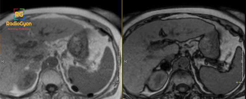
Chemical Shift Imaging (CSI) can distinguish benign adrenal adenomas from malignant lesions due to intracytoplasmic fat in adrenal adenomas. The signal intensity of lesions containing intracytoplasmic fat, such as adrenal adenomas, will “drop” signal on out-of-phase (also known as opposed phase) images. This can be subjectively observed in most cases, but in some cases, the signal drop may be subtle. In these cases, this adrenal MRI calculator can be used to detect signal loss on opposed phase images.
What formulas are used for adrenal signal dropout, and what are the cut-off values?
Adrenal-to-spleen CSI ratio = (Adrenal nodule SI out-phase / Spleen SI out-of-phase) / (Adrenal nodule SI in-phase / Spleen SI in-phase).
This calculates the drop in signal for the adrenal lesion and compares it with the drop in the spleen signal. Adrenal-to-spleen CSI ratio of less than 0.71 suggests a benign adrenal adenoma. In one study, the CSI ratio is better than the adrenal signal intensity index for lipid-rich adenomas. However, metastases were not included.
Adrenal signal intensity index (ASII) = (Adrenal nodule SI in-phase – Adrenal nodule SI out-of-phase) /Adrenal nodule SI in-phase X 100
This calculates the percentage drop-out in the lesion and is independent of the spleen signal drop-out. Adrenal signal intensity index (ASII) >16.5% at 1.5 Tesla (or >1.7% at 3 Tesla), suggests a benign adrenal adenoma.
What if these values are indeterminate?
If the Adrenal-to-spleen CSI ratio is greater than 0.71 or the Adrenal signal intensity index (ASII) is less than 16.5 %, the next best step is a dedicated adrenal protocol CT.
What are the limitations of this method in diagnosing adrenal adenomas?
- Other adrenal lesions containing intracytoplasmic fat, such as metastases from clear cell renal and hepatocellular carcinomas, can cause signal dropout on opposed phase images.
- The spleen can be unreliable in patients with iron deposition. In that case, kidneys can be used as a reference method. Liver and muscle are not ideal references as heterogeneous fatty infiltration can render these unreliable.
- Adrenal washout calculation on CT is better than chemical shift imaging.
- The signal-intensity index threshold of 16.5% is validated for 1.5 T. The threshold at 3T is 1.7%
What is adrenal mass protocol MRI?
Adrenal mass MRI protocol is an MRI examination of the abdomen tailored for evaluation of adrenal lesions. The most common indication is to distinguish a benign adrenal adenoma from other malignant lesions.
What is the key sequence for adrenal mass protocol MRI?
Chemical shift imaging (CSI) using in-phase and opposed phase images. Contrast enhanced sequences may not be needed if the lesion can be characterized on CSI imaging.
Disclaimer: The author makes no claims of the accuracy of the information contained herein; this information is for educational purposes only and is not a substitute for clinical judgment.
References:
- Schieda N, Siegelman E. Update on CT and MRI of adrenal nodules. AJR Am J Roentgenol 2017; 208:1-12
- Israel GM, Korobkin M, Hecht EN et al. Comparison of unenhanced CT and chemical shift MRI in evaluating lipid-rich adrenal adenomas. Am J Roentgenol 2004; 183:215-219
- Seo JM, Park BK, Park SY, Kim CK. Characterization of lipid-poor adrenal adenoma: chemical-shift MRI and washout CT. Am J Roentgenol 2014; 202:1043-50
- Blake MA, Cronin CG, Boland GW. Adrenal imaging. AJR Am J Roentgenol. 2010; 194 (6): 1450-60.
- Adam SZ, Nikolaidis P, Horowitz JM, et al. Chemical Shift MR Imaging of the Adrenal Gland: Principles, Pitfalls, and Applications. Radiographics. 2016;36(2):414-432. doi:10.1148/rg.2016150139

Improve Content And Help Your Colleagues!
Found an error in the post? Please let us know using our contact page and we will update it with due credits!
If you wish to contribute radiological images for this case (or any other article on the website), you can submit them here and you will be duly credited: Submit radiology case
About the Author
Dr. Amar Udare, MD, DNB
Dr. Udare holds an MBBS and MD degree, and his expertise lies in the field of radiology. He has authored multiple peer-reviewed publications, contributing significantly to the medical field. His works can be accessed on PubMed and Google Scholar.
In addition to his academic and professional achievements, Dr. Udare is an avid reader and enjoys exploring the latest advancements in medical technology. His commitment to making complex medical knowledge accessible to patients and the general public aligns with our mission at RadioGyan.com.
For any further questions or clarifications, feel free to reach out to Dr. Udare via the contact form.